General Topography
-
T1 Thoracic Vertebra – Highest part of sloping Thoracic Inlet
-
Cervical column rises above gently convex forwards
-
Extensor muscles lie behind vertebrae
- Supplied segmentally by Posterior Rami of Cervical Nerves emerging from Intervertebral Foramina
-
Flexor muscles (smaller amount)
- Lies in front of the Vertebrae
- Covered by Prevertebral Fascia
- Lies behind the Pharynx
- Supplied segmentally by Anterior Rami
-
C1 Vertebra level:
- Hard Palate
-
C2 Vertebra level: Tip of Chin/Jaw
-
Between C2 and C3:
- Lower border of the Mandible
-
C4 Vertebra level: Bifurcation of Common Carotid Artery
-
Pharynx extends from Base of Skull to level of Cricoid Cartilage (C6)
- Then continues on as Oesophagus
-
C6 Vertebra level: Omohyoid crossing Common Carotid
-
Larynx and Trachea:
- Anterior to lower Pharynx and upper Oesophagus
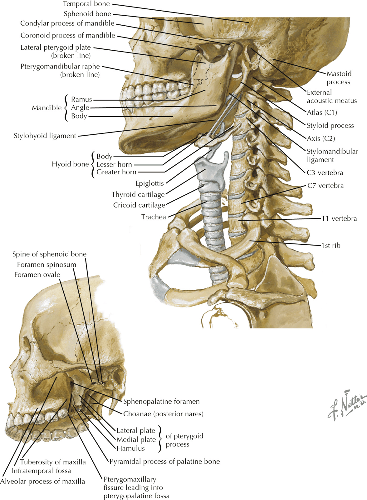
- Hyoid Bone lies above Larynx at C3 Vertebra level
- Connected to Mandible via Mylohyoid muscles (at the upper limit of anterior part of neck and form floor of mouth)
- Connected to Mandible via Mylohyoid muscles (at the upper limit of anterior part of neck and form floor of mouth)
-
Carotid Sheath:
- On each side of the Pharynx
- Contains Common and Internal Carotid Arteries, Internal Jugular Vein with Cervical Sympathetic Trunk behind
- External Carotid Artery lies outside the Sheath
-
- 9th, 10th, 11th and 12th Cranial Nerves descend into the neck
- 9th and 12th pass forwards to Oropharynx and Tongue
- 11th runs backwards to Sternocleidomastoid and Trapezius muscles
- 10th continues down in Carotid Sheath
Fascia of the neck
Superficial cervical fascia
- The contents of the superficial cervical fascia will vary depending upon location but include:
- Platysma (and cervical branch of facial nerve that innervate the muscle)
- Cutaneous nerves of the cervical plexus:
- Lesser occipital
- Great auricular
- Transverse cervical (largest sensory contribution)
- Supraclavicular
- Deep to the superficial cervical fascia is the investing (or superficial) layer of deep cervical fascia
Superficial Structures
Platysma
- Lies superficial to Investing Layer of Deep Cervical Fascia
- Extends from fascia over upper parts of Pectoralis Major and Deltoid to lower border of Mandible
- Some fibres extend into face blending with fascial expression muscles
- Covers External and Anterior Jugular Veins
- Separated below but converge above towards midline just beneath chin
- Nerve Supply:
- Cervical Branch of Facial Nerve
- Afferent (proprioceptive) fibres run with Transverse Cervical Nerve
- Action:
- Involved in facial expression
- May assist in mouth opening
Anterior Jugular Veins
- Commence beneath chin from the confluence of the submental and submandibular veins
- Side by side beneath Platysma
- Reaches Suprasternal region
- Pierce Deep Fascia and come to lie in Suprasternal Space (often connected by a short anastomotic vein - the jugular venous arch)
- Angles laterally to pass deep to Sternocleidomastoid but superficial to Strap muscles
- Opens into the External Jugular Vein near its termination
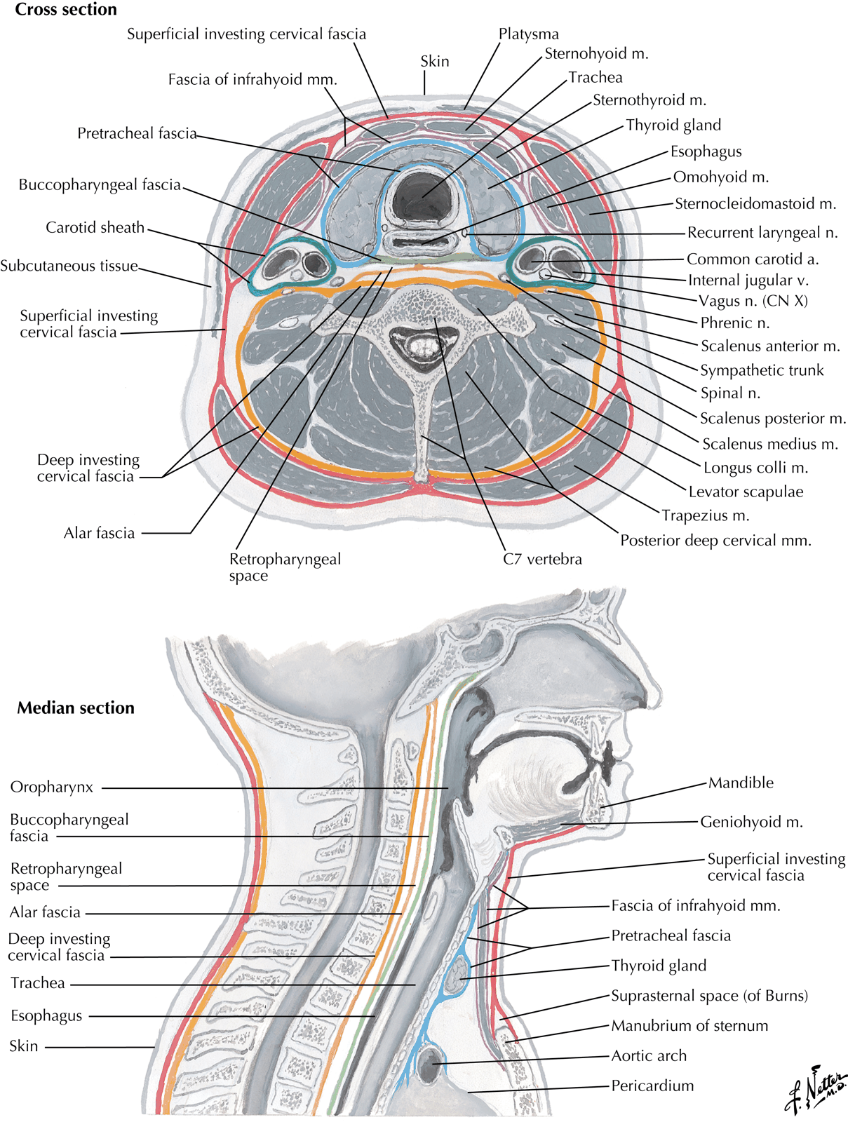
Deep Cervical Fascia
- Four parts:
- Superficial (Investing) Layer
- Pretracheal Fascia
- Posterior portion called the buccopharyngeal fascia - technically not part of the the pretracheal fascia but is continuous with it
- Prevertebral Fascia
- Alar fascia is a delamination of the prevertebral fascia that swings forward to attach to the buccopharyngeal fascia approximately at the C7 vertebral level.
- Carotid Sheath
1. Investing Layer
- Superficial investing fascia of the deep cervical fascia - confusing as there is also a superficial cervical fascia (above)
- Surrounds neck like a collar
- Splits around Sternocleidomastoid and Trapezius
- NB: Investing layer does NOT enclose the infra-hyoid muscles
- Posteriorly, blends with Ligamentum Nuchae (attached to spines of Cervical Vertebrae)
- Anteriorly – attached to Hyoid bone
- Above – attached to lower border of the Mandible
- Also attached to:
- Superior Nuchal Line
- External Occipital Protuberance at Base of Skull
- Encloses Parotid Gland between the Angle of the Mandible and the Tip of the Mastoid Process
2. Prevertebral Fascia
- Lies in front of the Prevertebral muscles
- Extends from Base of Skull downwards to blend with Anterior Longitudinal Ligament of T4 Vertebral Body in the Superior Mediastinum
- Alar fascia is a delamination of the prevertebral fascia that swings forward to attach to the buccopharyngeal fascia approximately at the C7 vertebral level.
3. Pretracheal Fascia
- Deep to Infrahyoid Strap muscles (Sternothyroid, Sternohyoid, Omohyoid)
- Upward attachment limited by respective attachments of those muscles
- I.e. Body of Hyoid Bone and Oblique Line of Thyroid Cartilage
- Splits to enclose Thyroid Gland
- Not adherent to Thyroid except:
- At back of Isthmus – attached to 2nd, 3rd and 4th Tracheal Rings
- Laterally – fuses with front of Carotid Sheath on deep surface of Sternocleidomastoid
- Inferiorly – passes behind Brachiocephalic Veins to blend with Adventitia of Aortic Arch and Fibrous Pericardium
- Thus, neck infections in front of the Pretracheal Fascia is directed into the anterior mediastinum
- Part of Cervical Visceral Fascia surrounding Pharynx, Oesophagus, Larynx and Trachea
- This is the buccopharyngeal fascia
4. Carotid Sheath
- Feltwork of areolar tissue
- Surrounds Common and Internal Carotid Arteries, Internal Jugular Vein, Vagus Nerve, and Deep Cervical Lymph Nodes
- Common Carotid Artery is medial to the Internal Jugular Vein
- Vagus Nerve deeply place between the two vessels
- Common Carotid Artery is medial to the Internal Jugular Vein
Strap Muscles
- a.k.a. Infrahyoid Muscles
- Consist of Sternohyoid, Omohyoid, Sternothyroid, Thyrohyoid
| Muscle | Origin | Insertion | Actions | Nerve Supply |
|---|---|---|---|---|
| Omohyoid (superior and inferior bellies connected by a tendon) | Superior border of the scapula | Body of the hyoid | Depresses the hyoid | Ansa cervicalis |
| Sternohyoid | Manubrium | Body of the hyoid | Depresses the hyoid | |
| Sternothyroid | Manubrium | Oblique line of the thyroid cartilage | Depresses the larynx | |
| Thyrohyoid | Oblique line of the thyroid cartilage | Greater cornu (horn) of the hyoid | Depresses the hyoid | C1 (ventral ramus, which follows the hypoglossal n.) |
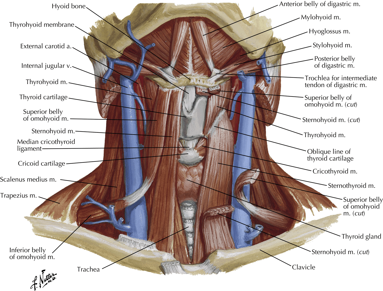
Sternohyoid
- Attachments:
- Back of upper part of Manubrium and adjoining Sternoclavicular Joint and Clavicle
- Lower border of Body of Hyoid
- Lie edge-to-edge at Hyoid bone and diverge from each other below
- Nerve supply:
- Branch from Ansa Cervicalis
- Enters lower part of muscle
Omohyoid
- Attachments:
- Lateral part of inferior border of Hyoid
- Complex course as detailed below
- Upper border of Scapula and Transverse Scapular Ligament
- Course:
- Lies edge-to-edge with Sternohyoid at its attachment
- Diverges away from Sternohyoid as it descends
- Passes deep to Sternocleidomastoid
- Lie over Carotid Sheath
- Muscle fibres replaced by flat tendon where it lies over the Internal Jugular Vein
- Inferior Belly runs almost horizontally above the Clavicle
- Intermediate Tendon and Supraclavicular portion of muscle bound down close to Clavicle by fascial sling derived from deep layer of the Investing Layer of the Deep Cervical Fascia
- Nerve supply:
- Superior Belly: Superior Root of the Ansa Cervicalis
- Inferior Belly: Ansa Cervicalis
Thyrohyoid
- Lies under cover of upper ends of Sternohyoid and Omohyoid
- Attachments:
- Greater Horn of Hyoid bone
- Oblique Line of Thyroid Cartilage (alongside Sternothyroid)
- Nerve supply:
- Branch of Hypoglossal Nerve – but fibres are all ‘hitch-hiking’ from C1
Sternothyroid
- Lies deep to Sternohyoid
- Attachments:
- Posterior surface of Manubrium (lower down than Sternohyoid) and adjacent 1st Costal Cartilage
- Oblique Line of Thyroid Cartilage
- Nerve supply:
- Ansa Cervicalis – gives a branch to lower part of muscle
Actions of Infrahyoid Muscles
- Depress Larynx
- Sternothyroid acts directly on Thyroid Cartilage
- Others act indirectly via Hyoid
- Increases volume of resonating chambers during phonation and affects quality of voice
- Oppose Larynx elevators
- I.e. Mylohyoid, Palatopharyngeus, Stylopharyngeus, Salpingopharyngeus
- Enables them to act progressively and gradually
- Prevent ascent of Hyoid Bone when Digastric and Geniohyoid lower Mandible
Suprahyoid Muscles
| Muscle | Origin | Insertion | Actions | Nerve Supply |
|---|---|---|---|---|
| Stylohyoid | Styloid process | Body of the hyoid | Elevates the hyoid Retracts the hyoid | Facial n. |
| Mylohyoid | Mylohyoid line of the mandible | Mylohyoid of opposite side at the raphe Body of the hyoid | Elevates the hyoid Elevates the floor of the oral cavity | Trigeminal n. (mandibular division) |
| Digastric Muscle (posterior and anterior bellies connected by a tendon attached to the hyoid) | Mastoid process | Digastric fossa of the mandible | Elevates hyoid Helps depress and retract the mandible | Facial n. (posterior belly) Trigeminal n. (anterior belly—mandibular division) |
| Geniohyoid | Inferior genial tubercle | Body of the hyoid | Helps move the hyoid and tongue anteriorly | C1 (ventral ramus, which follows the hypoglossal n.) |
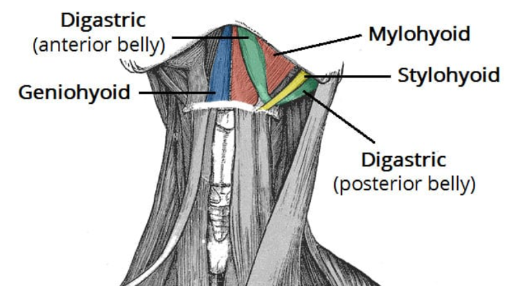
Digastric Muscle
- Arises as the Posterior Belly from the Digastric Groove on medial surface of base of Mastoid Process
- Tapers down to Intermediate Tendon held beneath a fibrous sling attached to junction of body and Greater Horn of Hyoid bone
- Anterior Belly lies on inferior surface of Mylohyoid and connects to the Digastric Fossa on the inner surface of the mandible near the midline
- Nerve Supply:
- Posterior Belly – Facial Nerve (by a branch arising between the Stylomastoid Foramen and Parotid Gland)
- Anterior Belly – Nerve to Mylohyoid
- Action: Depress and retract the chin, and assist Lateral Pterygoid in opening mouth
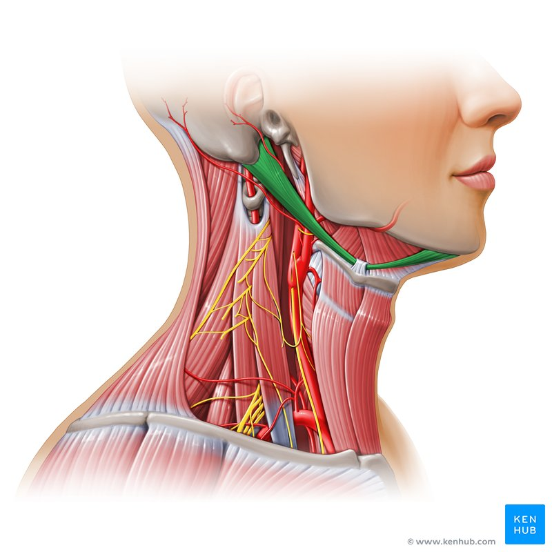 The posterior digastric is supplied by facial nerve and crosses (from posterior to anterior) the following:
 - Accessory nerve
- Transverse process of the atlas
- Internal jugular/internal carotid
- Hypoglossal nerve
- External carotid
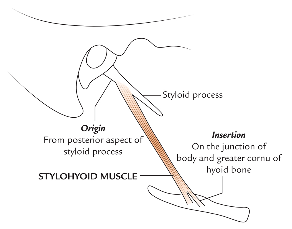
Stylohyoid
- Arises from back of Styloid Process
- Lower end divides to embrace the Digastric Tendon
- Inserted by 2 slips into the junction of the Greater Horn and Body of Hyoid
- Nerve Supply: Facial nerve
- Action: Retract and elevate Hyoid when swallowing
Mylohyoid
- Forms diaphragm of floor of the mouth
- Arises from Mylohyoid Line on inner aspect of Mandible
- Posterior quarter of each side inserts onto body of Hyoid
- Anterior three quarters interdigitate in a midline raphe extending from chin to Hyoid bone
- Nerve Supply: Own nerve – Branch of the Inferior Alveolar Nerve (From Mandibular Division of Trigeminal Nerve) – Also supplies anterior belly of Digastric
- Action: Contraction elevates the ‘gutter’ of the mouth causing tongue and hyoid bone elevation
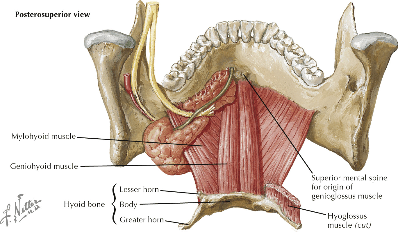
Geniohyoid
- Extends from Inferior Mental Spine (Genial Tubercle) of Mandible to body of Hyoid Bone
- Nerve Supply: Branch from Hypoglossal Nerve (consisting of fibres from C1)
- Action: Protract and elevate Hyoid Bone in swallowing or, if the Hyoid is fixed, to depress the Mandible
Triangles of the Neck
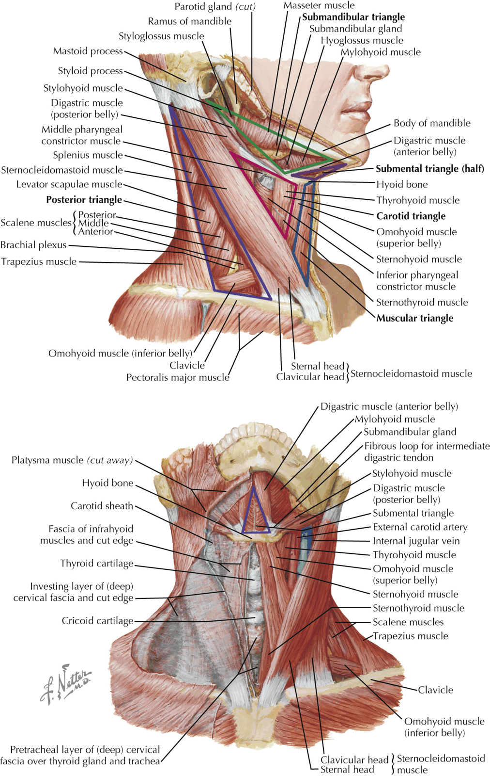
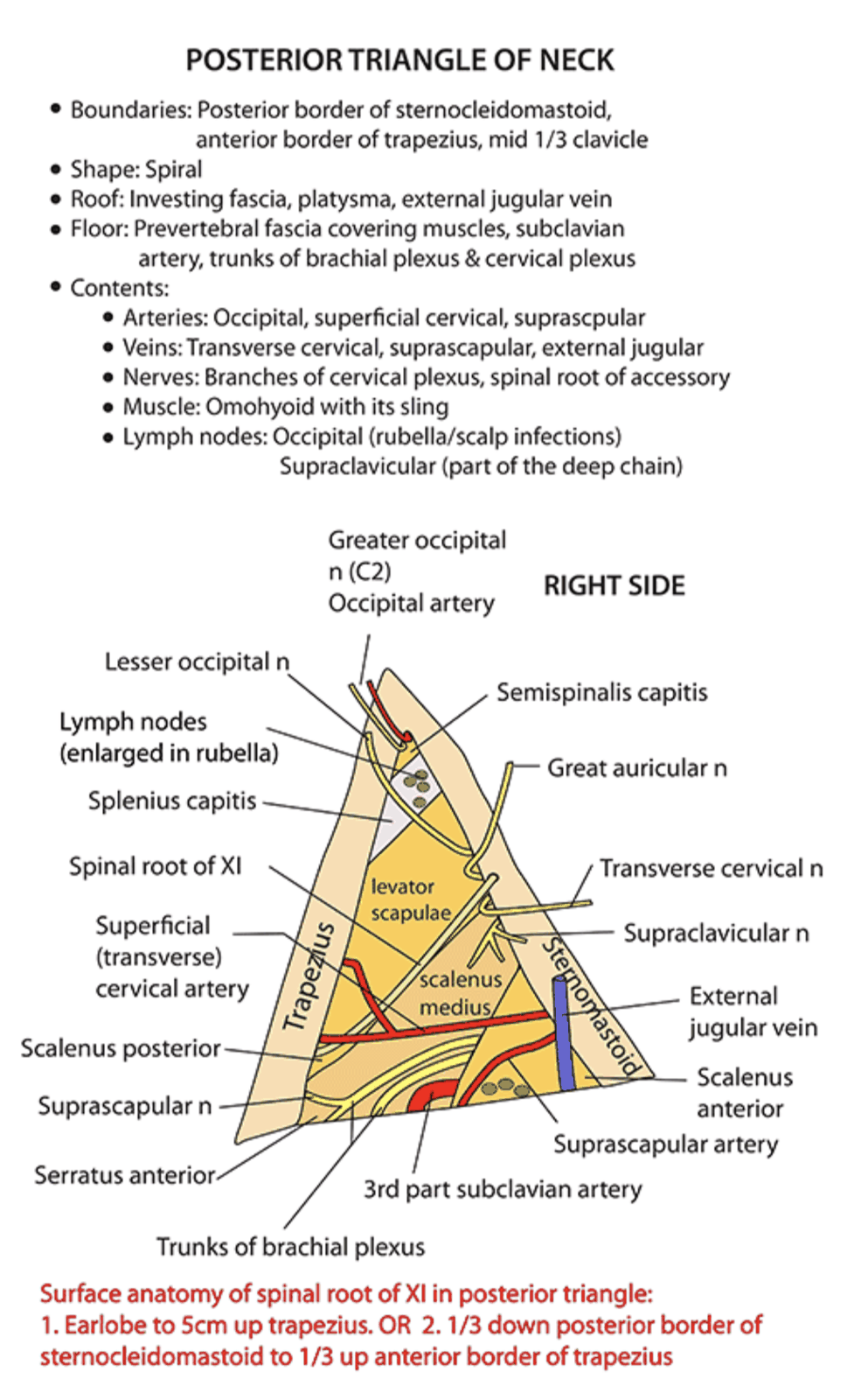
Posterior Triangle
- Boundaries:
- Posterior border of Sternocleidomastoid
- Anterior border of Trapezius
- Clavicle
- Roof:
- Investing Layer of the Deep Cervical Fascia
- Floor:
- Prevertebral Fascia lying on (from above downwards):
- Splenius
- Levator Scapulae
- Scalenus Medius
- Scalenus Anterior and the 1st digitation of Serratus Anterior may contribute to the floor
- Prevertebral Fascia lying on (from above downwards):
- Apex lies high up at the back of the skull on the Superior Nuchal Line
- Contents:
- Nerves
- [[Spinal Accessory Nerve
- Cervical Plexus (Branches)
- Lesser Occipital Nerve
- Greater Auricular Nerve
- Transverse Cervical Nerve
- Supraclavicular Nerve
- Deep to the Prevertebral fascia
- Brachial Plexus (Three Trunks)
- Arteries
- Subclavian Artery (3rd part)
- Transverse Cervical
- Suprascapular artery
- Occipital Artery
- Lymph Nodes
- Lie between the roof and floor
- 2-3 Occipital Nodes in the subcutaneous tissue at the apex
- Several Supraclavicular Nodes above the clavicle (Really are outlying members of the lower group of Deep Cervical Nodes)
- Muscles
- Omohyoid Muscle (Inferior Belly)
- Anterior, middle and posterior scalene
- Levator scapulae
- Vein
- External Jugular Vein
- Nerves
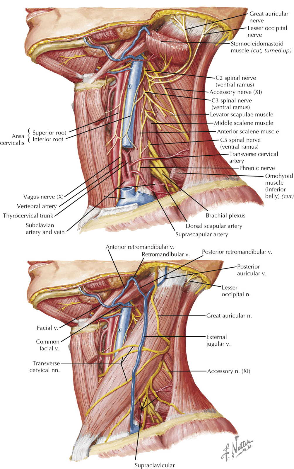
Anterior Triangle
- Boundaries:
- Anterior border of Sternocleidomastoid
- Lower border of the Mandible
- The midline
- Subdivided into Carotid Triangle, Digastric Triangle, Submental Triangle, and Muscular Triangle
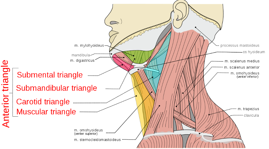
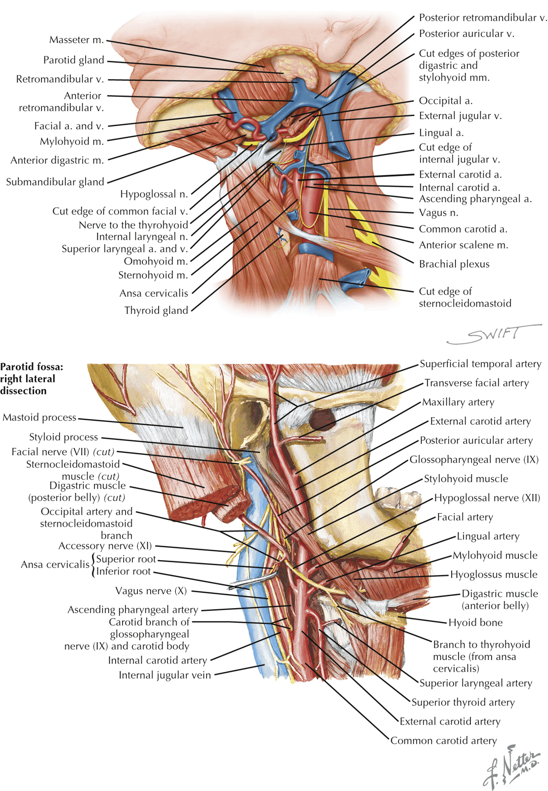
Carotid Triangle
- Boundaries:
- Sternocleidomastoid
- Posterior Belly of Digastric Muscle
- Superior Belly of Omohyoid
- Contents:
- Artery
- Bifurcation of Common Carotid Artery into internal and external
- Branches of External Carotid Artery (except Posterior Auricular)
- Veins
- Internal jugular vein
- Lingual Vein
- Facial Vein
- Superior Thyroid Veins
- Nerves:
- Hypoglossal Nerve
- Vagus Nerve
- Internal and External Laryngeal Nerves
- Superior Root of the Ansa Cervicalis
- Spinal accessory nerve
- Sympathetic trunk
- Lymph Nodes
- Artery
Submandibular Triangle
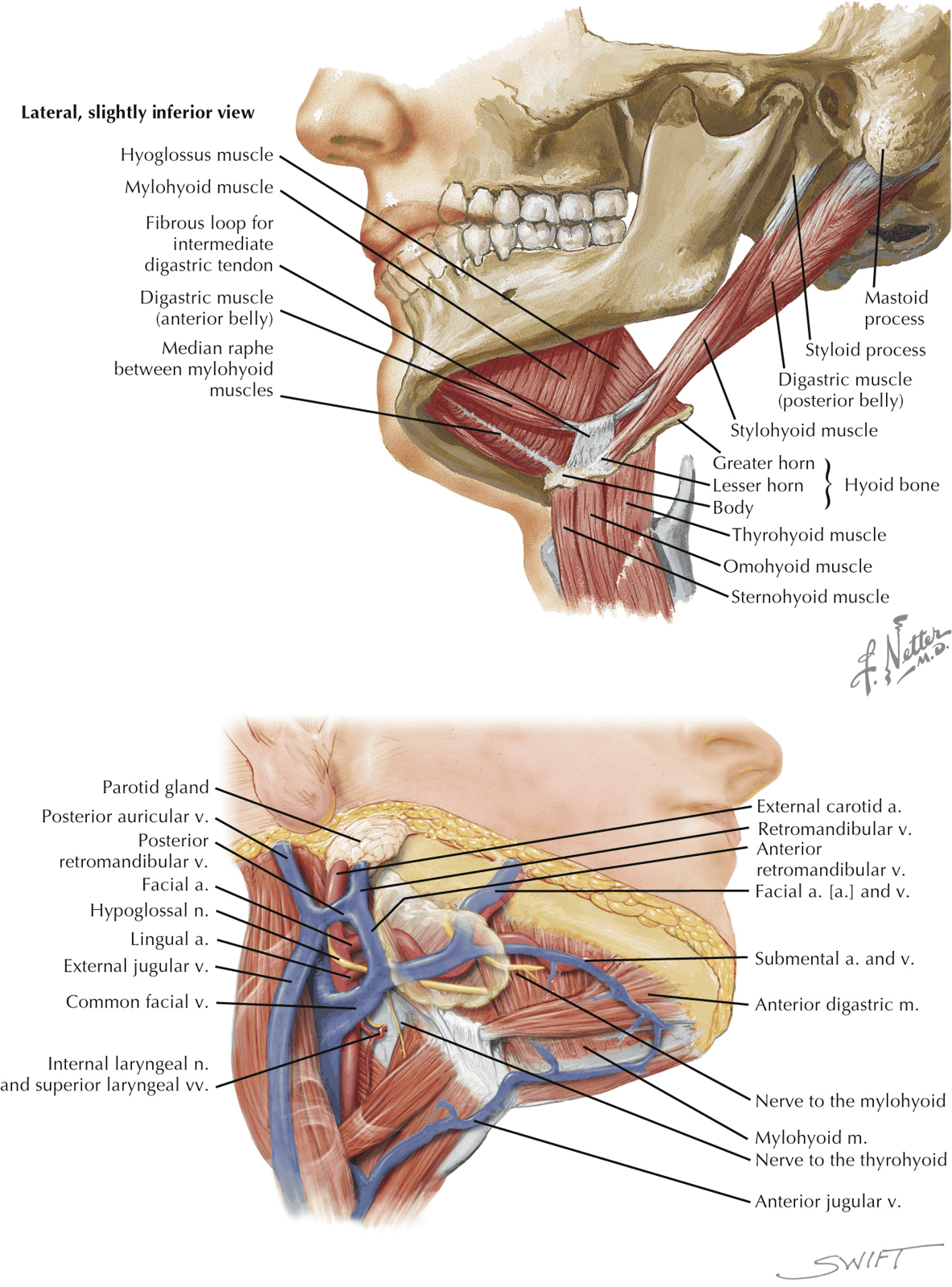
- Boundaries:
- Mandible
- Anterior and Posterior Bellies of Digastric
- Contents:
- Submandibular Gland
- Submandibular Lymph Nodes
- Facial artery and vein
- Submental Vessels
- Mylohyoid Vessels
- Hypoglossal Nerve
- Mylohyoid Nerve
Submental Triangle
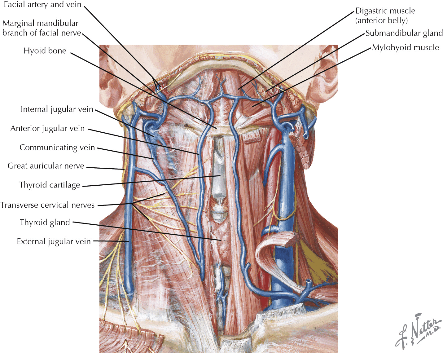
- Boundaries:
- Anterior Bellies of Digastric
- Body of Hyoid (Triangle crosses midline)
- Contents:
- Anterior Jugular Veins
- Lymph Nodes
Muscular Triangle
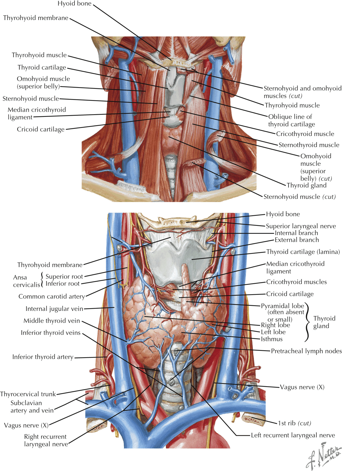
- Boundaries:
- Sternocleidomastoid
- Superior Belly of Omohyoid
- Midline from Hyoid to Jugular Notch
- Contents:
- Parts of Larynx
- Trachea
- Pharynx
- Oesophagus
- Thyroid and Parathyroid Glands
- Their Vessels
- Their Nerves
- Lymph Nodes
Surgical Lymph Node Levels of the Neck
- Neck Lymph Nodes are divided into 7 levels
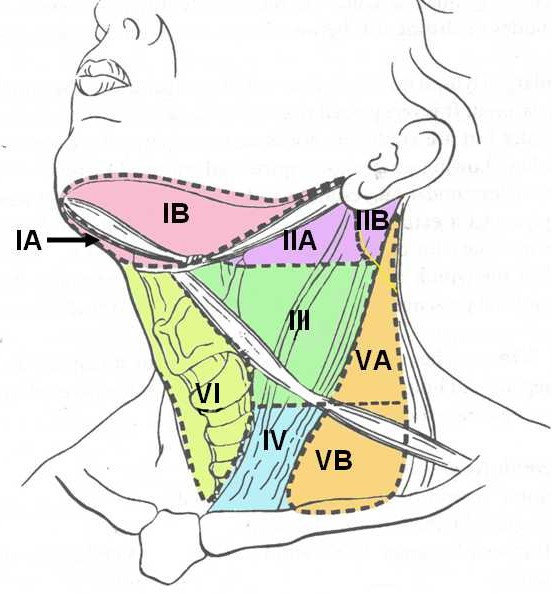
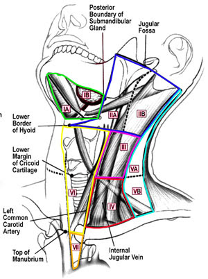
Level 1: Submental and Submandibular Nodes
- Boundaries:
- Level IA (Submental): (in the midline)
- Lateral (bilaterally): Anterior Digastric muscles
- Inferior: Hyoid
- Superior: Mandible
- Level IB (Submandibular):
- Anterior: Anterior Digastric muscle
- Posterior: Posterior Digastric muscle
- Lateral: Posterior edge of Submandibular Glands
- Superior: Mandible
- Level IA (Submental): (in the midline)
- Important Structures/Relationships:
- Lingual Nerve
- Hypoglossal Nerve
- Submandibular Duct
- Facial Artery
- Facial Vein
- Only structure found lateral to the Posterior Belly of Digastric
- Marginal Mandibular Branch of Facial Nerve
- Found in the fascia overlying the Submandibular Gland superficial to the Facial Artery and Vein
- Sites Draining:
- Floor of mouth
- Anterior Tongue
- Lower Lip
- Submandibular Gland
- Nasopharynx
Level 2: Upper Jugular Nodes
- Boundaries:
- Anterior: Posterior edge of Submandibular Glands
- Posterior: posterior border of the sternocleidomastoid muscle
- Superior: skull base
- Inferior: Level of the hyoid bone (clinical), carotid bifurcation (surgical)
- Level II is divided by the accessory nerve into levels IIa (anteriorly) and IIB (posteriorly).
- Important Structures
- Spinal Accessory Nerve (CN XI)
- Runs deep to Posterior Digastric and Occipital Artery
- Runs superficial to IJV for majority of the time
- Often gives small branch to Trapezius before entering SCM
- Spinal Accessory Nerve (CN XI)
- Sites Draining:
- Oropharynx
- Nasopharynx
- Nasal Cavity
- Larynx
- Parotid Gland
Level 3: Middle Jugular Nodes
- Boundaries:
- Lateral: Posterior border of SCM
- Medial: Lateral border of Sternohyoid
- Superior: Hyoid Bone
- Inferior: Inferior border of Cricoid Cartilage
- Important Structures:
- Phrenic Nerve
- Runs deep to the cervical rootlets and superficial to the Anterior Scalene muscle beneath the Deep layer of the Deep Cervical Fascia
- Phrenic Nerve
- Sites Draining:
- Oral Cavity
- Nasopharynx
- Hypopharynx
- Larynx
Level 4: Lower Jugular Nodes
- Boundaries:
- Lateral: Posterior border of SCM
- Medial: Lateral border of Sternohyoid
- Superior: Inferior border of Cricoid Cartilage
- Inferior: Clavicle
- Important Structures:
- From superficial to deep, the structures encountered:
- Omohyoid
- Carotid Sheath
- Thoracic Duct (more commonly seen in the left neck)
- Transverse Cervical Artery
- Phrenic Nerve
- Anterior Scalene muscle
- Lung Apices may also be present at inferior aspect of Level 4
- From superficial to deep, the structures encountered:
- Sites Draining:
- Larynx
- Hypopharynx
- Thyroid
- Cervical Oesophagus
Level 5: Posterior Triangle Nodes
- Boundaries:
- Lateral: Trapezius
- Medial: Lateral border of SCM
- Inferior: Clavicle
- Horizontal line extended from inferior border of Cricoid divides this level into 5A (superior to Cricoid) and 5B (inferior to Cricoid)
- Important Structures:
- Phrenic Nerve
- Embedded in fascia overlying Anterior Scalenes
- Protected by staying superficial to this plane
- Brachial Plexus
- Courses on the floor of level 5B between Anterior and Middle Scalene
- Phrenic Nerve
- Sites Draining:
- Nasopharynx
- Oropharynx
- Skin of Posterior Scalp and Neck
- Virchow’s Node
Level 6: Central Compartment Nodes
- Boundaries:
- Lateral: lateral border of the sternohyoid muscle
- Superior: Hyoid Bone
- Inferior: Sternum
- Divided into left and right by the Trachea
- Important Structures:
- Recurrent Laryngeal Nerve
- Thyroid Gland
- Parathyroid Glands
- Sites Draining:
- Thyroid
- Subglottic Larynx
- Cervical Trachea
- Hypopharynx
- Cervical Oesophagus
Level 7: Superior Mediastinal Nodes
- Boundaries:
- Superior: Inferior border of Suprasternal Notch
- Inferior: Innominate Artery/Brachiocephalic Artery
Root of the Neck Anatomy
| Muscle | Insertion | Origin | Innervation | Action |
|---|---|---|---|---|
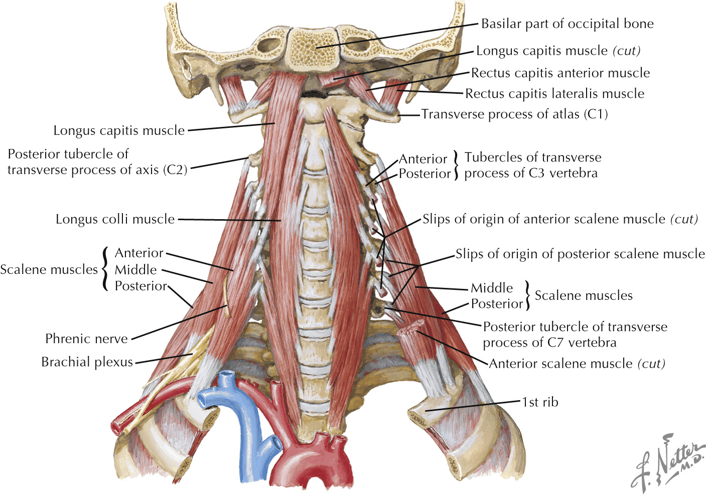
| Posterior scalene | 2nd rib | Posterior tubercles of transverse processes of C4-C6 | C5-C8 | Flexes neck laterally; elevates 2nd rib |
| Middle scalene | 1st rib | Posterior tubercles of transverse processes of C2-C7 | C3-C7 | Flexes neck laterally; elevates 1st rib |
| Anterior scalene | 1st rib | Anterior tubercles of transverse processes of C3-C6 | C5-C8 | Flexes neck laterally; elevates 1st rib |
| Overview |
- Root of Neck (a.k.a. Thoracic Outlet)
- Bounded by T1 Vertebrae, 1st pair of Ribs and their Cartilage, and the Manubrium of the Sternum
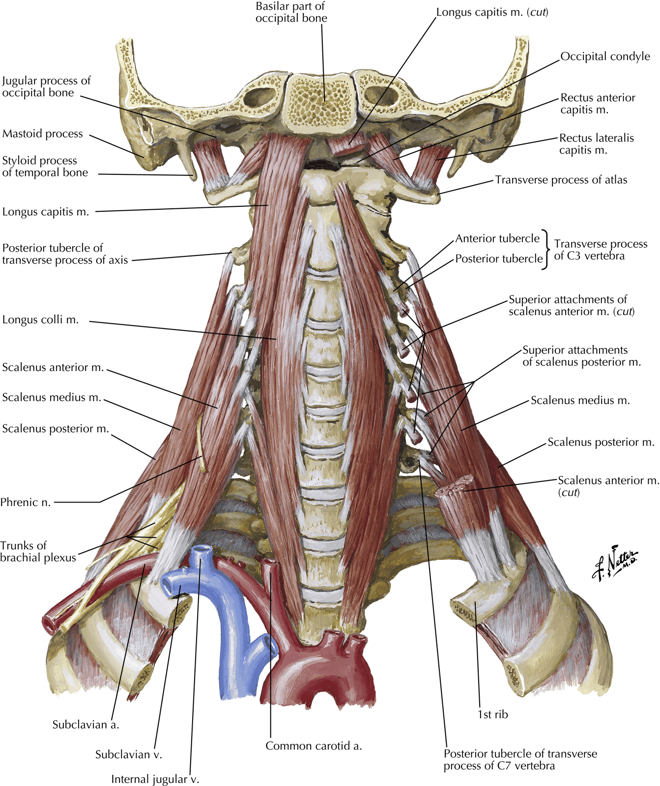
Scalenus Anterior
- Origin:
- Anterior Tubercles of the four ‘typical’ Cervical Vertebrae (C3 – C6) by 4 slender tendons of origin which lie end to end with those of Longus Capitis
- Muscle passes forwards, laterally and downwards to end in a narrow tendon
- Insertion:
- Scalene Tubercle and adjacent ridge on the inner border and upper surface of the 1st Rib
- Nerve Supply:
- Separate branches from the Anterior Rami of C4-C6 nerves
- Actions:
- Assists in Flexion and Rotation of the Neck
- Helps to stabilize the 1st Rib
- Shows some electromyographic activity even in quiet respiration
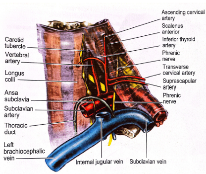
Anterior Relations
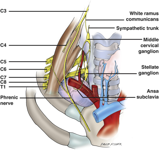
- Phrenic Nerve
- Passes vertically down across the obliquity of the muscle
- Plastered on the muscle by the Prevertebral Fascia and a pad of fat lies in front of the Prevertebral Fascia
- Nerve leaves the medial border of the muscle low down
- Crosses anterior to the Subclavian Artery and its Internal Thoracic Artery Branch
- Behind the Subclavian Vein
- Occasionally, the Phrenic Nerve may pass in front of the Subclavian Vein or posterior to the Internal Thoracic Artery
- Lies on the Suprapleural membrane
- Passes medial to the Apex of the Lung
- Crosses in front of the Vagus Nerve as it enters the Superior Mediastinum
- Ascending Cervical Artery
- Branch of the Inferior Thyroid Artery or the Thyrocervical Trunk
- Runs up on the Prevertebral Fascia medial to the Phrenic Nerve
- Transverse Cervical Artery and Suprascapular Artery
- Anterior to the Prevertebral Fascia
- Lie between the Anterior Scalene and the Carotid Sheath (Internal Jugular Vein)
- Subclavian Vein
- Lies in a groove on the 1st Rib
- Due to the slope, lies at a lower level than the insertion of Scalenus Anterior
- Runs medially
- Joins the Internal Jugular Vein at the medial border of Scalenus Anterior to form the Brachiocephalic Vein
- Thoracic Ducton the left and the Right Lymphatic Duct on the right enters the angle of confluence of the 2 veins
- Used for central venous lines:
- Infraclavicular approach – from a point 2cm below the midpoint of the Clavicle along a line that passes behind the Clavicle towards the Jugular Notch of the Sternum
- Needle pierces the Clavipectoral Fascia and enters the vein just behind the Fascia
- Lies in a groove on the 1st Rib
Medial Relations
- Medial edge of Scalenus Anterior makes a pyramidal shape with the lateral border of the lower part of Longus Colli
- Prevertebral Fascia in front of these muscles attached to bone at their opposing margins
- No fascial roof across the Pyramidal Space
- Base of space: Subclavian Artery (1st Part) (lying on Suprapleural Membrane)
- Apex: Carotid (Chassaignac’s) Tubercle on the Transverse Process of C6
- Common Carotid Artery:
- Medial to the Internal Jugular Vein:
- Lies deep to Sternocleidomastoid immediately anterior to the Pyramidal Space
- Inferior Cervical Sympathetic (or Stellate ganglion):
- Posterior to the Common Carotid Artery (and Carotid Sheath)
- Vertebral Artery and Vein anterior to the ganglion
- Inferior Thyroid Artery
- Arches medially in a bold curve whose upper convexity lies in front of the Apex of the Pyramidal Space (C6 Level)
- Sympathetic Chain (usually the Middle Ganglion) in front of the Artery
- Thoracic Duct (or Right Lymphatic Duct)
- At a lower level and further anterior
- Makes a similar convexity behind the Carotid Sheath
- Arches over the Lung Apex and Subclavian Artery
- Enters the confluence of the Subclavian Vein and Internal Jugular Vein
- Vagus Nerve (slightly medial - can also be considered anterior)
- Within the Carotid Sheath
- Passes down anterior to the Subclavian Artery
- On the right side, gives off the Right Recurrent Laryngeal Nerve
- Hooks around the artery and passes upwards
- On the right side, gives off the Right Recurrent Laryngeal Nerve
- Inclines posteriorly and run on the medial surface of the Apex of the Lung to enter the Superior Mediastinum
- Internal Jugular Vein has Deep Cervical Lymph Nodes closely adjacent to it
Posterior Relations
- Separated from the Scalenus Medius by:
- Subclavian Artery
- Anterior rami of the lower Cervical and 1st Thoracic Nerves, i.e. Roots of Brachial Plexus
- Subclavian Artery(2nd Part)
Lateral Relations
- Trunks of the Brachial Plexus:
- Emerge from the lateral border of Anterior Scalene
- Subclavian Artery (3rd Part) – see below
- Both the above lie behind the Prevertebral Fascia on the floor of the Posterior Triangle
Subclavian Artery
- See notes
Cervical Plexus
- See notes