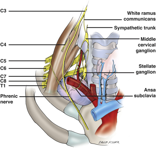
- Passes vertically down across the obliquity of the Scalenus Anterior
- Plastered on the muscle by the Prevertebral Fascia and a pad of fat lies in front of the Prevertebral Fascia
- Nerve leaves the medial border of the muscle low down
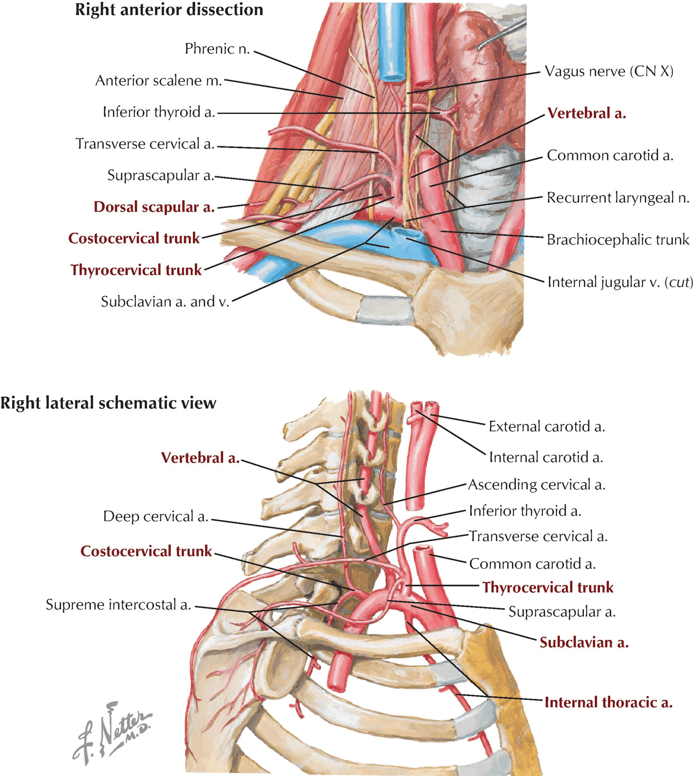
- Crosses anterior to the Subclavian Artery and its Internal Thoracic Artery Branch
- Behind the Subclavian Vein
- Occasionally, the Phrenic Nerve may pass in front of the Subclavian Vein or posterior to the Internal Thoracic Artery
- Lies on the Suprapleural membrane
- Passes medial to the Apex of the Lung
- Crosses in front of the Vagus Nerve as it enters the Superior Mediastinum
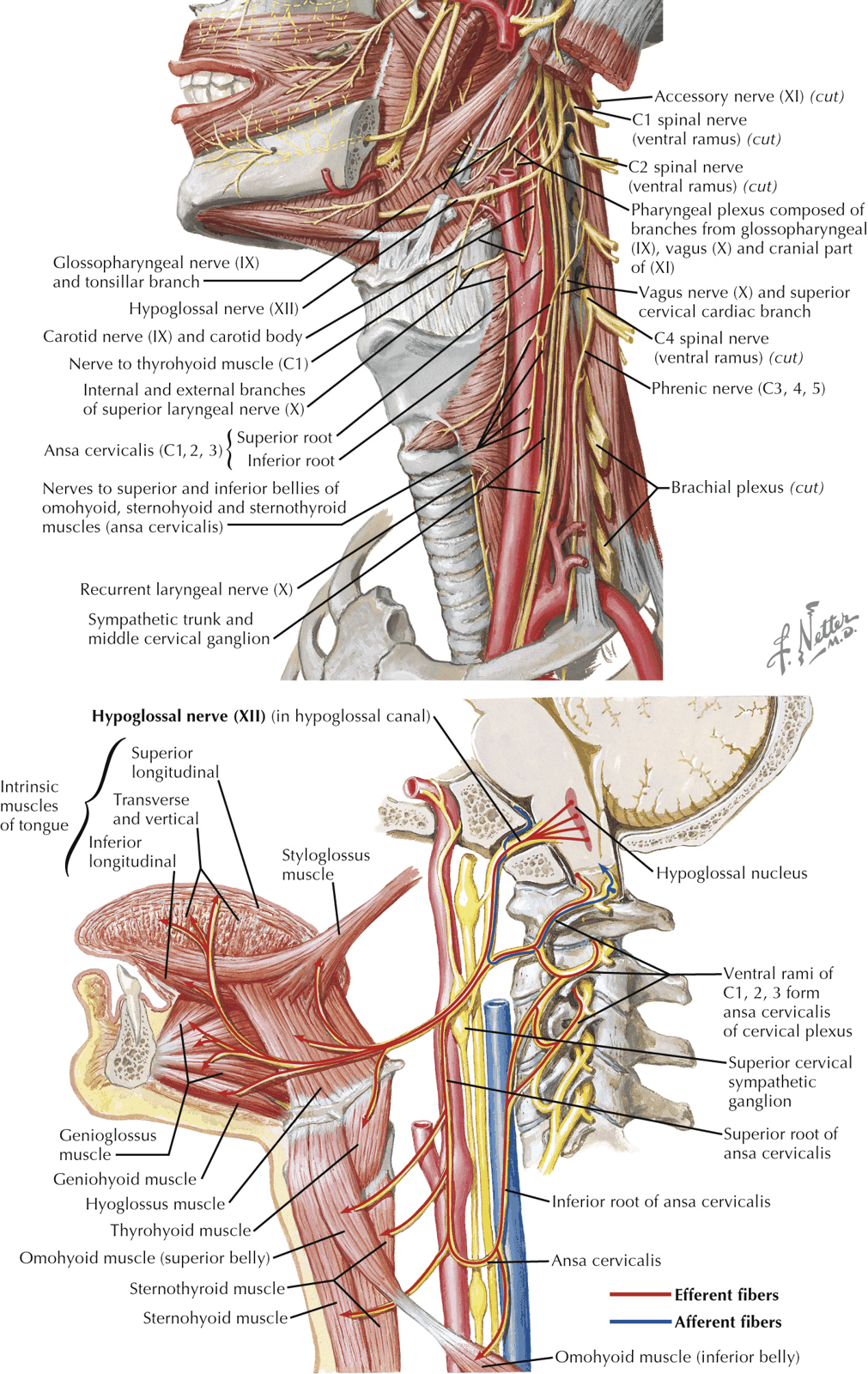
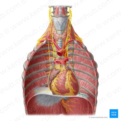
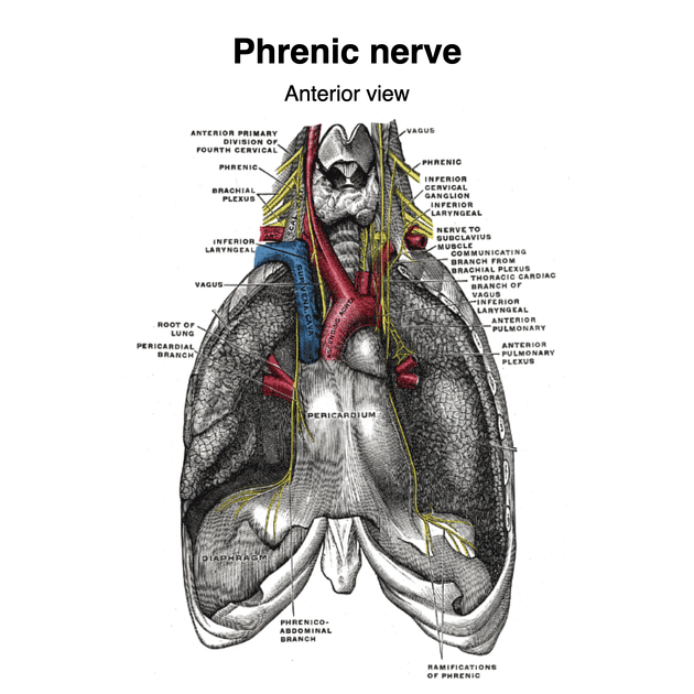
- Arrises from the Cervical Plexus
- Left and Right Phrenic Nerves (C3, 4, 5 but predominantly C4) – sole motor nerve supply
- Course of Phrenic Nerves:
- Arise principally from C4 in the neck
- Passes down over the Anterior Scalene muscle
- Deep to the Prevertebral Fascia
- Passes from lateral to medial over the Anterior Scalene surface
- Crosses in front of the Subclavian Artery and its Internal Thoracic Artery branch
- Across dome of the pleura behind the Subclavian Vein
- Can sometimes pass anterior or even through the Subclavian Vein
- May contain C5 fibres that pass anterior to the Subclavian Vein
- Can sometimes pass posterior to the Internal Thoracic Artery
- Lies lateral to the Ascending Cervical Artery (branch of the Inferior Thyroid Artery or Thyrocervical Trunk)
- Ascending Cervical Artery runs up on the Prevertebral Fascia
- Crosses anterior to the Vagus
- Runs through the mediastinum in front of the lung root
- Each nerve is in contact laterally with the mediastinal pleura throughout the entire course
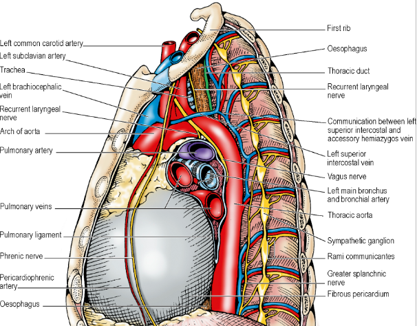
- Right Phrenic Nerve
- Related medially with venous structures throughout its thoracic course
- Right Brachiocephalic Vein, Superior Vena Cava, Pericardium over the Right Atrium, and the Inferior Vena Cava
- i.e. The Right Phrenic Nerve is lateral to the above structures
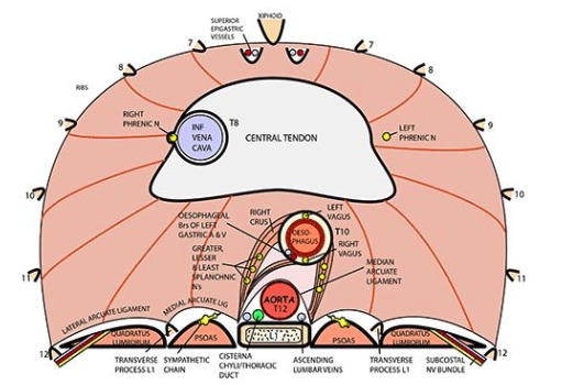
- Reaches under-surface of Diaphragm by piercing the Central Tendon adjacent to the Caval Opening
- Related medially with venous structures throughout its thoracic course
- Course of Phrenic Nerves:
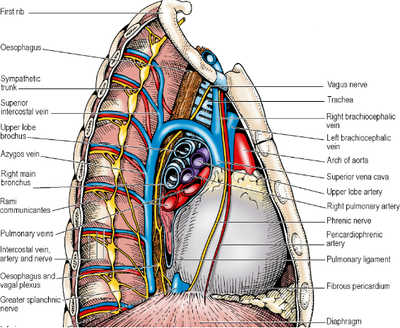
- Left Phrenic Nerve
- Related medially to arterial structures throughout its thoracic course
- Left Common Carotid Artery, Left Subclavian Artery
- Crosses the arch lateral to the Superior Intercostal Vein; Anterior to the Vagus Nerve
- Runs laterally down the pericardium over the Left Ventricle towards the Apex of the heart
- Reaches under-surface of Diaphragm by piercing the muscular part just to the left of the pericardium
- Related medially to arterial structures throughout its thoracic course
- Both
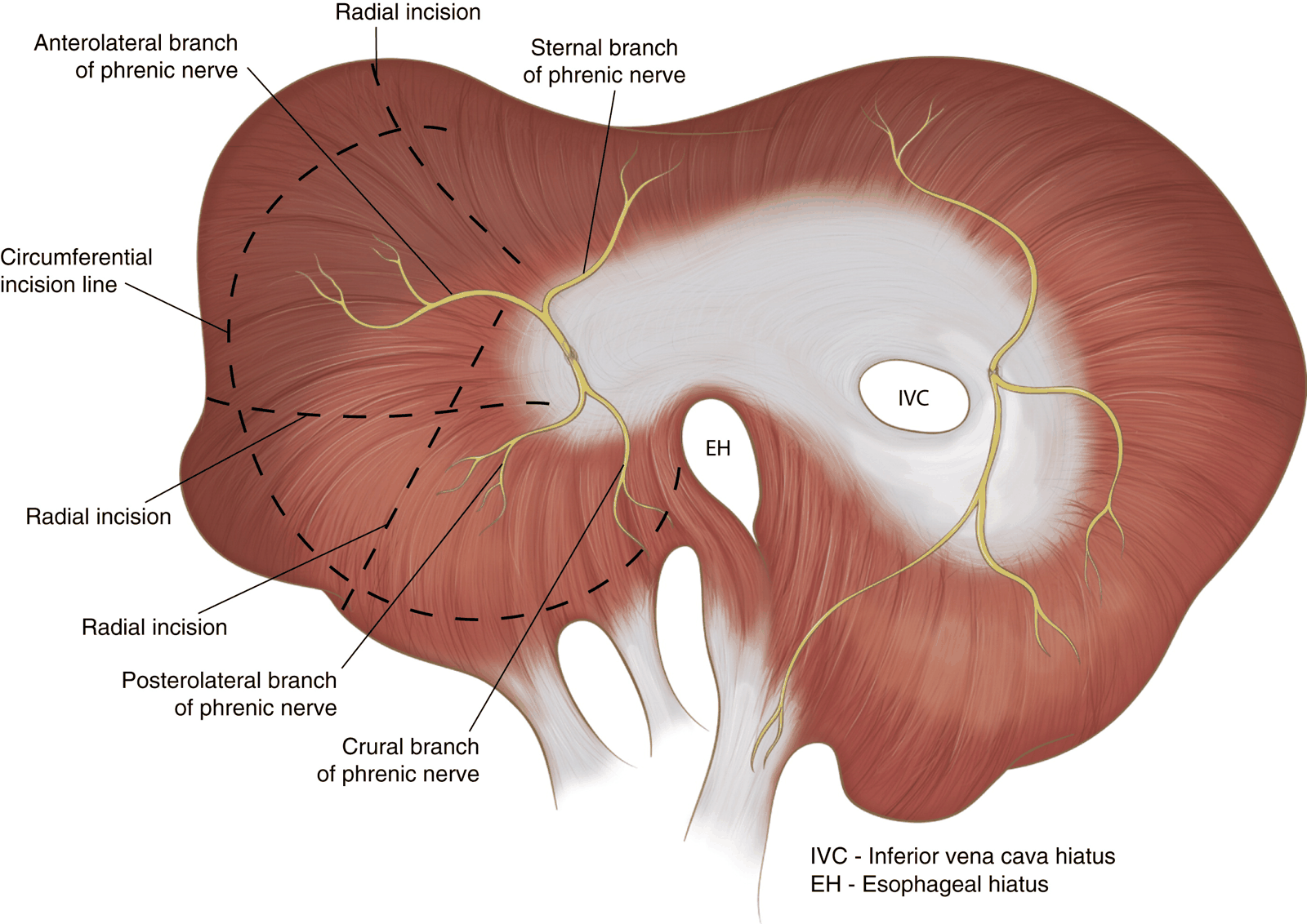
- On reaching abdominal surface of Diaphragm, divides into Anterior, Lateral and Posterior branches which run radially giving off branches that enter the muscle from below
- Fibres of the Right Crus that loop to the left around the Oesophageal Opening: supplied by the Left Phrenic Nerve
- Lower intercostal nerves give proprioceptive fibres to Diaphragm periphery
- Phrenic Nerves – proprioception to central part
- About 2/3rd of the Phrenic Nerve fibres are motor to the Diaphragm
- Rest are sensory to the:
- Diaphragm (except the most peripheral parts, which receive intercostal afferent fibres)
- Mediastinal pleura
- Fibrous pericardium
- Parietal layer of the serous pericardium
- Central parts of the diaphragmatic pleura and peritoneum
- Pain from diaphragmatic part of peritoneum classically felt in shoulder tip (C4)
- Pain from thoracic surfaces supplied by the Phrenic Nerve (pleura and pericardium) is usually only vaguely located there
- Rest are sensory to the:
- Diaphragm moves in respiration under central control of cell groups in the region of the Nucleus of the Tractus Solitarius