• Origin:
    • Anterior Tubercles of the four ‘typical’ Cervical Vertebrae (C3 – C6) by 4 slender tendons of origin which lie end to end with those of Longus Capitis
  • Muscle passes forwards, laterally and downwards to end in a narrow tendon
  • Insertion:
    • Scalene Tubercle and adjacent ridge on the inner border and upper surface of the 1st Rib
  • Nerve Supply:
    • Separate branches from the Anterior Rami of C4-C6 nerves
  • Actions:
    • Assists in Flexion and Rotation of the Neck
    • Helps to stabilize the 1st Rib
    • Shows some electromyographic activity even in quiet respiration

Anterior Relations

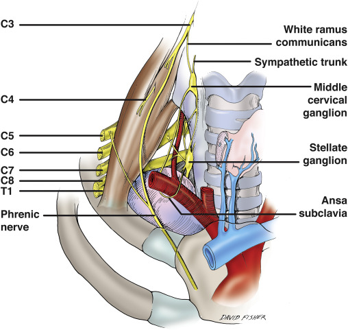
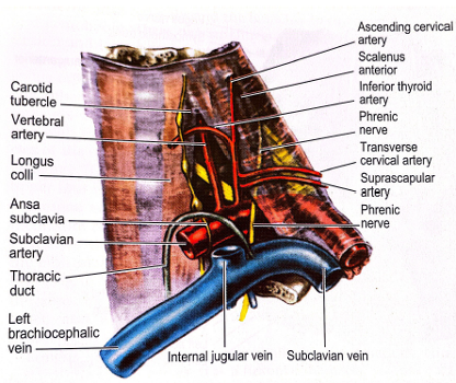
  • Phrenic Nerve
    • Passes vertically down across the obliquity of the muscle
    • Plastered on the muscle by the Prevertebral Fascia and a pad of fat lies in front of the Prevertebral Fascia
    • Nerve leaves the medial border of the muscle low down
    • Crosses anterior to the Subclavian Artery and its Internal Thoracic Artery Branch
    • Behind the Subclavian Vein
      • Occasionally, the Phrenic Nerve may pass in front of the Subclavian Vein or posterior to the Internal Thoracic Artery
    • Lies on the Suprapleural membrane
    • Passes medial to the Apex of the Lung
    • Crosses in front of the Vagus Nerve as it enters the Superior Mediastinum
  • Ascending Cervical Artery
  • Transverse Cervical Artery and Suprascapular Artery
    • Anterior to the Prevertebral Fascia
    • Lie between the Anterior Scalene and the Carotid Sheath (Internal Jugular Vein)
  • Subclavian Vein
    • Lies in a groove on the 1st Rib
      • Due to the slope, lies at a lower level than the insertion of Scalenus Anterior
    • Runs medially
    • Joins the Internal Jugular Vein at the medial border of Scalenus Anterior to form the Brachiocephalic Vein
      • Thoracic Ducton the left and the Right Lymphatic Duct on the right enters the angle of confluence of the 2 veins
    • Used for central venous lines:
      • Infraclavicular approach – from a point 2cm below the midpoint of the Clavicle along a line that passes behind the Clavicle towards the Jugular Notch of the Sternum
      • Needle pierces the Clavipectoral Fascia and enters the vein just behind the Fascia

Medial Relations

  • Medial edge of Scalenus Anterior makes a pyramidal shape with the lateral border of the lower part of Longus Colli
  • Prevertebral Fascia in front of these muscles attached to bone at their opposing margins
  • No fascial roof across the Pyramidal Space
  • Base of space: Subclavian Artery (1st Part) (lying on Suprapleural Membrane)
  • Apex: Carotid (Chassaignac’s) Tubercle on the Transverse Process of C6
  • Common Carotid Artery:
    • Medial to the Internal Jugular Vein:
    • Lies deep to Sternocleidomastoid immediately anterior to the Pyramidal Space
  • Inferior Cervical Sympathetic (or Stellate ganglion):
    • Posterior to the Common Carotid Artery (and Carotid Sheath)
    • Vertebral Artery and Vein anterior to the ganglion
  • Inferior Thyroid Artery
    • Arches medially in a bold curve whose upper convexity lies in front of the Apex of the Pyramidal Space (C6 Level)
    • Sympathetic Chain (usually the Middle Ganglion) in front of the Artery
  • Thoracic Duct (or Right Lymphatic Duct)
    • At a lower level and further anterior
    • Makes a similar convexity behind the Carotid Sheath
    • Arches over the Lung Apex and Subclavian Artery
    • Enters the confluence of the Subclavian Vein and Internal Jugular Vein
  • Vagus Nerve (slightly medial - can also be considered anterior)
    • Within the Carotid Sheath
    • Passes down anterior to the Subclavian Artery
    • Inclines posteriorly and run on the medial surface of the Apex of the Lung to enter the Superior Mediastinum
    • Internal Jugular Vein has Deep Cervical Lymph Nodes closely adjacent to it

Posterior Relations

  • Separated from the Scalenus Medius by:
    • Subclavian Artery
    • Anterior rami of the lower Cervical and 1st Thoracic Nerves, i.e. Roots of Brachial Plexus
  • Subclavian Artery(2nd Part)

Lateral Relations

  • Trunks of the Brachial Plexus:
    • Emerge from the lateral border of Anterior Scalene
  • Subclavian Artery (3rd Part) – see below
  • Both the above lie behind the Prevertebral Fascia on the floor of the Posterior Triangle