Originates from the Vagus Nerve in the Superior Mediastinum
Left Recurrent Laryngeal Nerve
Originates from the Vagus Nerve in the Superior Mediastinum
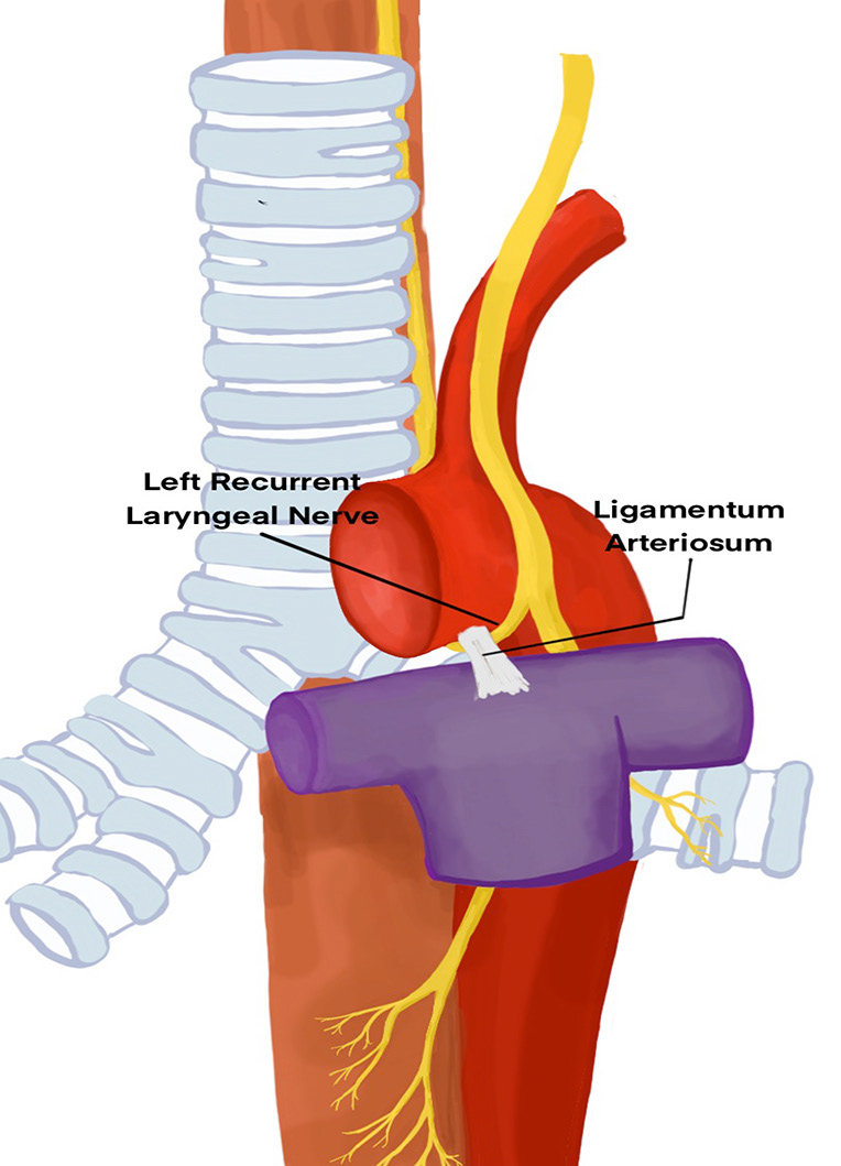
Recurves around Arch of Aorta in Superior Mediastinum
Hooks around the Ligamentum Arteriosum to pass upwards on the right- hand side of the Arch
Hooks around posterior to the Ligamentum Arteriosum, not anterior
Passes into root of neck moving from left towards the midline to enter trachea- oesophageal groove
More likely to have entered the groove and lie posterior to Inferior Thyroid Artery
Right Recurrent Laryngeal Nerve
Originates from the Vagus Nerve in the neck
Recurves around Right Subclavian Artery at Root of the Neck
NB: This is NOT in the Superior Mediastinum
May be more lateral to the Trachea
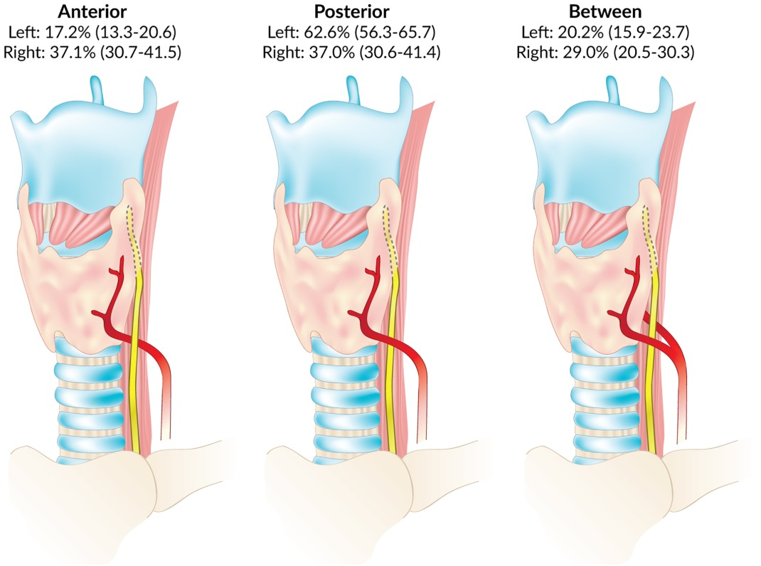
Passes anterior or posterior to Inferior Thyroid Artery or in between its branches
More vertical course than the left
Each nerve is behind the Pretracheal Fascia
Runs medial or lateral or through a thickening of the fascia attached to the Cricoid Cartilage and upper Tracheal Rings (the Suspensory Ligament of Berry)
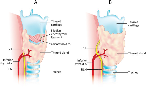
Both nerves pass behind the lateral lobe of the Thyroid in close association with the Inferior Thyroid Artery in the region of the Tubercle of Zuckerkandl
Variable relationship to ITA - most commonly posterior, then anterior then in-between branches. Posterior is more common on the left
Nerve runs behind the Cricothyroid joint
Passes upwards under cover of the Inferior Constrictor
NB: May also be seen disappearing below the inferior border of the Cricopharyngeus muscle
May supply a branch to the Inferior Constrictor Muscle before entering the larynx
Reaches the lower part of the Piriform Recess and penetrates the Laryngeal wall
At level of upper border of Isthmus, nerve often divides into two
If so, anterior (larger) branch is motor branch to laryngeal muscles and posterior branch sensory only
Note: Relationships are not constant with considerable anatomical variation however, the Recurrent Laryngeal Nerve usually passes close to and behind the Tubercle of Zuckerkandl, under Berry ligaments and is infront, inbetween or behind the inferior thyroid artery
Recurrent Laryngeal Nerve branches outside of the Larynx in 1/3 of pts
Posterior branch is sensory
Anterior branch (more vulnerable) is motor
Innervation
All muscles of the Larynx (except Cricothyroid – supplied by the External Laryngeal Nerve – Branch of Superior Laryngeal Nerve)
All motor fibres in both nerves are from cell bodies in the Nucleus Ambiguus derived mainly via the cranial part of the accessory nerve
Supplies mucous membrane of the folds and Larynx below the level of the Vocal Folds