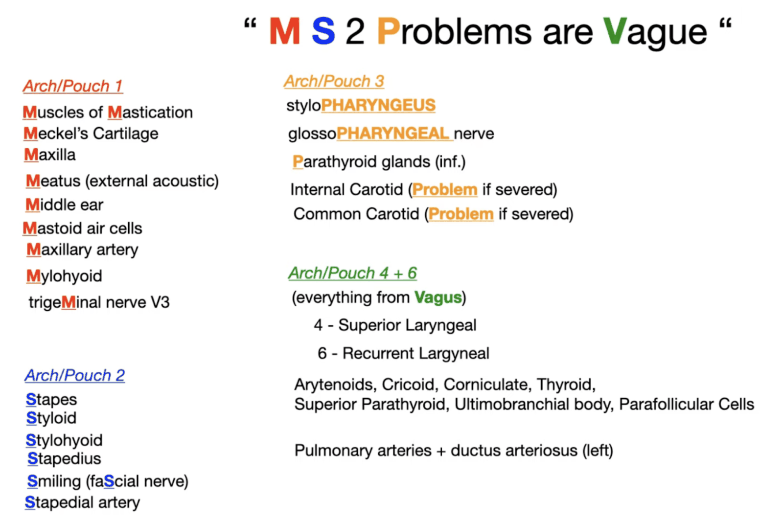
General Information
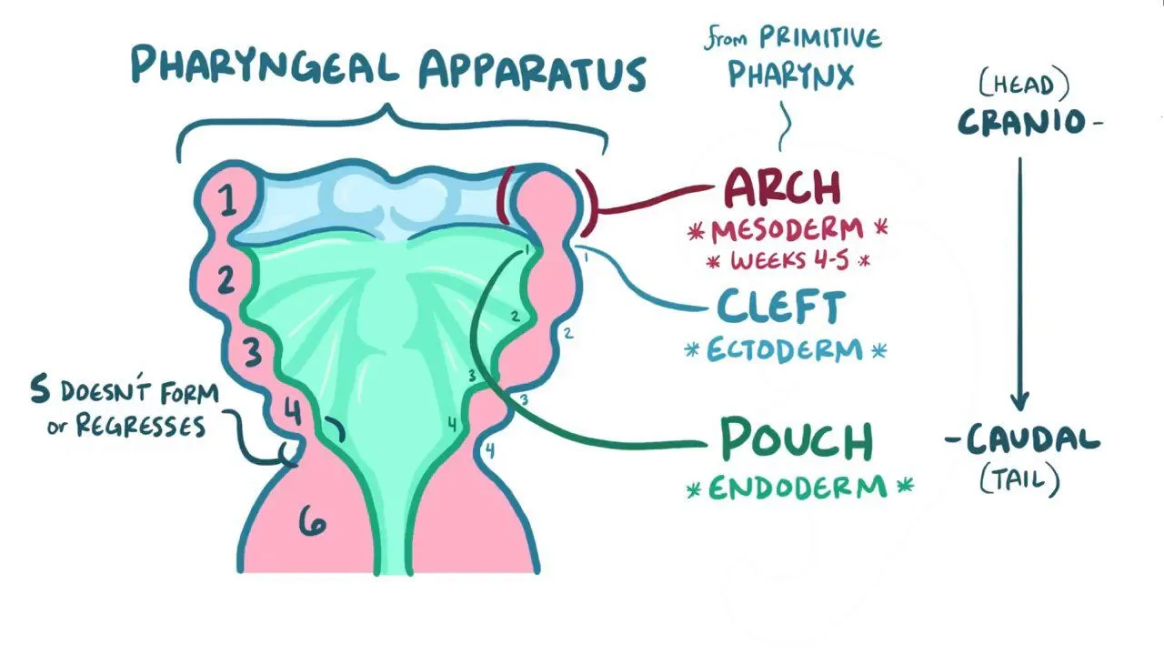
- The pharyngeal arches, pouches, and clefts are paired structures that appear during the 4th week of human embryonic development
- Branchial Arches form Clefts externally and Pouches internally (separated by pharyngeal membranes)
- Pouches lined by endoderm
- Arch derivatives lined by mesoderm
- Cleft derivatives lined by ectoderm
- Each arch associated with:
- Cartilaginous bar
- Carotid artery derivative
- Cranial nerve
- Vein
Pharyngeal Arches
- The pharyngeal arches are five paired structures (1st, 2nd, 3rd, 4th, and 6th) visible externally as bulges in the developing embryo.
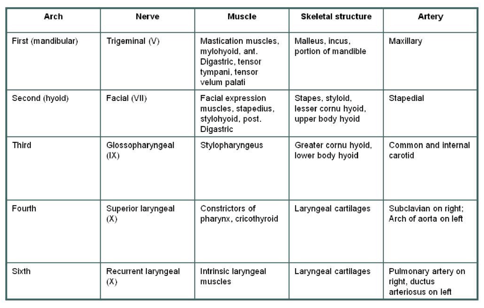
- Each arch contains:
- Mesodermal core: Develops into muscles and blood vessels.
- Neural crest cells: Contribute to bones, cartilage, and connective tissues.
- Cranial nerve: Specific to each arch.
- Aortic arch artery: Forms part of the vascular system.
Key Derivatives:
- 1st Arch (Mandibular):
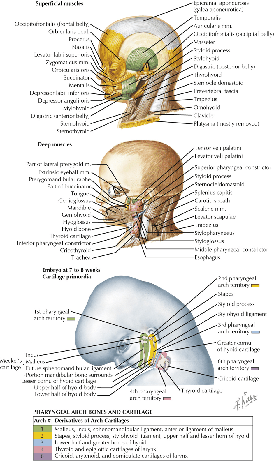
- Bones: Maxilla, mandible, malleus, and incus.
- Muscles: Mastication muscles (e.g., temporalis, masseter), anterior belly of digastric.
- Nerve: Mandibular division of the Trigeminal nerve (CN V3).
- 2nd Arch (Hyoid):
- Bones: Stapes, styloid process, lesser horn of hyoid.
- Muscles: Facial expression muscles, posterior belly of digastric.
- Nerve: Facial Nerve (CN VII).
- 3rd Arch:
- Bones: Greater horn of hyoid.
- Muscles: Stylopharyngeus.
- Nerve: Glossopharyngeal nerve (CN IX).
- 4th Arch:
- Cartilage: Thyroid and cricoid cartilage.
- Muscles: Pharyngeal constrictors, cricothyroid.
- Nerve: Superior laryngeal branch of vagus nerve (CN X).
- 6th Arch:
- Cartilage: Laryngeal cartilages.
- Muscles: Intrinsic laryngeal muscles (except cricothyroid).
- Nerve: Recurrent laryngeal branch of vagus nerve (CN X).
Pharyngeal Pouch
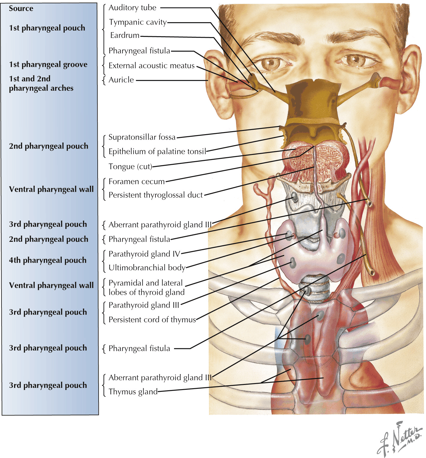
The pouches are internal structures made of endoderm, located between the pharyngeal arches.
- 1st Pouch:
- Forms the Eustachian tube and middle ear cavity.
- 2nd Pouch:
- Forms the palatine tonsils.
- 3rd Pouch:
- Dorsal part: Inferior parathyroid glands.
- Ventral part: Thymus.
- 4th Pouch:
- Dorsal part: Superior parathyroid glands.
- Ventral part: Ultimopharyngeal body (forms parafollicular cells of the thyroid).
| Pouch | Location | Embryonic Structure | Adult Structure |
|---|---|---|---|
| 1 | Opposite the 1st pharyngeal cleft, separated by the 1st pharyngeal membrane | Tubotympanic recess | - Epithelium of the (pharyngotympanic auditory) tube - Tympanic cavity |
| 2 | Opposite the 2nd pharyngeal cleft, separated by the 2nd pharyngeal membrane | Primordial palatine tonsils | - Tonsillar (sinus fossa) - Epithelium of the palatine tonsil |
| 3 | Opposite the 3rd pharyngeal cleft, separated by the 3rd pharyngeal membrane | - Divides into a dorsal and a ventral part - Dorsal part migrates inferiorly toward the thorax | - Inferior parathyroid gland (from the dorsal part) - Thymus (from the ventral part) |
| 4 | Opposite the 4th pharyngeal cleft, separated by the 4th pharyngeal membrane | - Divides into a dorsal and a ventral part - Ventral part is invaded by neural crest to form the parafollicular or C cells | - Superior parathyroid gland (from the dorsal part) - Ultimobranchial body (from the ventral part) |
Pharyngeal Cleft
- 1st Cleft:
- Forms the external auditory meatus.
- 2nd–4th Clefts:
- These are overgrown by the 2nd arch, forming the cervical sinus, which later obliterates. Failure to obliterate can lead to branchial cysts.
| Cleft | Location | Adult Structure |
|---|---|---|
| 1 | A groove between the 1st and 2nd pharyngeal arches | External acoustic meatus |
| 2 | A groove between the 2nd and 3rd pharyngeal arches | Obliterated cervical sinus by the 2nd pharyngeal arch, which grows over the clefts |
| 3 | A groove between the 3rd and 4th pharyngeal arches | ” |
| 4 | A groove between the 4th and 6th pharyngeal arches | ” |
Summary