s- VII
Summary
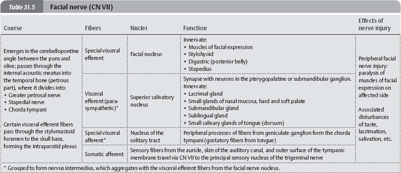
- Somatic Afferents
- External auditory meatus
- Auricle – Posterior auricular nerve
- Special Afferents
- Taste to anterior 2/3 tongue
- Visceral Efferents
- Lacrimal Gland
- Submandibular Gland
- Sublingual gland
- Somatic Efferents
- Muscles of Facial expression
- Stylohyoid
- Stapedius
- Posterior belly of Digastric
Origin
- Main Facial Nerve: Lower border of the Pons above the Olive
- Nervus Intermedius: Between pons and Inferior Cerebellar Peduncle – near the Vestibulocochlear Nerve
Course
- Arises in the pons with a motor and a sensory root
- Enter Internal Acoustic Meatus
- Near the Middle Ear, the nerve makes a sharp posterior bend (the Geniculum)
- Enlarged by the cell bodies of the Afferent taste fibres to form the Geniculate Ganglion
- Nerve runs back in the medial wall of the Middle Ear
- Near the Middle Ear, the nerve makes a sharp posterior bend (the Geniculum)
- Passes vertically down the Facial Canal
- Branches of Facial Nerve within Petrous Bone:
- Greater Petrosal Nerve
- Nerve to Stapedius
- Chorda Tympani
- Consists of Nervus Intermedius Fibres
- Leaves Facial nerve in Facial Canal 6mm above Stylomastoid Foramen
- Passes through Posterior Wall of Middle Ear
- Joins the Lingual Nerve in the Infratemporal Fossa
- Taste fibres taken to the anterior part of the tongue
- Secretomotor fibres relay in the Submandibular Ganglion for the Submandibular Gland
- Branches of Facial Nerve within Petrous Bone:
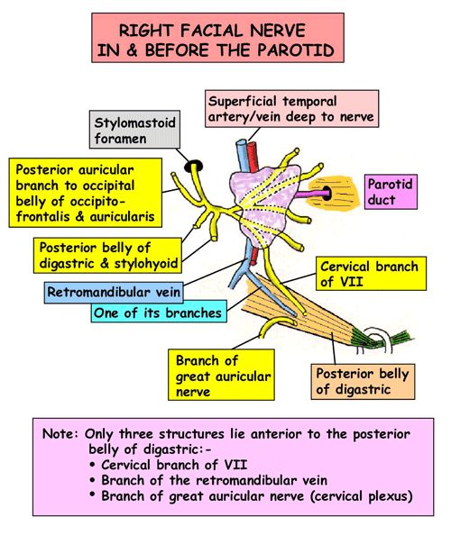
- Emerges from the Stylomastoid Foramen
- Near origin of the Posterior Belly of Digastric
- Immediately gives off Posterior Auricular Nerve
- Passes upwards behind the ear to supply Auricularis Posterior and Occipital Belly of Occipitofrontalis
- Muscular Branch next given off which divides to supply
- Posterior Belly of Digastric
- Stylohyoid muscles
- Nerve approaches posteromedial surface of Parotid Gland
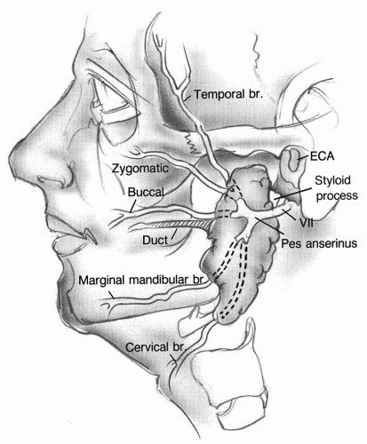
- Just before entering or within the gland, it divides into an upper Temporofacial and a lower Cervicofacial division
- Within substance of Parotid Gland, each division divides and rejoins to divide again and finally emerge from the Parotid Gland in 5 main groups of branches
- Pes anserinus (this plexiform arrangement) lies in the gland superficial to the Retromandibular Vein and the External Carotid Artery
- Branches emerge from behind the anterior border of the gland
- No branches emerge from the superficial aspect of the gland
Muscular branches
Temporal Branches
- Emerge from upper border of gland
- Lies in front of Superficial Temporal Artery
- Cross Zygomatic Arch
- Supply:
- Auricularis anterior and superior
- Part of Frontalis
- Action:
- Wrinkling forehead
- Consequence of injury: Brow asymmetry and brow ptosis, inability to wrinkle forehead
Zygomatic Branches
- Cross the Zygomatic Arch and Zygomatic Bone
- Lies directly on the periosteum
- May be damaged in fractures or operations in this region
- Supply:
- Obicularis Oculi
- Action:
- Blinking of the eye allowing precorneal film of tears to be spread
- If damaged, precorneal film of tears no longer spread ⇒ Dry cornea ulcerates ⇒ Resultant scar impairs vision
- The most serious consequence of impaired Facial Nerve Function
Buccal Branches
- Run forwards close to the Parotid Duct in parallel
- Often one above and one below the Duct
- Supply:
- Buccinator
- Muscle fibres of the Nose
- Muscle fibres of Upper Lip
- Action:
- Emptying of the cheek pouch
- If damaged, bolus lodges in cheek pouch and cannot be returned to molar teeth
- Chewing has to be performed on the other side
Marginal Mandibular Branch
- Frequently single
- Runs forwards above, along or below the lower border of the Mandible
- From below Mandible, it crosses the inferior border of the bone to reach the face just beyond the anterior border of the Masseter
- Passes superficial to the Facial Artery and Facial Vein
- Small node lies here
- Deep to platysma
- Nerve in danger when an incision is made at or near the lower border of the Mandible
- Nerve does not communicate with a Buccal Branch
- Supplies:
- Depressors of the Lower Lip and Mouth Angle
- If damaged, detectable paralysis of the depressors will be seen – asymmetry of the mouth when speaking or smiling
Cervical Branch
- Passes downwards from the lower border of the Parotid Gland
- Supplies Platysma
- Consequence of injury: Asymmetry of the motion of the corner of the mouth
Identification of Facial Nerve During Parotid Surgery
- Main trunk emerges from Stylomastoid Foramen
- Winds lateral to the Styloid Process just above the tendon of the Stylohyoid Muscle
- Relationship of Stylomastoid Foramen and the Tympano-mastoid Fissure useful landmark
- Main trunk < 1cm deep to Tympano-mastoid suture line at the level of the Digastric Muscle
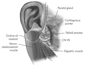
- Facial Nerve approached along a plane in front of the anterior margin of the cartilage
- Just deep to the junction of the cartilaginous and bony parts of the External Meatus
- Cartilage in this region has an arrow-headed projection pointing downwards to the nerve trunk
- a.k.a. Tragal Pointer of Conley
- About 1cm above and medial to the upper end of the Posterior Belly of Digastric
- Stylomastoid branch of Posterior Auricular Artery is superficial to the Facial Nerve and is a guide to its proximity
- Alternative approach: find a Facial Nerve Branch as it leaves the gland and follow it in a centripetal manner back to the Trunk and other branches
- Marginal Mandibular Branch may be identified as it lies superficial to the Retromandibular Vein or its Anterior branch – aided by the colour contrast between the white nerve and the dark vein
- Occasionally, this nerve will pass behind the vein
- Alternatively, the Cervical Branch may be followed up from its communication with the Ascending Branch of the Transverse Cervical Nerve