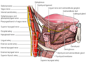
Secretomotor fibres – Cell bodies in the Submandibular Ganglion
Hangs suspended from the Lingual Nerve on the surface of Hyoglossus
Pre-ganglionic fibres pass from cell bodies in the Superior Salivary Nucleus in the Pons by way of: Nervus Intermedius ⇒ Facial Nerve ⇒ Chorda Tympani ⇒ Lingual Nerve
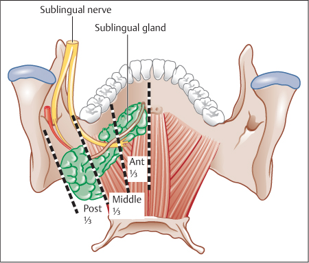
Post-ganglionic fibres pass to Submandibular Gland and also to Lingual Nerve for transmission to the Sublingual Gland
Sympathetic (Vasoconstrictor) fibres – Plexus around the Facial Artery
Development
Ectodermal groove in the floor of the mouth becomes converted into a tunnel whose blind end proliferates to form the secreting acini.
Develops during 6th week of gestation (after Parotid Gland)
Summary of Important Anatomical Relations
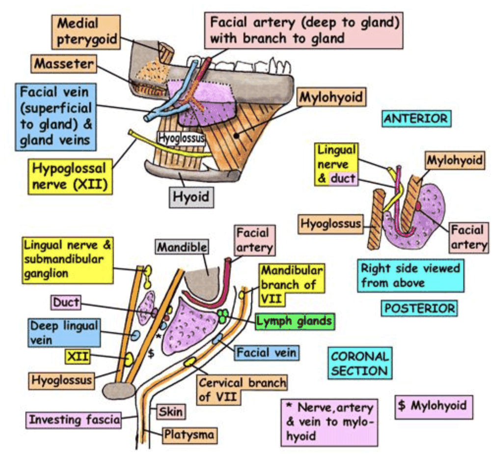
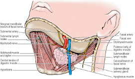
Superficial: Mandibular and Cervical Branches of the Facial Nerve
Mandibular and Cervical branches emerge from Parotid Gland near angle of Mandible
Mandibular branch runs downwards and forwards deep to Platysma
Curves upwards to cross Mandible close to Facial Artery
Mandibular nerve protected during surgery by making incision 4cm below Mandible
On the gland: Facial Artery and Vein
Facial Artery
Originates from External Carotid Artery
Passes upwards
Identified at the posterior aspect of the Submandibular Gland
Passes forwards above the gland
Emerges beneath the lower border of the Mandible
Curls upwards along the face
Facial Vein
Descends along the face from anterior to posterior
Crosses the Mandible about 2cm in front of the angle
Runs along the external surface of Submandibular Gland and receives the Submental Vein forming the Common Facial Vein
Continues to drain into the Internal Jugular Vein
Facial Artery and Vein usually encountered after identification and preservation of the Mandibular Nerve
Usually ligated at this point and again at the posterior aspect of the gland
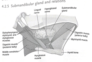
Deep: Lingual and Hypoglossal Nerves and Submandibular Ganglion
Lingual Nerve:
Deep to Mylohyoid
Above the deep part of the Submandibular Gland
Runs forward between Mylohyoid and Hyoglossus
Passes around the Submandibular Duct by initially passing from medial to lateral above the duct then curving underneath the duct from Lateral to medial, emerging at the medial side of the duct
During ligation of Submandibular Duct, care must be taken to identify the lingual nerve so that it is not damaged
More proximally, the Lingual Nerve is identified by retracting the Submandibular Gland laterally and the Mylohyoid muscle anteriorly
Lingual Nerve is freed from the Submandibular Gland by dividing between the nerve and the Submandibular Ganglion
Hypoglossal Nerve:
Lies deep and below the Submandibular Gland
Runs between the Mylohyoid and Hyoglossus muscles
Nerve identified after posterior border of the gland is mobilised and retracted upwards
Mylohyoid also retracted anteriorly to expose the nerve as it emerges from the Posterior Belly of Digastric