See: Submandibular gland anatomy
Indications
- Non-neoplastic
- Sialolithiasis
- Sialadenitis
- Neoplastic
- Benign
- Malignant
- Primary
- Secondary
Prep
- Supine with neck extension and head turned away on a head ring
Operation
-
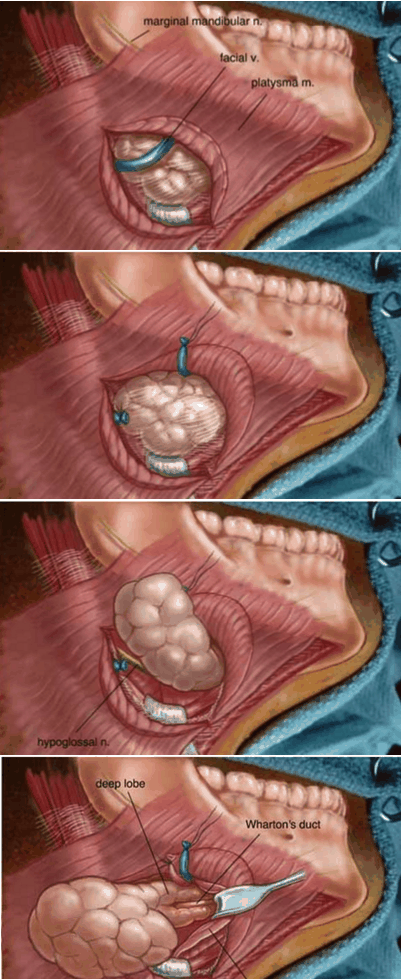
Incision
- From 2cm lateral to midline and extending to angle of mandible
- 3cm inferior to lower border of mandible
- Protect MMN
- NO FLAPS - avoid injury to MMN
-
Inferior border bluntly dissected free
-
Facial vein ligated (on superficial surface)
- Hayes Martin Manoeuvre - elevate a flap under the FV which protects the MM nerve
-
Posterior border dissected
- Facial A ligated when encountered
-
Superior border dissected
- Facial A ligated again (as it loops around inferior border of mandible)
-
Superficial lobe dissected off mylohyoid muscle
-
Operator retracts it laterally
-
Assistant retracts mylohyoid medially
-
Deep lobe (between mylohyoid and hyoglossus) also bluntly dissected
- CN12 on hyoglossus inferiorly is protected
-
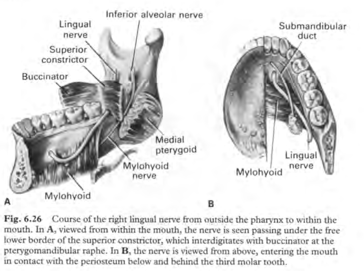
Duct closely related to lingual nerve
- Superior to duct
- Lingual nerve retracted inferiorly with the gland
-
Submandibular ganglion fibres attaching the nerve to the duct are divided
-
Lingual nerve is free and retracted superiorly
-
Duct ligated with vicryl ties and divided
-
Gland and duct excised
-
Layered closure over a 10F drain
-
If being done for malignancy
- Need to raise flaps but the subplatysmal flaps are made with care to ID the MMN which is just deep to the superior flap
- Then retract it superiorly
- Excise all tissue in submandibular triangle (between digastric bellies and mandible)
- ID and preserve CN 12 and lingual N if possible
Summary of Important Anatomical Relations
- Superficial: Mandibular and Cervical Branches of the Facial Nerve
- Mandibular and Cervical branches emerge from Parotid Gland near angle of Mandible
- Mandibular branch runs downwards and forwards deep to Platysma
- Curves upwards to cross Mandible close to Facial Artery
- Mandibular nerve protected during surgery by making incision 4cm below Mandible
- On the gland: Facial Artery and Vein
- Facial Artery
- Originates from External Carotid Artery
- Passes upwards
- Identified at the posterior aspect of the Submandibular Gland
- Passes forwards above the gland
- Emerges beneath the lower border of the Mandible
- Curls upwards along the face
- Facial Vein
- Descends along the face from anterior to posterior
- Crosses the Mandible about 2cm in front of the angle
- Runs along the external surface of Submandibular Gland and receives the Submental Vein forming the Common Facial Vein
- Continues to drain into the Internal Jugular Vein
- Facial Artery and Vein usually encountered after identification and preservation of the Mandibular Nerve
- Usually ligated at this point and again at the posterior aspect of the gland
- Facial Artery
- Deep: Lingual and Hypoglossal Nerves and Submandibular Ganglion
- Lingual Nerve:
- Deep to Mylohyoid
- Above the deep part of the Submandibular Gland
- Runs forward between Mylohyoid and Hyoglossus
- The nerve lies against the periosteum of the mandible at the posterior border of the third molar tooth, and runs forward on the upper surface of mylohyoid muscle.
- The lingual nerve then dips under the submandibular duct and runs forward on the surface of hyoglossus above the level of the duct
- During ligation of Submandibular Duct, care must be taken to identify the lingual nerve so that it is not damaged
- More proximally, the Lingual Nerve is identified by retracting the Submandibular Gland laterally and the Mylohyoid muscle anteriorly
- Lingual Nerve is freed from the Submandibular Gland by dividing between the nerve and the Submandibular Ganglion
- Hypoglossal Nerve:
- Lies deep and below the Submandibular Gland
- Runs between the Mylohyoid and Hyoglossus muscles
- Nerve identified after posterior border of the gland is mobilised and retracted upwards
- Mylohyoid also retracted anteriorly to expose the nerve as it emerges from the Posterior Belly of Digastric
- Lingual Nerve: