Physiology
Histology
- Secretory acini opening onto arborized ducts which empty into the oral cavity via the parotid duct
- Myoepithelial cells surround each acinus for secretion
- How to differentiate from others
- Parotid
- Mainly serous acini/ducts
- Many ducts
- Fat between acini and ducts
- Submandibular
- Mixed serous and mucinous acini/ducts
- Few ducts
- Sublingual
- Mucous acini
- Few ducts
- Parotid
Function
- Saliva produced in acini
- Altered into mixture of electrolytes and macromolecules
- Active transport into ducts
- Hypotonic in mouth but flow rates can alter this
- Carbohydrate digestion with amylase
Submandibular gland anatomy
Overview
- Mixed mucous and serous gland
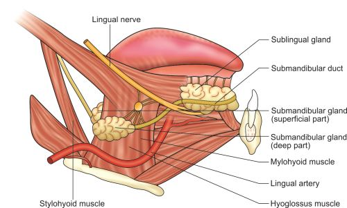
- Large superficial part and a small deep part
- Continuous with one another round the free posterior margin of the Mylohyoid muscle
- Lies in the Digastric Triangle
- Anterior border: Anterior Belly of the Digastric
- Posterior border: Posterior Belly of the Digastric
- Lateral border: Lower border of the Mandible and Medial Pterygoid Muscle
- Floor: Hyoglossus and Mylohyoid
- Roof: Skin, Platysma, Investing Layer of Cervical Fascia
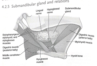
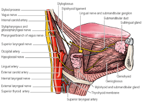
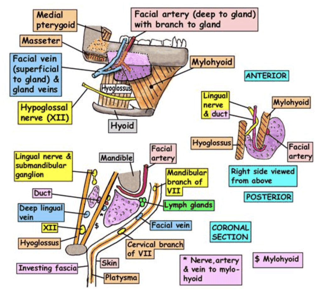
Superficial part
- Three surfaces – lateral, inferior and medial
- Lateral surface:
- Lies against the Submandibular Fossa of the Mandible
- Overlaps the front of the Medial Pterygoid insertion
- Deeply grooved posteriorly by the Facial Artery
- Facial Artery hooks under the Mandible to reach the face at the front of the Masseter muscle
- Inferior/Superficial surface:
- Covered by skin, Platysma and Investing Fascia
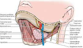
- Crossed by the Facial Vein and Cervical Branch of the Facial Nerve
- Crossed sometimes by the Marginal Mandibular Branch of the Facial Nerve
- Nerves lie outside the Investing Fascia
- Submandibular Lymph Nodes lie in contact with the surface of the gland and within its substance
- Thus, the need to remove the gland as well as nodes in the operation of radical neck dissection
- Medial surface:
- Lies against Mylohyoid
- Behind Mylohyoid, it is related to:
- Hyoglossus
- Lingual Nerve
- Hypoglossal Nerve
- Accompanying veins
- Facial Artery initially deep to the gland and then grooves the posterosuperior part as it hooks over the top of the gland on its lateral surface
Deep part
- Extends forwards for a variable distance
- Lies between Mylohyoid and Hyoglossus
- Below the Lingual Nerve
- Above the Hypoglossal Nerve
Summary of Relations
- Superficial Part
- Lateral - mandible, facial artery
- Inferior - skin/platysma/facial vein
- Medial - mylohyoid, hyoglossus, lingual N, CN12, facial A
- Deep Part
- Between mylohyoid and hyoglossus
- Below lingual nerve and above CN12
Submandibular Duct (of Wharton)
- Length: 5cm – same length as Parotid Duct
- Emerges from medial surface of the Deep Part of the Submandibular Gland
- Near the posterior border of Mylohyoid
- Runs with the deep part – forwards and slightly upwards
- First between Mylohyoid and Hyoglossus
- Then between Sublingual Gland (lateral) and Genioglossus (medial)
- Opens into floor of mouth on the Sublingual Papilla beside the Frenulum of the Tongue
- As it lies on Hyoglossus, the duct is crossed laterally by the Lingual Nerve
- Which then turns under the duct to pass medially to the Tongue
- Consequences of damaging Lingual Nerve:
- Altered salivary secretion on the affected side
- Loss of taste to anterior 2/3rd of tongue
- Sensory changes to the anterior 2/3rd of tongue and floor of mouth
- Consequences of damaging Hypoglossal Nerve:
- Loss of motor innervation to all intrinsic muscles of tongue except Palatoglossus
- Consequences of damaging Marginal Mandibular Nerve:
- Facial asymmetry and drooling
- Supplies Depressor Anguli Oris, Depressor Labii Inferioris, Risorius, and part of Obicularis Oris
Blood Supply
- Facial Artery
- Lingual Artery
- Veins drain into Lingual and Facial Veins
Lymph Drainage
- Submandibular Lymph Nodes
Nerve Supply
Summary
-
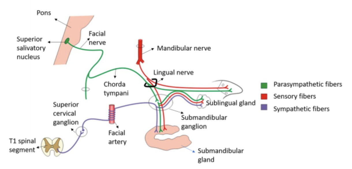
PNS (Secretomotor)
- Via facial N branch - Chorda tympani
- Travels in lingual N to submandibular ganglion
-
SNS (Vasomotor)
- Superior cervical sympathetic ganglion
- Travels with the facial A
-
Sensory
- In lingual N
-
Secretomotor fibres – Cell bodies in the Submandibular Ganglion
- Hangs suspended from the Lingual Nerve on the surface of Hyoglossus
- Pre-ganglionic fibres pass from cell bodies in the Superior Salivary Nucleus in the Pons by way of: Nervus Intermedius ⇒ Facial Nerve ⇒ Chorda Tympani ⇒ Lingual Nerve
- Post-ganglionic fibres pass to Submandibular Gland and also to Lingual Nerve for transmission to the Sublingual Gland
-
Sympathetic (Vasoconstrictor) fibres – Plexus around the Facial Artery
Development
- Ectodermal groove in the floor of the mouth becomes converted into a tunnel whose blind end proliferates to form the secreting acini.
- Develops during 6th week of gestation (after Parotid Gland)
See also: Submandibular gland excision
Sublingual Gland
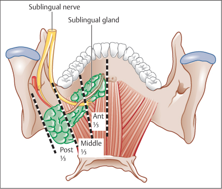
- Smallest of paired glands
- Contributed 3-5% of salivary volume
- Boarders
- Lie on the floor of the oral cavity.
- Underneath the tongue,
- Lateral border
- Mandible
- Shallow groove on the medial surface of the mandible known as the sublingual fossa.
- Mandible
- Medial
- Genioglossus muscle of the tongue
- Relations
- Submandibular duct and Lingual Nerve pass alongside the medial aspect of the sublingual gland.
- Duct
- Drainage via multiple ducts of rivinus - open onto sublingual folds which run oblique to the lingual frenulum
- And a larger sublingual duct of Bartholin which drains into Whartons duct and into floor of mouth on either side of lingual frenulum
- Innervation
- Same as Submandibular gland anatomy
- PNS (secretomotor)
- Facial N (CN7) via chorda tympani (CN7 br)
- Travels with lingual N (V3 br) to synapse at submandibular ganglion
- Postganglionic fibres direct to sublingual gland - ACh
- SNS
- Preganglionic from T1 lateral horn to superior cervical ganglion
- Through but not synapsing in the submandibular ganglion
- Along Facial A
- To sublingual gland
- Sensory
- Via lingual N
- Via lingual N
Parotid Gland
Overview
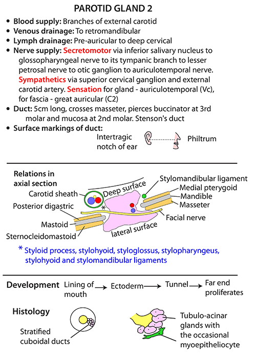
- Mainly serous gland with only a few scattered mucous acini
- Wedge-shaped on transverse section
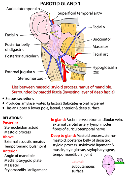
- Extends from Zygomatic Arch to the upper part of the neck
- Overlaps the Posterior Belly of Digastric and the anterior border of Sternocleidomastoid
- Anteriorly, gland overlaps Masseter
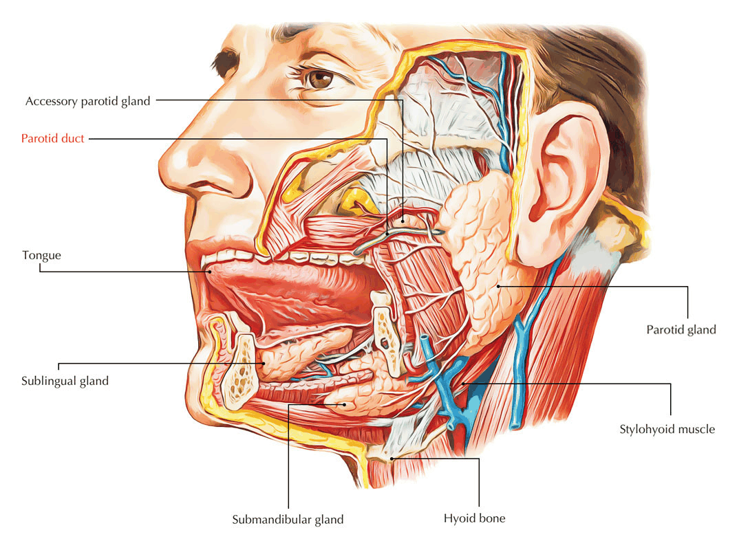
- Accessory Parotid – small, usually detached gland lies above the Parotid Duct on the aponeurotic part of the Masseter
- Extends below the External Acoustic Meatus posteriorly onto the Mastoid Process
- Occupies gap between the Ramus of the Mandible and the Mastoid and Styloid Processes of the Temporal Bone
- Reaches close to the lateral wall of the Oropharynx
- Thus, there is a need to look at the region of the Fauces when examining a patient with a Parotid Mass
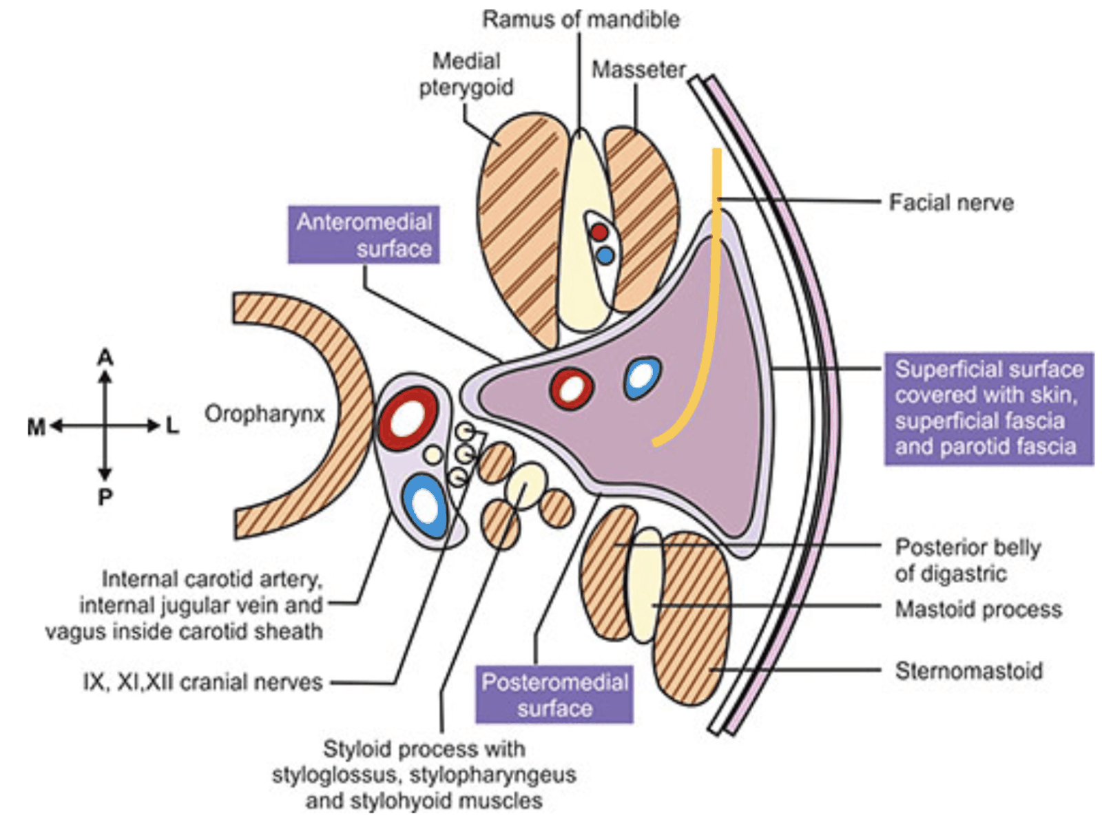
Relationships
- Lateral (Superficial) Surface:
- Covered by skin and Superficial Fascia
- Investing Layer of Deep Cervical Fascia splits to envelope the gland
- a.k.a. Parotid Sheath
- Inner leaf passes up to the Base of the Skull
- Condensation of the fascia known as the Stylomandibular Ligament attaches from the Styloid Process to the Styloid Process of the Mandible
- This partitions the Parotid Gland from the posterior aspect of the Submandibular Gland
- Outer leaf extends superiorly as the Parotidomasseteric Fascia
- Reaches the Zygomatic Arch
- On the gland, fascia termed Parotid Capsule or Masseteric Fascia more anteriorly
- Superficial Muscular Aponeurotic System (SMAS) overlies the gland
- Anteromedial Surface
- Grooved by the posterior border of the Mandibular Ramus
- Related to the Masseter and Medial Pterygoid muscles which are attached to the Ramus
- Gland wrapped around the Capsule of the Temporomandibular Joint
- Anterior edge of this surface meets the lateral surface over, as well as below, the Masseter forming the irregularly convex anterior border of the gland
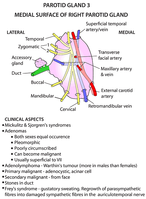
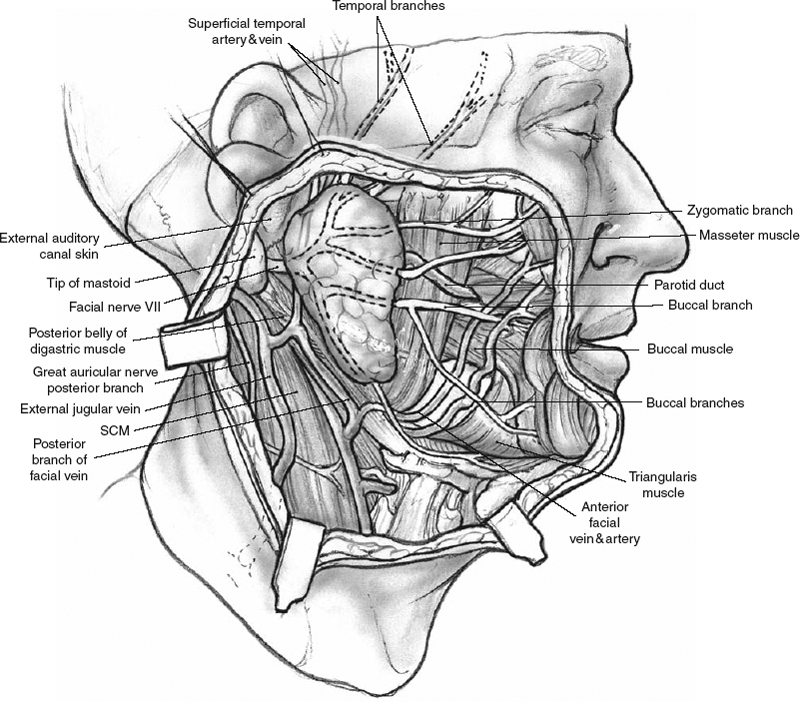
- Parotid Duct and Facial Nerve branches emerge from the anteromedial surface and run forwards deep to the anterior border
- Terminal branches of External Carotid Artery (i.e. Superficial Temporal and Maxillary Arteries) leave this surface further back
- Posteromedial Surface
- In contact with the Mastoid Process with its attached Sternocleidomastoid and Posterior Belly of Digastric muscles
- SCM attached to superficial surface
- Posterior Belly of Digastric attached to deep surface
- More medially, the Styloid Process and its attached muscles (i.e. Stylohyoid, Stylopharyngeus, and Styloglossus) separate the gland from the Carotid Sheath
- External Carotid Artery enters the gland through the lower part of this surface
- Facial Nerve trunk (or its Temporofacial and Cervicofacial divisions) enter the gland between the Mastoid and Styloid Processes
- In contact with the Mastoid Process with its attached Sternocleidomastoid and Posterior Belly of Digastric muscles
- Within
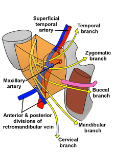
- Facial Nerve
- Branches run in different directions in different (superficial to deep) planes
- Surgeons consider the gland to have a superficial and deep part separated by the plane of the Facial Nerve
- No specific, developmentally determined plane in which the Facial Nerve branches pass between Superficial and Deep Lobes of the gland; the Parotid is an integral gland not divided into lobes
- Within the gland, nerve branches communicate with each other forming a plexiform arrangement
- Lies superficial to the Retromandibular Vein (which is superficial to the External Carotid Artery)
- Branches run in different directions in different (superficial to deep) planes
- Retromandibular Vein
- Formed within the Parotid Gland by the confluence of the Superficial Temporal and Maxillary Veins
- Emerges from the lower part (pole) of the Parotid Gland
- Divides into anterior and posterior branches
- Anterior branch joins the Facial Vein to form the Common Facial Vein
- Posterior branch joins the Posterior Auricular Vein to form the External Jugular Vein, which then drains into the Subclavian Vein
- Division may occur within the Parotid Gland and the two branches emerge from the lower pole
- External Carotid Artery
- Pierces the Parotid Fascia to enter the Parotid Gland
- Divides within the gland behind the neck of the Mandible into Maxillary Artery and Superficial Temporal Artery
- As it lies within the Parotid Gland, it is separated from the Internal Carotid by the deep part of the Gland, its fascia, Styloid Process and it’s continuation the Stylohyoid Ligament, Styloglossus and the ‘Pharyngeal Structures’
- Stylopharyngeus muscle
- Glossopharyngeal Nerve
- Pharyngeal Branch of the Vagus Nerve
- Lymph Nodes
- Off the Preauricular (Parotid) group lie on or deep to the fascial capsule of the Parotid, as well as within the gland
- Facial Nerve
Parotid Duct (of Stensen)
- Length: 5cm
- Course:
- Passes forwards across the Masseter
- Turns around anterior border of Masseter
- Pass through Buccal fat pad
- Pierce Buccinator
- Opens on the mucous membrane of the check opposite the 2nd upper molar tooth
- Pierces Buccinator further back and runs forwards beneath the mucous membrane
- When intraoral pressure is raised, the submucous part of the duct is compressed between the buccinator and the mucous membrane, preventing inflation of the gland
- Surface marking: Lies on the middle third of a line between the Intertragic Notch of the Auricle and the Midpoint of the Philtrum
- Palpable on the clenched Masseter
Accessory Parotid Gland
- Usually lies on the Masseter between the Parotid Duct and the Zygomatic Arch
- Several small ducts open into the Parotid Duct
- The Accessory Parotid Gland and the Parotid Duct both lie on the aponeurotic part of the surface of the Masseter
Blood Supply
- Branches from the External Carotid Artery
- Venous return via Retromandibular Vein
Lymph Drainage
- Drains to Pre-auricular (Parotid) lymph nodes ⇒ Nodes of the upper group of Deep Cervical Nodes
Nerve Supply
- ANS (saliva production)
- PNS (increase saliva production - rest and digest)
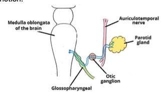
- Begins with the glossopharyngeal nerve (cranial nerve IX)
- Synapses with the otic ganglion
- Auriculotemporal nerve (branch of mandibular nerve V3) then carries parasympathetic fibres from the otic ganglion to the parotid gland
- Traverses
- Emerges from superior surface, behind superficial temporal artery and vein
- Communicates with facial nerve within gland
- Parasympathetic stimulation causes an increase in saliva production.
- SNS (reduce saliva production via vasoconstriction)
- Superior cervical ganglion (symp chain)
- Fibres from it travel along ECA and middle meningeal A to reach parotid gland
- PNS (increase saliva production - rest and digest)
- Sensory
- Auriculotemporal nerve - parotid gland parenchyma
- Great auricular nerve - parotid sheath (capsule)
Development
- A groove that appears in the Ectoderm of the mouth pit (Stomodeum) becomes converted into a tunnel
- Cells from the blind end proliferate to form the Parotid Gland
- Development starts in the 5th week of gestation (first salivary gland to develop)