Neoplastic (EXCEPT benign lymphoepithelial cysts and parotid lymphoma)
Benign tumours
Malignant tumours
Primary
Secondary
LN dissection for cutaneous malignancies of face and scalp
Preoperatively
Assess all branches of facial nerve
Consent
General
Bleeding
Infection
Scar - keloid, hypertrophic
GA risks - DVT/PE/MI/death
Specific 5 F’s
Facial nerve and branch injury
Frey’s syndrome (post-op gustatory sweating and flushing)
Abhorrent nerve regeneration after damage to parasymp supply to parotid
Normally the auriculotemporal nerve (branch of V3) has post ganglionic secretory fibres running with it from the otic ganglion (CN9) to the parotid
PNS fibres around parotid are damaged and regenerate by inadvertently joining SNS fibres to sweat glands and BV ok skin over parotid
Gustatory stimulus causes sweating and flushing instead of salivation
Summary - gustatory sweating and flushing due to ANS damage during parotidectomy. Parasympathetic regeneration an inappropriate innervation of sweat glands and blood vessels of the skin
Investigations
Corn starch test and Iodine test = paint, powder and sweat
Paint face with iodine and let it dry
Powder with cornstarch
Sweat will turn blue
Management
Botox to affected skin area
Blocks ACh release from NMJ
Topical antiperspirants (aluminium based)
Fistula (from parotid duct)
Mx - botox to the gland
Blocks ACh release from parasymp secretomotor neurons
Formication (sensation of insects crawling on skin)
Paraesthesia
Damage to V3
Flap necrosis
Setup
GA with no muscle relaxant so facial nerve can be tested
Supine, shoulder roll for neck extension
Exposure - ear, neck, parotid, corner of mouth, corner of eye
NIM probe
Operation
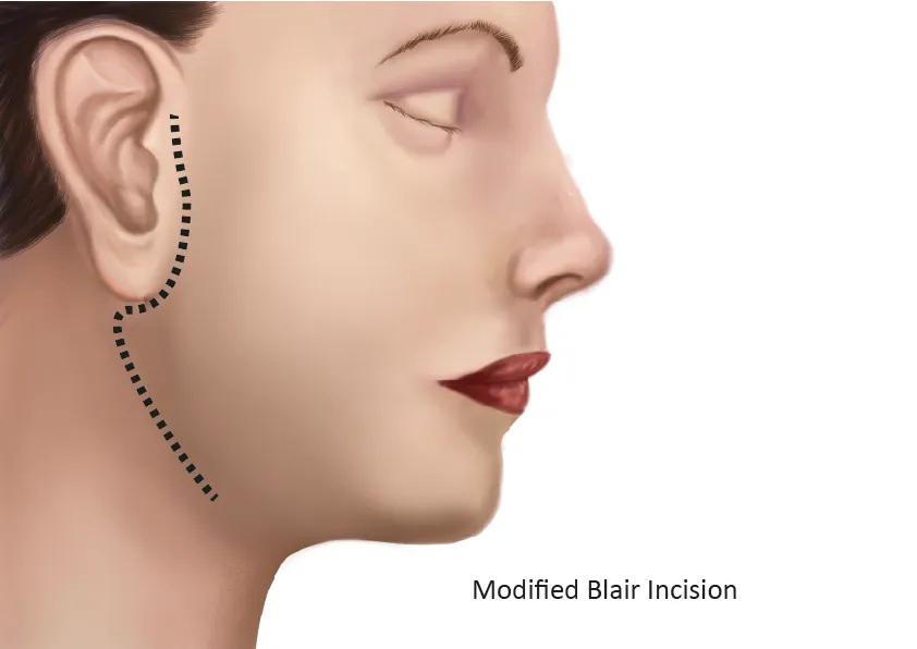
Modified Blair incision
S-shaped incision from front of ear to mastoid process, then down and forward 2cm below angle of mandible
Layers
Skin
Subcut fat
Superficial fascia (platysma, ant br of greater auricular N, pre-auricular lymphatics)
Parotid fascia
Key points
Wide flaps for good exposure
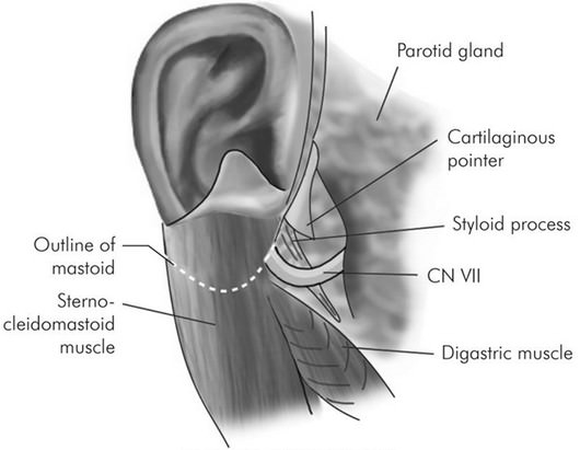
Clear the border of SCM muscle and identify the posterior belly of digastric and the auriculotemporal nerve (preserve it)
Retract back of gland forward
Identify posterior belly of digastric, styloid process and external aud meatus
ID main nerve trunk
Styloid process - nerve lies LATERAL to styloid process base
Tragal pointer - 1cm INFEROMEDIAL to ridge of bone joining cartilaginous and bony external auditory meatus
Tympanomastoid suture by palpation (the most consistent landmark) (approximately 2mm inferior to suture)
Digastric muscle (nerve is immediately superior to digastric)
Dissect superficial to the nerve
Under vision the whole time
Haemostats pointed up
Dissect main branch until it divides
Into 2 branches
Temporozygomatic
Cervicofacial
Dissect all 5 branches carefully
Then superficial parotid will come off
Use NIM to stimulate the main nerve - prove all branches are intact
2 layer monocryl closure
10Fr blakes drain, don’t place it on any part of the nerve
Landmarks for Facial Nerve TPTS
Inferomedial, superomedial, medial, anterolateral
Tragal pointer
1cm INFEROMEDIAL to tip of tragal pointer
Ridge where bony and cartilaginous external auditory meatus meet
Posterior belly of digastric
1cm SUPEROMEDIAL to upper end of posterior digastric belly
Tympanomastoid suture
Part of the temporal bone - divides it tympanic part from mastoid process
5-10mm MEDIALto the TMS
Styloid process
Passes ANTEROLATERAL to the base of the styloid process