Section: Head and neck Curriculum: Curriculum, page 42
Incidence
- Larger salivary glands more likely to have benign pathology
- Smaller glands get, higher the malignant incidence
- Parotid - 20%
- Submandibular - 50%
- Sublingual - 80%
Classification
- (Summary of WHO 2017 classification)
- Benign
- Pleomorphic Adenoma
- Warthins Tumour
- Lipoma
- Myoepithelioma
- Basal cell adenoma
- Oncocytoma
- Malignant
- Mucoepidermoid carcinoma
- Adenoid cystic carcinoma
- Carcinoma ex pleomorphic adenoma
- Secondary
- SCC
- Melanoma
- Lymphoma
Risk Factors
- Radiation exposure
- Viral infection - HPV, EBV, HIV
- Environmental exposure - rubber manufacturing, hair dresser, nickel compounds
- Smoking (Warthins Tumour)
Clinical
Local
- Major gland
- Painless mass/swelling
- Facial nerve involvement suggests malignancy
- Minor gland
- Painless submucosal mass or mucosal ulceration
- Palate, lips, buccal mucosa
- Looks like SCC
- Location and size driven
- Nasal obstruction and congestion
- Visual change
- Trismus
- Nasopharynx - invasion of skull base with CN involvement
- Painless submucosal mass or mucosal ulceration
Regional
- Regional LN mets dependent on which gland is involved
- Parotid - level 2
- Submandibular - level 1
- Sublingual - level 1
- Minors - retropharyngeal
Distant metastasis
- Lung, bone and liver usual spots
- Adenoid cystic carcinoma can have distant mets even 20yrs after tx
Investigations
- Helps to determine if benign vs malignant, location in relation to the gland, local invasion and extent
- Imaging
- Goals of imaging
- Confirm dx
- Location
- TNM staging
- USS
- Good for 3 big glands and can guide FNA/core
- CT
- Bony involvement, distant mets
- MRI
- Sublingual location (high malignancy risk)
- Soft tissue invasion
- PNI
- Intracranial extension
- Goals of imaging
- Biopsy - FNA or core
- FNA 80% sens and spec, lower risk
- Core 100% sens and spec, higher risk
- All benign’s cardinal feature
- Circumscribed growth
- Absence of infiltrative growth pattern
Stage
TNM AJCC 8th Staging Salivary Gland Tumours
| T1 | <2cm, no extraparenchymal invasion (clinical/MACROscopic) |
| T2 | 2-4cm , no extraparenchymal invasion |
| T3 | >4cm AND/OR extraparenchymal invasion |
| T4a | Moderately advanced Invades skin, mandible, ear canal and/or facial nerve |
| T4b | Very advanced Invades skull base and/or pterygoid plates and/orencases carotid artery |
| N1 | 1x ipsilateral LN, 3cm or less, no ENE (ExtraNodal Extension) |
| N2a | 1x ipsilateral LN, 3-6cm, no ENE |
| N2b | >1 ipsilateral LN, all <6cm, no ENE |
| N2c | >1 bilateral/contralateral LN, al <6cm, no ENE |
| N3a | Any >6cm, no ENE |
| N3b | Any ENE + |
| M1 | Distant mets |
Management
Overview
- Benign
- Complete excision for histology or if symptomatic
- Consider surveillance if unclear or asymptomatic
- Malignant
- Excision with clear margins +/ RTx +/- nec dissection
Principles
- Excise tumour with clear margins
- Preserve facial nerve function if possible
- Reduce risk of complications
- Reduce risk of local recurrence
- Options
- Surgery
- Radiation
Surgery
Parotid Gland
- Superficial parotidectomy
- For benign tumours
- For malignant tumours that are
- Low grade
- High grade and peripheral
- Total parotidectomy
- High grade central/deep tumours
- Wide resection
- T4 tumours may need resection of
- Bone - mandible, zygoma, temporal
- Skin
- Soft tissue/muscle
- Nerve if involved
- Nerve reconstruction or tx of facial droop
- Nerve interposition graft
Submandibular Gland
- Sialoadenectomy
- Considerations
- Facial artery and vein
- MMN (CN7)
- CN12 and lingual N
- Submandibular duct
- Approach
- Transcervical → STANDARD FOR MALIGNANCY
- Intraoral
- Endoscopic
- Malignant resection
- En bloc with affected skin, soft tissue
- Aforementioned nerves are preserved unless ENCASED
Sublingual Gland
- Ipsilateral submandibular gland should also be resected
- Even for small tumours
- Due to duct involvement
Minor Salivary Gland
- Surgical resection
- Post op RTX as per same guidelines as SCC
Neck Dissection
- Clinically positive pre op
- If having surgery → do a neck dissection - level 2+3 definitely
- Level 1,4 and 5 when indicated
- Then adjuvant RTX
- If having definitive RTX - thats it → NO NECK DISSECTION
- Clinically negative - RTX and neck dissection equivalent
- If having surgery for parotid → elective neck dissection if has high risk features
- Locally advanced
- High grade pathology
- Facial nerve involvement
- Elective neck dissection if cancer is sublingual or minor salivary gland
- If having surgery for parotid → elective neck dissection if has high risk features
- Summary of knowledge for salivary gland tumour neck dissections
- Clinically positive
- Neck dissection (level 2+3 +/- 1,4 and 5) if doing surgery anyways
- RTX if definitively RTX treatment
- Clinically negative
- Elective neck dissection if
- High risk and operating anyway
- Locally advanced, high grade or facial nerve involved
- High risk and operating anyway
- Elective neck dissection if
- Sublingual/minor salivary gland tumour
- NOTE RADICAL = 1-5 + IJV, SCM and CN11
- Clinically positive
Radiation
- Goal
- Reduce locoregional recurrence
- Dose
- 60Gy 2Gy/fraction - tumour bed and LN
- Indications
- Adjuvant
- High grade
- Locally advanced
- T3
- Large >4cm
- Skin/nerve/bone/soft tissue invasion
- Located where wide resection not feasible e.g. nasopharynx
- Adenoid cystic carcinoma
- All non-parotid tumours
- Definitive
- Unresectable
- Not fit for surgery
- Adjuvant
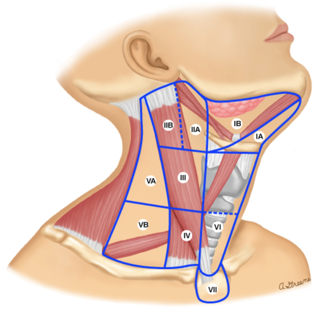
Neck Levels
I - submental and submandibular
Ia - submental - anteromedial to ant belly digastric
Ib - submandibular - posterolateral to ant belly digastric
II - Upper IJ chain (deep cervical)
IIa - anterior to IJV
IIb - posterior to IJV
III - Middle IJ chain (deep cervical)
Note I, II and III are all anterior triangle and above hyoid bone
IV - Lower IJ chain (deep cervical)
V - Posterior triangle
VI - Central (anterior compartment)
VII - inominates to the jugular notch
Prognosis
- Poor prognostic features from malignant salivary tumours
- Demography/lifestyle - older, male, smoker
- Pre-op facial nerve involvement
- Positive LN
- High grade, PNI, positive margins
Types
Benign
Pleomorphic Adenoma
Definition
- Benign salivary gland tumour
Epidemiology
- Rare <1%
- F>M 2:1
- Age 30-60
- Most common salivary gland tumour
- Most commonly affects parotid gland
Location
- Mainly parotid 80% but can be in any other
- Majority are superficial parotid
- Malignancy risk
- Low at 2-5%
- Carcinoma ex pleomorphic adenoma
- Can be within them therefore all should be excised
Risk Factors
- Previous head and neck irradiation
- Exposure - rubber, nickel
Histology
- Proliferation of
- Epithelial cells
- Myoepithelial cells
- Stromal/mesenchymal cells
- Pseudopod extensions
- Don’t enucleate - risk of recurrence higher
- Benign
- Circumscribed growth pattern without infiltration
Clinical
- Slow growing painless mass
Investigations
- USS - hypoechoic, lobulated, posterior acoustic enhancement
- CT - lobulated mass, homogenous
- MRI - soft tissue involvement
- USG - FNA or core biopsy for histology
Management
- Surgery
- Parotid - Superficial Parotidectomy facial nerve preservation
- Enucleation has high recurrence rates up to 50%
- Total parotidectomy does NOT reduce recurrence risk
- BUT does increase risk of nerve damage
- Submandibular - gland excision
- Parotid - Superficial Parotidectomy facial nerve preservation
- Radiation
- Indications
- Positive margins (should really re-resect if possible)
- Multiple recurrence
- Inoperable primary or recurrence
- Indications
- Recurrence
- Why does it occur?
- Pseudopod formation
- Incomplete capsule
- Close proximity to facial nerve and branches
- Why does it occur?
- Complications of Parotidectomy
- 5 F’s
- Frey’s syndrome - prevention → thick flaps (SCM), post op RTX, botox
- Formication
- Flap necrosis
- Fistula
- Facial N injury
- 5 F’s
Spiel on pleomorphic adenoma
- Most common benign salivary gland tumour
- Typified by epithelial, myoepithelial and stromal proliferation
- Mainly parotid (80%)
- RFs same as other tumours - environmental exposure - rubber, nickel compounds, hairdresser. prior radiation, viral - HIV, HPV, EBV
- Important due to small risk of harboring carcinoma ex pleomorphic adenoma 5%
- Painless neck lump
- Diagnosed with CT/MRI and biopsy (FNA or core)
- Mx is with superficial parotidectomy with facial nerve preservation
- Risks of surgery include Freys, formication, fistula and facial nerve injury
- Recurrence rate low with above, higher with enucleation
- Can be given RTX for recurrence but prefer surgery if possible
- Risk increases with recurrent surgery
Warthins Tumour
Definition
- Benign salivary gland tumour
Epidemiology
- Rare <1%
- 2nd most common benign parotid gland tumour
- M>F
Risk Factor
- SMOKING (only salivary gland tumour associated to smoking)
- Others
- Radiation
- EBV
Location
- Parotid (almost exclusively)
Features
- Commonly cystic and bilateral
Pathology
- Macro
- Well circumscribed mass
- Solid and cystic components
- Micro
- Bilayered oncocytic epithelium
- Cyst formation
- Lymphoid stroma
Clinical
- Painless lump near angle of mandible
Investigations
- Assess both sides - bilaterality and multiple
- USS - well defined mass, anechoic areas, post acoustic shadowin
- CT - bony involvement
- MRI - soft tissue involvement, encapsulated, hypo T1
- Biopsy - FNA or core
Management
- Usually superficial parotidectomy
- Surveillance if biopsy proven and not changing, asymptomatic
- Low recurrence rate and minimal malignant potential
Spiel on Warthin’s tumour
- 2nd most common benign salivary tumour
- Affects almost exclusively the parotid gland
- Typified by bilayered oncocytic epithelium, cyst formation and lymphoid stroma
- RF - SMOKING, prior RTX, viral (EBV/HPV/HIV) and exposures - nickel, rubber, hairdresser
- Minimal risk of malignant transformation
- Can avoid surgery and monitor if no sx
- Presents with painless lump
- Work up with USS/CT/MRI and FNA/core
- Mx is superficial parotidectomy and facial nerve preservation
- Minimal benefit for doing total even if deep component is involved
Malignant
Histology
- Features of malignancy
- Invasion
- PNI/LVI
- Necrosis
Theories of Development
Multicellular hypothesis - Arise from mature epithelium in various parts of salivary unit
- Reserve cell hypothesis
- Arise from undifferentiated or pluripotent cells
Types
- Mucoepidermoid carcinoma
- Adenoid cystic carcinoma
- Carcinoma ex pleomorphic adenoma
Grades
- Help to determine type of resection
- Low grade = only low grade mucoepidermoid carcinoma
- Superficial parotidectomy
- High grade = high grade mucoepidermoid carcinoma, adenoid cystic carcinoma and carcinoma ex pleomorphic adenoma
- Total parotidectomy UNLESS
- Located peripherally → superficial parotidectomy
- Low grade = only low grade mucoepidermoid carcinoma
Mucoepidermoid carcinoma
Definition
- Malignant neoplasm of the parotid gland and other salivary glands
Epidemiology
- F=M, age 40-50
- Most common salivary gland malignancy
- 1% of all cancers
- 5% of all H+N cancers
Risk Factors
- Radiation (ionizing radiation and H+N irradiation)
- Viral - EBV, HIV
- Immunosuppression
- Exposure - rubber and nickel
Pathology
- Macro
- Poorly circumscribed mass with incomplete capsule
- Cystic components
- Micro
- Mucous, intermediate and epidermoid cells
- Mixed features
Clinical
- Painless swelling
- Facial nerve palsy - invasion
- Fixed, solid
Investigations
- USS - well circ, hypoechoic, cystic component
- CT - same + higher grade = solid with infiltration
- MRI - solid high grade hypo T2, poor defined margins, PNI
- FNA/Core - core for grading is better
Staging
- Distant mets to lung, bone and liver so need CT CAP
Management
- Resectable, no distant mets and fit
- Surgical resection with R0 resection
- Superficial vs total and facial nerve
- Neck dissection
- Clinically positive - neck dissection
- Clinically negative - elective neck dissection if high risk
- Locally advanced, high grade, facial nerve involvement, LVI
- Adjuvant radiation - high risk
- Surgical resection with R0 resection
- Unresectable, distant mets, unfit
- Primary radiation
- Palliative chemo (poor response)
Prognosis
- High risk features - + margins, cervical mets, aggressive histo/grade, PNI/LVI, extraglandular extension
- Determined by - stage, site, margins
- 60% 5YS adults
Adenoid Cystic Carcinoma
- Definition
- Same - Malignant neoplasm of the parotid gland and other salivary glands
- Epidemiology
- Same, age 50-60
- Risk Factors
- Same
- Pathology
- Macro - infiltrative firm mass
- Micro - biphasic with ductal and myoepithelial differentiation, tubular/cribriform/solid
- Clinical, investigations and management
- Same
- Prognosis
- 5YS 80%
- 15YS 30%