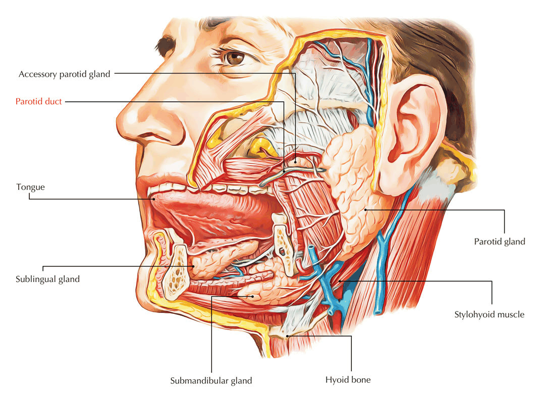
Overview
- Mainly serous gland with only a few scattered mucous acini
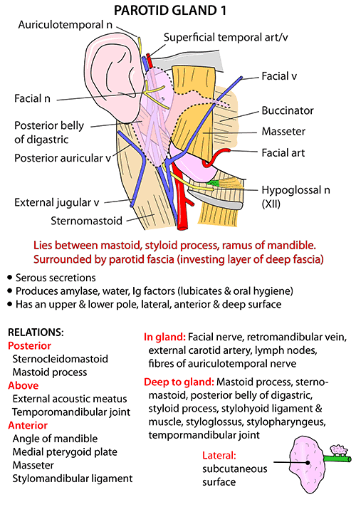
- Wedge-shaped on transverse section
- Extends from Zygomatic Arch to the upper part of the neck
- Overlaps the Posterior Belly of Digastric and the anterior border of Sternocleidomastoid
- Anteriorly, gland overlaps Masseter
- Accessory Parotid – small, usually detached gland lies above the Parotid Duct on the aponeurotic part of the Masseter
- Extends below the External Acoustic Meatus posteriorly onto the Mastoid Process
- Occupies gap between the Ramus of the Mandible and the Mastoid and Styloid Processes of the Temporal Bone
- Reaches close to the lateral wall of the Oropharynx
- Thus, there is a need to look at the region of the Fauces when examining a patient with a Parotid Mass
Relationships
- Lateral (Superficial) Surface:
- Covered by skin and Superficial Fascia
- Investing Layer of Deep Cervical Fascia splits to envelope the gland
- a.k.a. Parotid Sheath
- Inner leaf passes up to the Base of the Skull
- Condensation of the fascia known as the Stylomandibular Ligament attaches from the Styloid Process to the Styloid Process of the Mandible
- This partitions the Parotid Gland from the posterior aspect of the Submandibular Gland
- Outer leaf extends superiorly as the Parotidomasseteric Fascia
- Reaches the Zygomatic Arch
- On the gland, fascia termed Parotid Capsule or Masseteric Fascia more anteriorly
- Superficial Muscular Aponeurotic System (SMAS) overlies the gland
- Anteromedial Surface
- Grooved by the posterior border of the Mandibular Ramus
- Related to the Masseter and Medial Pterygoid muscles which are attached to the Ramus
- Gland wrapped around the Capsule of the Temporomandibular Joint
- Anterior edge of this surface meets the lateral surface over, as well as below, the Masseter forming the irregularly convex anterior border of the gland
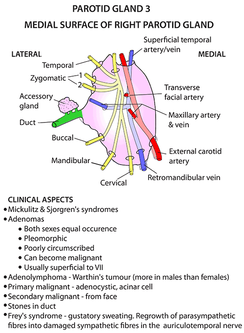
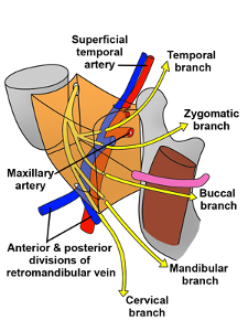
- Parotid Duct and Facial Nerve branches emerge from the anteromedial surface and run forwards deep to the anterior border
- Terminal branches of External Carotid Artery (i.e. Superficial Temporal and Maxillary Arteries) leave this surface further back
- Posteromedial Surface
- In contact with the Mastoid Process with its attached Sternocleidomastoid and Posterior Belly of Digastric muscles
- SCM attached to superficial surface
- Posterior Belly of Digastric attached to deep surface
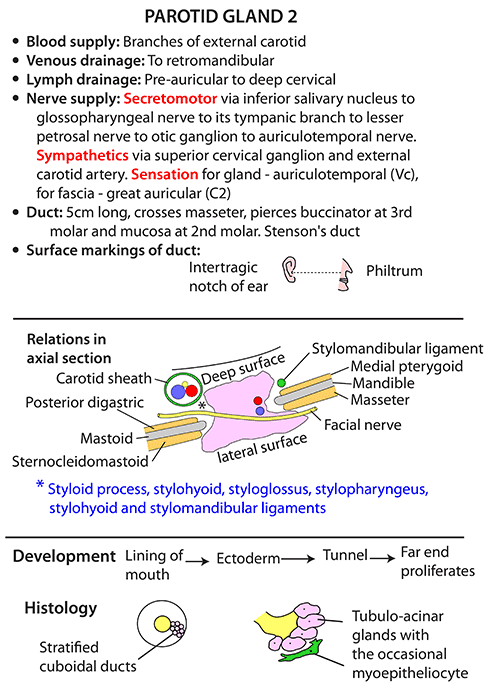
- More medially, the Styloid Process and its attached muscles (i.e. Stylohyoid, Stylopharyngeus, and Styloglossus) separate the gland from the Carotid Sheath
- External Carotid Artery enters the gland through the lower part of this surface
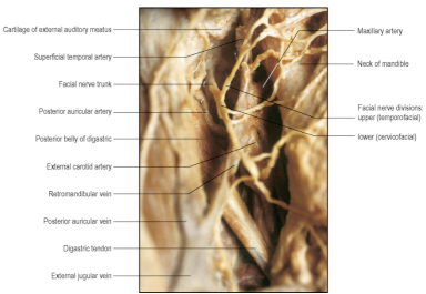
- Facial Nerve trunk (or its Temporofacial and Cervicofacial divisions) enter the gland between the Mastoid and Styloid Processes
- In contact with the Mastoid Process with its attached Sternocleidomastoid and Posterior Belly of Digastric muscles
- Within
- Facial Nerve
- Branches run in different directions in different (superficial to deep) planes
- Surgeons consider the gland to have a superficial and deep part separated by the plane of the Facial Nerve
- No specific, developmentally determined plane in which the Facial Nerve branches pass between Superficial and Deep Lobes of the gland; the Parotid is an integral gland not divided into lobes
- Within the gland, nerve branches communicate with each other forming a plexiform arrangement
- Lies superficial to the Retromandibular Vein (which is superficial to the External Carotid Artery)
- Branches run in different directions in different (superficial to deep) planes
- Retromandibular Vein
- Formed within the Parotid Gland by the confluence of the Superficial Temporal and Maxillary Veins
- Emerges from the lower part (pole) of the Parotid Gland
- Divides into anterior and posterior branches
- Anterior branch joins the Facial Vein to form the Common Facial Vein
- Posterior branch joins the Posterior Auricular Vein to form the External Jugular Vein, which then drains into the Subclavian Vein
- Division may occur within the Parotid Gland and the two branches emerge from the lower pole
- External Carotid Artery
- Pierces the Parotid Fascia to enter the Parotid Gland
- Divides within the gland behind the neck of the Mandible into Maxillary Artery and Superficial Temporal Artery
- As it lies within the Parotid Gland, it is separated from the Internal Carotid by the deep part of the Gland, its fascia, Styloid Process and it’s continuation the Stylohyoid Ligament, Styloglossus and the ‘Pharyngeal Structures’
- Stylopharyngeus muscle
- Glossopharyngeal Nerve
- Pharyngeal Branch of the Vagus Nerve
- Lymph Nodes
- Off the Preauricular (Parotid) group lie on or deep to the fascial capsule of the Parotid, as well as within the gland
- Facial Nerve
Parotid Duct (of Stensen)
- Length: 5cm
- Course:
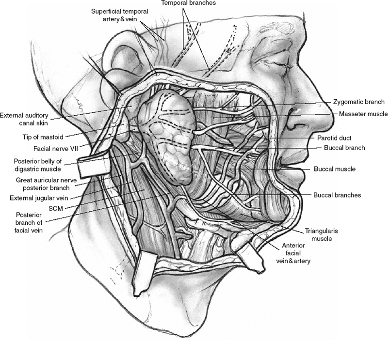
- Passes forwards across the Masseter
- Turns around anterior border of Masseter
- Pass through Buccal fat pad
- Pierce Buccinator
- Opens on the mucous membrane of the check opposite the 2nd upper molar tooth
- Pierces Buccinator further back and runs forwards beneath the mucous membrane
- When intraoral pressure is raised, the submucous part of the duct is compressed between the buccinator and the mucous membrane, preventing inflation of the gland
- Surface marking: Lies on the middle third of a line between the Intertragic Notch of the Auricle and the Midpoint of the Philtrum
- Palpable on the clenched Masseter
Accessory Parotid Gland
- Usually lies on the Masseter between the Parotid Duct and the Zygomatic Arch
- Several small ducts open into the Parotid Duct
- The Accessory Parotid Gland and the Parotid Duct both lie on the aponeurotic part of the surface of the Masseter
Blood Supply
- Branches from the External Carotid Artery
- Venous return via Retromandibular Vein
Lymph Drainage
- Drains to Pre-auricular (Parotid) lymph nodes ⇒ Nodes of the upper group of Deep Cervical Nodes
Nerve Supply
-
Secretomotor fibres:
- From cell bodies in the Otic Ganglion
- Reach the gland by hitch-hiking along the Auriculotemporal Nerve
- Auriculotemporal Nerve – branch of the Mandibular division of the Trigeminal Nerve
- Nerve is in contact with the anteromedial surface of the Parotid Gland as it passes backwards along the Mandibular Neck and ascends behind the Temporomandibular Joint
- Pre-Ganglionic fibres arise from Cell bodies in the Inferior Salivary Nucleus in the Medulla
- Travel by way of:
- Glossopharyngeal Nerve ⇒ Tympanic Branch of Glossopharyngeal Nerve - → Tympanic Plexus (on the Promontory of the Cochlear in the Middle Ear) and Lesser Petrosal Nerve ⇒ Otic Ganglion ⇒ Auriculotemporal Nerve
-
Sympathetic (vasoconstrictor) Fibres
- Reach gland from Superior Cervical Ganglion via plexus on External Carotid and Middle Meningeal Arteries
Development
- A groove that appears in the Ectoderm of the mouth pit (Stomodeum) becomes converted into a tunnel
- Cells from the blind end proliferate to form the Parotid Gland
- Development starts in the 5th week of gestation (first salivary gland to develop)
Surgical Approach
- Most common neoplasm of the Parotid Gland: Pleomorphic Adenoma (mixed Parotid tumour)
- Requires removal with a margin of normal Parotid tissue, conserving the Facial Nerve and its branches
- Approached via S-shaped incision made from in front of the ear, backwards to the Mastoid Process and then downwards and forwards below the Angle of the Mandible
- Make incision in 3 parts – from below upwards first – avoids bleeding from the upper part of the wound obscuring your field lower down
- Gland retracted forwards from the Sternocleidomastoid to expose the Posterior Belly of Digastric and Stylohyoid and Cartilage of the External Meatus
- Facial Nerve approached along a plane in front of the anterior margin of the cartilage
- Trunk emerges from the Stylomastoid Foramen
- Just deep to the junction of the cartilaginous and bony parts of the External Meatus
- Cartilage in this region has an arrow-headed projection pointing downwards to the nerve trunk
- About 1cm above and medial to the upper end of the Posterior Belly of Digastric
- Stylomastoid branch of Posterior Auricular Artery is superficial to the Facial Nerve and is a guide to its proximity
- Once identified, Facial Nerve followed forwards into the Parotid Gland and the required amount of Parotid tissue is removed with preservation of Facial Nerve Branches
- Alternative approach: find a Facial Nerve Branch as it leaves the gland and follow it in a centripetal manner back to the Trunk and other branches
- Marginal Mandibular Branch may be identified as it lies superficial to the Retromandibular Vein or its Anterior branch – aided by the colour contrast between the white nerve and the dark vein
- Occasionally, this nerve will pass behind the vein
- Alternatively, the Cervical Branch may be followed up from its communication with the Ascending Branch of the Transverse Cervical Nerve
- Marginal Mandibular Branch may be identified as it lies superficial to the Retromandibular Vein or its Anterior branch – aided by the colour contrast between the white nerve and the dark vein
Post-Gustatory Sweating/Frey’s Syndrome
- Caused by aberrant regeneration of post-ganglionic parasympathetic nerve fibres from the Auriculotemporal Nerve supplying the Parotid Gland to severed Post-ganglionic sympathetic fibres which innervate the sweat glands of the face and blood vessels of the skin
Visible Structures following Superficial Parotidectomy
- Sternocleidomastoid along inferior border
- Anterior branch of Greater Auricular Nerve lying on SCM
- Branches of Facial Nerve
- Posterior Belly of Digastric attached to the inner aspect of Mastoid Process
Images