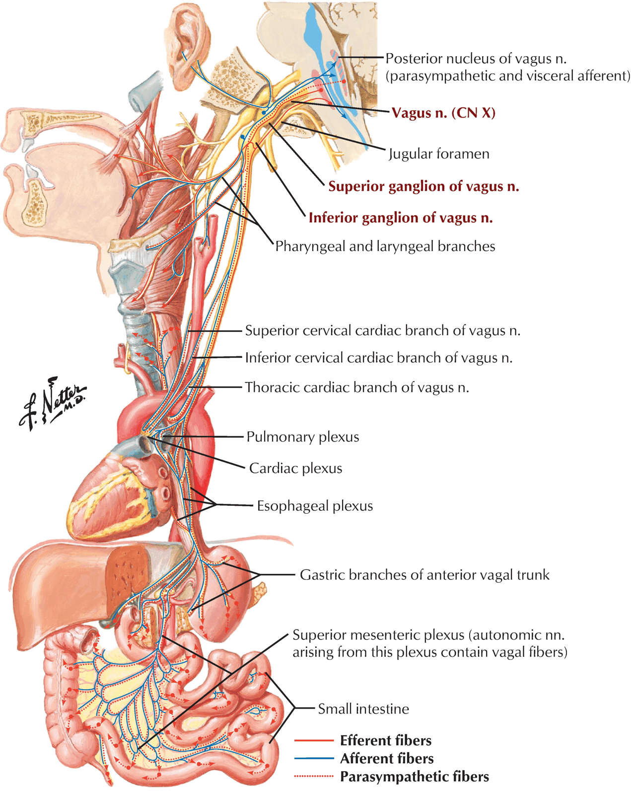
Emerge from Medulla in a series of rootlets below those of the Glossopharyngeal Nerve
Unite into a single nerve
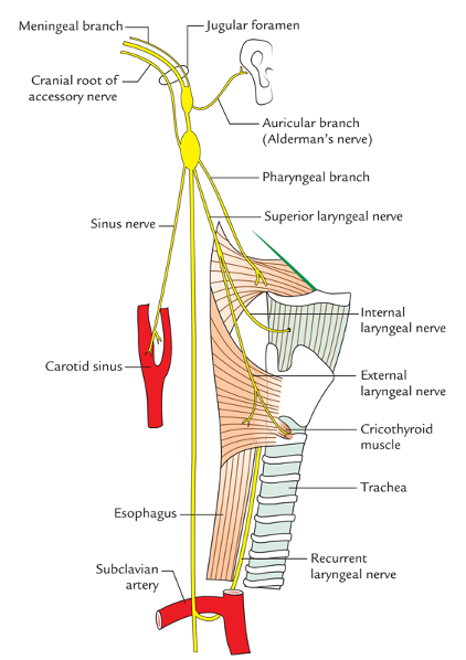
Enters the middle compartment of the Jugular Foramen (with the Accessory Nerve – sharing an arachnoid and dural sheath)
Small Superior Ganglion just above the long Inferior Ganglion – lies in the Jugular Fossa below skull base
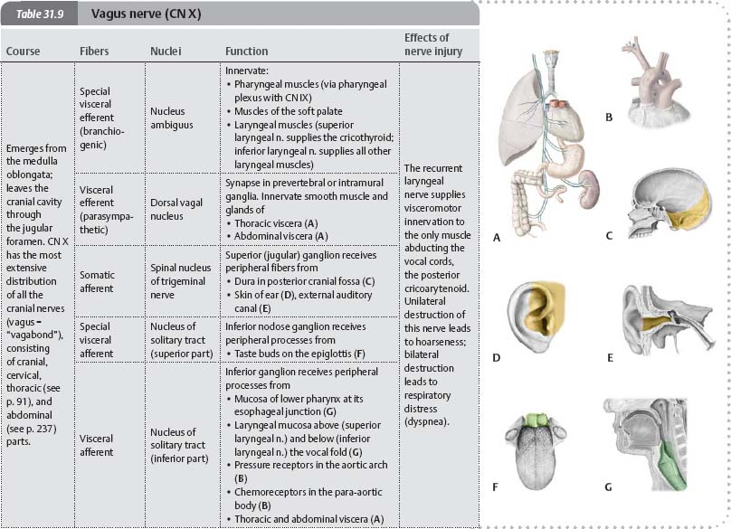
Superior Ganglion has cell bodies for the Auricular Branch
Inferior Ganglion has cell bodies of all other sensory fibres in the Vagus Nerve
Fibres of Cranial Root of Accessory Nerve join the Vagus just above the Inferior Ganglion – thereby, Accessory Nerve gives its Nucleus Ambiguus fibres to the Vagus
Neck
Lies vertical in the Carotid Sheath – deep in the gutter between the Internal and Common Carotid Artery and Internal Jugular Vein
Meningeal, Auricular and Carotid Body Branches are all afferent
Meningeal branch carries C1 and C2 fibres which join the Vagus at its exit from the skull
Supply dura mater of the posterior fossa below the tentorium
Auricular Branch (Arnold’s Nerve)
Runs laterally through a canaliculus in the lateral wall of the Jugular Fossa
Supplies the posteroinferior quadrant of the outer surface of the tympanic membrane and a small adjacent area of the skin of the External Acoustic Meatus and a small area of skin on the cranial auricular surface
Carotid Body Branch
Forms a plexus with the Carotid Sinus branch of the Glossopharyngeal Nerve
Branches in the neck
Pharyngeal Branches
Passes between the Internal and External Carotid Arteries
Provides both motor and sensory fibres for the Pharyngeal Plexus on the Middle Constrictor Muscle
Supply muscles of the Pharynx (except Stylopharyngeus)
Supply muscles of the Soft Palate (except Tensor Palati)
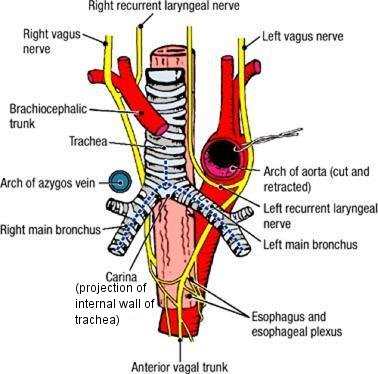
The right recurrent laryngeal nerve loops around the right subclavian artery around the T1–T2 vertebral level.
The left recurrent laryngeal nerve loops around the aortic arch around the T4–T5 vertebral level and ascends in tracheoesophageal groove.
Innervation
All intrinsic muscles of larynx except the cricothyroid muscle.
Sensation below the vocal fold
Cervical Cardiac branches
The cervical cardiac branches (sometimes ambiguously called superior cardiac branches) of vagus nerve, two or three in number, arise from the vagus, at the upper and lower parts of the neck.
The upper branches are small, and communicate with the cardiac branches of the sympathetic. They can be traced to the deep part of the cardiac plexus.
The lower branch arises at the root of the neck, just above the first rib. That from the right vagus passes in front or by the side of the innominate artery, and proceeds to the deep part of the cardiac plexus; that from the left runs down across the left side of the aortic arch, and joins the superficial part of the cardiac plexus.
Chest
Left Vagus:
Held away from trachea by the great vessels from the Aorta
Crosses Aortic Arch medial to the Left Superior Intercostal Vein
Left Vagus Nerve crossed by the Left Superior Intercostal Vein on the Arch
Left Superior Intercostal Vein passes forwards across the arch superficial to the Vagus, deep to the Phrenic, to empty into the Left Brachiocephalic Vein
Crossed by Left Phrenic Nerve just above the Arch of the Aorta
Right Vagus:
In contact with trachea
Lies on trachea medial to the arch of the Azygos Vein
Each Vagus:
Passes down behind/posterior to the lung root
Phrenic Nerve passes anterior to the lung root
Between them lie the Sympathetic and Vagus Branches to the Superficial Part of the Cardiac Plexus
Divides into branches which contribute to the pulmonary plexuses
Pass onwards to form plexus on oesophagus
Innervations in the Chest
Baroreceptors in the adventitial layer of the Arch of the Aorta
Aortic bodies – chemoreceptors in the region of the Ligamentum Arteriosum
Anterior and Posterior Vagi in the Abdomen
Through these branches, the Vagi supply the Foregut and Midgut and their derivates – Biliary Tract and Pancreas
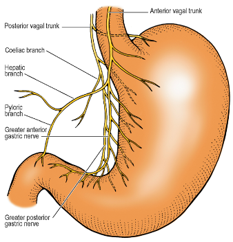
Anterior Vagal Trunk
Comprised mainly left vagal fibres from the Oesophageal Plexus in the posterior mediastinum
Lies in contact with anterior oesophageal wall (usually nearer to its right margin)
Also gives off several gastric branches which supply Fundus and Body
Gives off the Greater Anterior Gastric Nerve or Anterior Nerve of Latarget ⇒ Runs down in Lesser Omentum near the Lesser Curvature with the Left Gastric Artery ⇒ Subdivides in manner of Crow’s foot to supply Antrum and Pyloric Sphincter
Hepatic Branch – one or two branches derived from Anterior Vagal Trunk
Runs in upper part of Lesser Omentum to join the plexus on the Hepatic Artery and Portal Vein
Turns down in anterior wall of Epiploic Foramen to reach Pylorus
Posterior Vagal Trunk
Comprised mainly of right vagal fibres
Lies in loose tissue a little behind and to the right, not in contact with the posterior surface of the oesophagus
Gives Coeliac branches that run backwards along the Left Gastric Artery to the Coeliac Plexus
Coeliac Plexus supplies large bowel up to transverse colon
Gives Gastric branches to the Fundus and Body
Gives off the Greater Posterior Gastric Nerve or the Posterior Nerve of Latarget ⇒ Runs in the Lesser Omentum behind the Anterior Trunk to reach the Antrum (but NOT the pyloric sphincter)
Consequence of Nerve Division at Hiatus
A truncal vagotomy divides both vagal trunks 4cm proximal to the gastroesophageal junction
The consequences include:
Reduced gastric acid secretion due to removal of the acetylcholine-mediated secretion of acid from the parietal cells
Accelerated emptying of liquids due to removal of relaxation of the gastric fundus
Decreases the emptying of solids due to removal of relaxation of the pylorus
Summary
Special (visceral) efferent
Innervates the pharyngeal and laryngeal (branchiomeric) muscles