Anatomical Structure
The sympathetic nervous system consists of the following components:
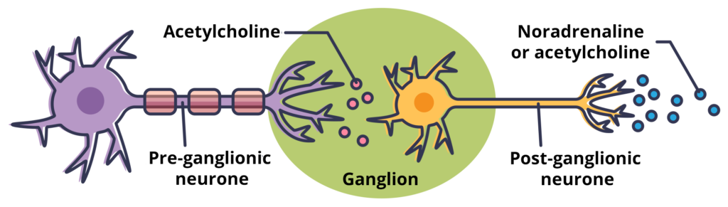
- Pre-ganglionic fibres – these originate within the thoracic and lumbar divisions of the spinal cord. They end by synapsing with post-ganglionic fibres at peripheral ganglia.
- Peripheral ganglia – groups of neuronal cell bodies where the pre-ganglionic and post-ganglionic fibres synapse.
- Post–ganglionic fibres – these begin at the peripheral ganglia and carry sympathetic fibres to the target organs.
Pre-Ganglionic Neurones
The pre-ganglionic neurones originate in the thoracic and lumbar segments of the spinal cord. They are short, myelinated, and release acetylcholine.
They exit the spinal cord via the anterior spinal nerves and then pass through white ramus communicantes to enter the sympathetic trunk.
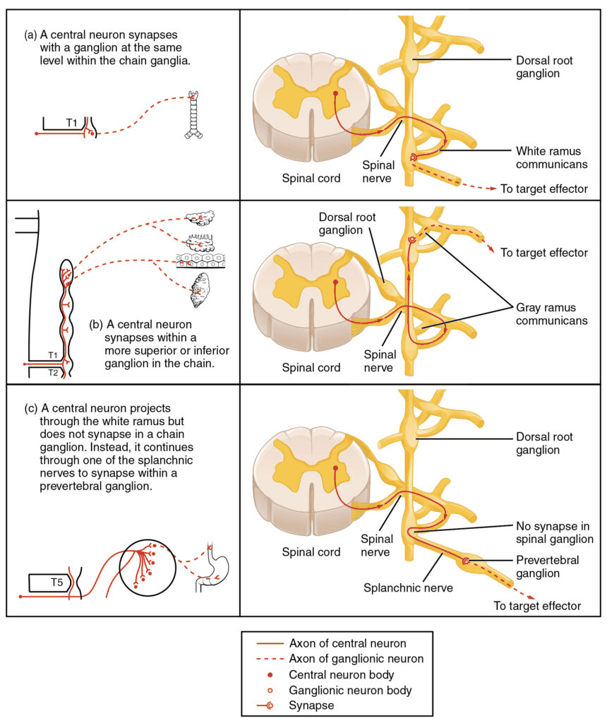
Within the sympathetic trunk, the sympathetic fibres may take one of three actions:
- Synapse with post-ganglionic neurones – occurring within a sympathetic ganglion at the same spinal level.
- Ascend or descend within the sympathetic chain – to synapse with post-ganglionic neurones at a higher or lower spinal level.
- Pass through the chain without synapsing – will later synapse at a prevertebral ganglion or the adrenal medulla (these fibres are known as splanchnic nerves)
Peripheral Ganglia
The peripheral ganglia are groups of neuronal cell bodies where the pre-ganglionic and post-ganglionic fibres synapse.
In the sympathetic nervous system, they are organised into two main structures:
- Sympathetic trunks – two chains of interconnected ganglia that lie either side of the vertebral column.
- Prevertebral ganglia – three ganglia arranged around the major branches of the abdominal aorta (coeliac, superior mesenteric and inferior mesenteric ganglia).
Sympathetic Chain
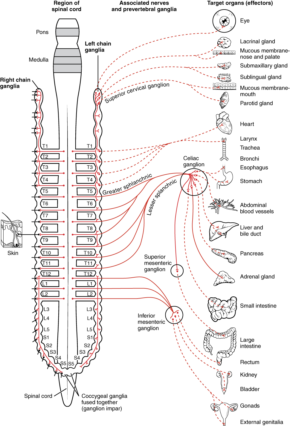
The sympathetic chain (sympathetic trunk or paravertebral ganglia) are 22 pairs of interconnected neuronal cell bodies which run parallel either side of the vertebral column – extending from the base of the skull to the coccyx.
The sympathetic chain may be broken down into regions, each of which has multiple ganglia:
- Cervical ganglia– three ganglia (superior/middle/inferior)
- Thoracic – eleven or twelve ganglia
- Lumbar – four ganglia
- Sacral – four or five ganglia
- Coccyx – a single, ganglion impar
Prevertebral Ganglia
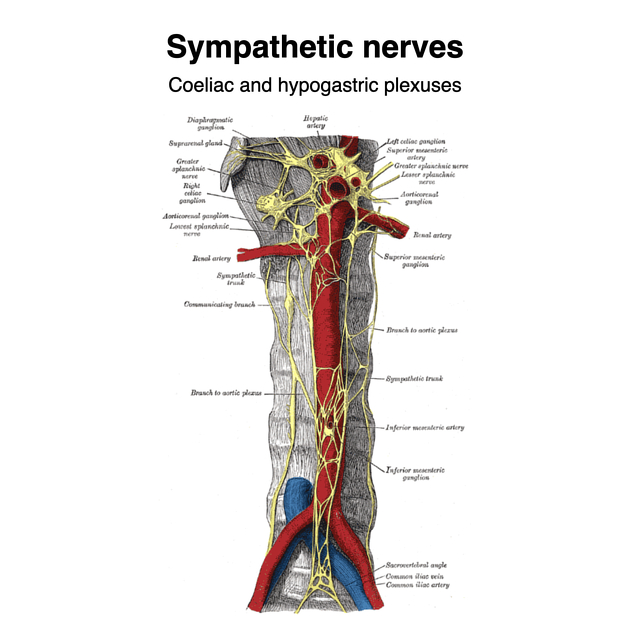
The prevertebral ganglia or plexus (pre-aortic ganglia or collateral ganglia) are a collection of cell bodies located anterior to the abdominal aorta.
- Thoracic
- Cardiac plexus
- Divided into deep and superficial
- Parasympathetic
- Cardiac branches of the Vagus Nerve
- Sympathetic
- Cervical ganglia
- Superior cervical cardiac nerve (a visceral branch of the Superior cervical ganglia)
- Middle cervical cardiac nerve (a visceral branch of the Middle cervical ganglia)
- Inferior cervical cardiac nerve (a visceral branch of the Inferior cervical ganglia or cervicothoracic ganglion; a.k.a. Stellate ganglion)
- Thoracic cardiac nerves (branches of the upper four or five thoracic sympathetic ganglia).
- Cervical and thoracic cardiac nerves contain post-ganglionic sympathetic neurons, which arise from the above-mentioned cervical and thoracic sympathetic ganglia.
- Cervical ganglia
- Pulmonary plexus
- Parasympathetic
- Pulmonic branches of the right and left Vagus Nerve
- Sympathethetic
- The post-ganglionic sympathetic neurons arise from the upper thoracic sympathetic ganglia.
- Whereas, the preganglionic parasympathetic neurons originate from the dorsal motor nucleus of vagus and arrive into the pulmonary plexus via the
- Parasympathetic
- Cardiac plexus
- Abdominopelvic
- Phrenic plexus
- Coeliac plexus
- Supply from Greater splanchnic nerve and Lesser splanchnic nerve
- Foregut - It supplies liver, gallbladder, stomach, spleen, pancreas, part pf duodenum
- Hepatic plexus
- Periarterial extension of the coeliac plexus along the common hepatic artery and portal vein
- Aorticorenal plexus
- Inferior perivascular extension of the larger coeliac plexus (Greater splanchnic nerve and Lesser splanchnic nerve)
- Direct innervation from the Lesser splanchnic nerve
- Supplies the kidneys, abdominal aorta and gonads
- Renal plexus
- Lateral perivascular extension of the aorticorenal plexus.
- Least splanchnic nerve
- Sympathetic supply of the kidneys
- Superior mesenteric plexus
- Formed by branches of the coeliac plexus
- Lesser splanchnic nerve
- Midgut
- Inferior mesenteric plexus
- Formed mainly from branches from the aorticorenal plexus
- Some contributions from the lumbar splanchnic nerves (sympathetic)
- Minor contributions arise via the left and right hypogastric nerves from the inferior and superior hypogastric plexus (parasympathetic)
- Superior hypogastric plexus
- Anterior to the aortic bifurcation extending inferiorly between the common iliac arteries and along the left common iliac vein and median sacral vessels
- Origin
- Lumbar splanchnic branches (sympathetic)
- Nerves from the aorticorenal plexus also contribute
- Gives origin to a complex network of small branches and the left and right hypogastric nerves which extend inferiorly to the inferior hypogastric plexuses
- Supplies large bowel distal to the left third of the transverse colon, ureters, gonads and the iliac vessels
- Inferior hypogastric plexus
- The plexuses and ganglia lie in the pelvis within the extraperitoneal connective tissue of the pelvic sidewalls anterolateral to the mesorectum
- Origin
- Formed mainly from Pelvic splanchnic nerves branches (parasympathetic) and sacral splanchnic branches (sympathetic)
- Some contributions arise via the left and right hypogastric nerves from the superior hypogastric plexus
- Branches and supply: gives origin to a complex network of small branches that supply the pelvic viscera
- Males: ductus deferens, seminal vesicles, prostate, accessory sex glands, penis
- Females: ovary, uterine tubes, uterus, cervix, vagina
- Both: urinary bladder
Splanchnic nerves
Adrenal Medulla
- The adrenal medulla is a modified sympathetic ganglion.
- Although a target organ, the pre-ganglionic fibres synapse directly in the medulla (having passed through the sympathetic chain and coeliac ganglion without synapsing).
- Upon sympathetic stimulation, the adrenal medulla secretes adrenaline and noradrenaline into the bloodstream – where they have a more longer lasting hormonal effect on target organs.
- Supplied by Greater splanchnic nerve
Post-Ganglionic Neurones
- The post-ganglionic neurones originate in the peripheral ganglia and carry signals to the target organs.
- They travel to their target organs either as distinct, named nerves, alongside blood vessels (arteries in particular) or within autonomic plexi.
- Their axons are typically long and unmyelinated. They release noradrenaline (with the exceptions of neurones synapsing at sweat glands and erector pili muscles in skin, which release acetylcholine).
| Pre-Ganglionic Fibre (cholinergic) | Post-Ganglionic Fibre (most adrenergic)* | |
|---|---|---|
| Length | Short | Long |
| Myelination | Yes | No |
| Neurotransmitter released | Acetylcholine | Noradrenaline* |
| * Exception of nerves to sweat glands and erector pili muscles where acetylcholine is released>) |