Overview
- Two symmetrical lobes
- United by an Isthmus anterior to 2nd, 3rd and 4th Tracheal Rings
- Lobes:
- Lie on either side of Trachea and Larynx
- Extends from Oblique Line of Thyroid Cartilage to 6th Tracheal Ring
- Weight: Approx 25g
- Enclosed by own capsule AND Pretracheal Fascia
Lateral Lobe
- Narrow upper pole and broader lower pole
- Triangular in cross section
- Lateral, medial and posterior surfaces
- Lateral surface:
- Deep to Sternothyroid and Sternohyoid
- Medial surface:
- Lies against lateral side of Larynx and upper Trachea
- Lower Pharynx and Upper Oesophagus immediately behind
- Related to:
- Cricothyroid Muscle of Larynx
- Inferior Constrictor of Pharynx
- External Laryngeal Nerve
- Recurrent Laryngeal Nerve
- Also related medially to Cricopharyngeus
- Posterior surface:
- Overlaps medial part of Carotid Sheath (i.e. the part containing the Common Carotid Artery)
- If enlarged, the lobe may extend across the more laterally placed Internal Jugular Vein
- In contact with Parathyroid Glands – between it and the Fascial Sheath
Tubercles of Zuckerkandl
- Lateral or posterior projection from the lateral lobe of the Thyroid
- Originates from the ventral portion of the Ultimobranchial Body
- In > 90% of cases, Recurrent Laryngeal Nerve runs posterior to the Tubercle, either in:
- Tracheo-oesophageal Groove (60%)
- Posterior (24%)
- Lateral (5%) to Trachea
- Only a small percentage (¬5%) of cases does the Recurrent Laryngeal Nerve run anterior to the Thyroid – usually re-op surgeries
- Relationship to Inferior Thyroid Artery variable:
- 85% - posterior and intertwined between branches
- 15% - anterior (more common on the right)
Recurrent Laryngeal Nerve
External Laryngeal Nerve
Cernea Classification
Categorizes relationship between External Laryngeal Nerve and Superior Thyroid Artery [WB] Type 1: - Nerve crosses the Superior Thyroid Vessels > 1cm above the upper pole of Thyroid Type 2a: - Crosses the Superior Thyroid vessels < 1cm above the upper pole of Thyroid Type 2b: - Crosses at or below level of upper pole of Thyroid
Isthmus
- Joins anterior parts of the lobes towards their lower poles
- Posterior surface firmly adherent to 2nd, 3rd and 4th Tracheal Rings
- Pretracheal Fascia here fixed between them
- This fixation and the investment of the whole gland by Pretracheal Fascia is responsible for the gland to move up and down with the Larynx during swallowing
- An anastomosis between the two Superior Thyroid Artery runs across the upper border of the Isthmus
- Tributaries of the Inferior Thyroid Veins emerge from its lower border
Pyramidal Lobe
- Small portion of gland substance projecting upwards from the Isthmus
- Generally to the left of midline
- Represents a development of glandular tissue from the caudal end of the Thyroglossal Duct
- May be attached to the inferior border of the Hyoid Bone by fibrous tissue
- Levator Glandulae Thyroidae – muscle fibres present within this fibrous tissue
- Nerve supply: External Laryngeal Nerve
- Levator Glandulae Thyroidae – muscle fibres present within this fibrous tissue
Accessory Thyroid Glands
- Not uncommonly found near:
- Hyoid
- In the Tongue
- In Superior Mediastinum
- Anywhere along the path of descent of the Thyroglossal Duct
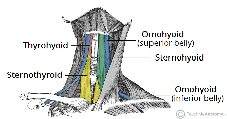
Strap Muscles
-
Consists of:
- Sternohyoid
- Omohyoid
- Sternothyroid
- Thyrohyoid
-
Nerve Supply:
- Sternohyoid – Anterior rami of spinal nerves (C1-C3) via Ansa Cervicalis
- Omohyoid – Superior root of Ansa Cervicalis (Superior belly) and Ansa Cervicalis (Inferior Belly)
- Sternothyroid – Anterior rami of C1-C3 via the Ansa Cervicalis
- Thyrohyoid – Branch of Hypoglossal Nerve (but all fibres are hitch-hiking from C1)
Blood Supply
Superior Thyroid Artery
- Origin
- Arises at commencement of External Carotid Artery
- 1st branch from the anterior aspect of the External Carotid Artery after giving off its Sternocleidomastoid and Superior Laryngeal branches
- Arises at commencement of External Carotid Artery
- Course
- Pierces Pretracheal Fascia
- Runs almost vertically downwards, with the vein, to the upper pole of Thyroid
- Runs with the External Laryngeal Nerve
- The Cernea Classification describes this relationship
- 4 branches prior to reaeching the upper pole
- A single vessel reaches summit of upper pole
- Artery divides on the gland into anterior and posterior glandular branches
- Anterior branch – runs down to Isthmus
- Posterior branch – runs down the back of the lobe and anastomoses with an ascending branch of the Inferior Thyroid Artery from the lower pole
- In thyroidectomy – artery ligated close to upper pole or its anterior and posterior branches are ligated instead – avoid damage to External Laryngeal Nerve
- Branches
- Before reaching Thyroid, it gives off
- Infrahyoid branch
- SCM brnach
- Superior Laryngeal branch
- Superior Laryngeal Artery pierces the Thyrohyoid Membrane with the Internal Laryngeal Nerve
- Cricothyroid branch
- Cricothyroid Artery crosses the upper part of the Cricothyroid membrane to anastomose with the contralateral artery
- Before reaching Thyroid, it gives off
Inferior Thyroid Artery
- From Thyrocervical Trunk
- Arches upwards and medially behind the Carotid Sheath
- Loops downwards to lower pole
- Divides outside Pretracheal Fascia into branches that pierce the fascia separately to reach the lower part of the gland
- Recurrent Laryngeal Nerve has variable relationship with artery
- BUT always lies behind the Pretracheal Fascia
- 85% - posterior and intertwined between branches
- 15% - anterior (more common on the right)
- To safeguard the nerve, ligate the Inferior Thyroid Artery well lateral to the gland or carefully ligate its small branches on the surface of the gland
- Branches
- Ascending Cervical Artery (or this can come off Thyrocervical Trunk)
- Small Pharyngeal branches
- Oesophageal branches
- Tracheal branches
- Inferior Laryngeal artery
- Inferior Laryngeal Artery ascends with the Recurrent Laryngeal Nerve
Thyroidea Ima Artery
- Arises from Brachiocephalic Trunk, Arch of the Aorta or the Right Common Carotid Artery
- Enters the lower part of the Isthmus in 3% of individuals
Venous Drainage
- Venous plexus on gland’s surface
Thyroid veins
- Superior thyroid vein
- Follows the Superior Thyroid Artery
- Enters either the Internal Jugular Vein or Facial Vein in equal proportions
- Middle thyroid vein
- Crosses anterior to Common Carotid Artery
- Drains into the Internal Jugular Vein
- Early ligature and division of this vein during thyroid surgery facilitates mobilisation of the gland
- Inferior Thyroid Vein
- Drain downwards mainly into Left Brachiocephalic Vein – one may enter the Right Brachiocephalic Vein
Lymph Drainage
- Mainly to Deep Cervical nodes
- Few pass into Pre-Laryngeal, Pre and Para-Tracheal Nodes and directly into the Thoracic Duct
Nerve Supply
- Sympathetic (vasoconstrictor) nerves from Superior, Middle and Inferior Cervical Ganglia – accompany Thyroid Arteries
Structure
- Thyroid made up of rounded follicles containing varying amounts of colloid produced by a single layer of Epithelial (Follicular) cells that form the walls of the follicles
- Only endocrine gland to store its secretions outside the cells
- Colloid is iodinated when in the follicle
- Reabsorbed by the cells before being discharged into blood capillaries
- Main hormonal products: Thyroxine (T4) and Triiodothyronine (T3)
- Parafollicular Cells or C-Cells - make up < 2% of the epithelial cells
- Secrete Calcitonin
- Scattered on outer aspects of follicles
- Do not reach the lumina of the follicles
Embryology of thyroid
- Develops as a proliferation of cells from the caudal end of the Thyroglossal Duct
- Thyroglossal Duct
- Originates from the Endoderm of the Floor of the Pharynx at the Foramen Caecum in the region of the developing tongue
- Then passes caudally in front of the Hyoid Bone, behind which it forms a recurrent loop
- Thyroid gland buds from the duct’s distal end
- Distal end, itself, may give rise to the Pyramidal Lobe
- Other remnants of the duct may persist as Accessory Thyroid Glands or Thyroglossal cyst
- Removal of Retrohyoid Duct Remnants usually requires excision of a segment from the middle of the Hyoid Bone]
- Failure of descent of the Thyroglossal Duct may result in the development of a Lingual Thyroid
- Parafollicular Calcitonin-producing cells develop from the Ultimobranchial Body (5th Pharyngeal Pouch) under influence of Neural Crest Cells
Surgical Approach
- Transverse incision in a low skin crease at the front of the neck
- 2 finger-breadths above the clavicle and between the sternocleidomastoids
- Layers divided: Skin ⇒ Subcutaneous Fat ⇒ Platysma ⇒ Develop superior platysmal flap by dissecting in subplatysmal plane ⇒ Raise upper flap as far as Thyroid Cartilage + Raise lower flap as far as Sternal Notch ⇒ Incise Investing Layer of Deep Cervical Fascia vertically ⇒ Incise Midline Raphe between strap muscles ⇒ Strap muscles retracted laterally ⇒ Incise Pretracheal Fascia
- Sternohyoid and Sternothyroid muscles retracted
- Or divided at a high level to safeguard their nerve supply from the Ansa Cervicalis and prevent adherence of their subsequent suture line to the closure of the skin incision
- Pretracheal Fascia divided to expose the gland proper
Structures Spared During Initial Exposure
- Strap muscles
- Anterior Jugular Veins
Structures Divided During Initial Exposure
- Platysma
- Midline Raphe