Types
- Minimal invasive
- Single gland
- Unilateral neck
- Localised to one side
- Should find a adenoma and a normal gland
- IF find two normal glands then need to stop or continue to bilateral
- Localised to one side
- Bilateral neck
- Indications
- No localisation
- 4 gland
- Two options
- Subtotal
- Leave 1/2 a node (inferior as easier access)
- Total
- With re implant
- SCN or BC
- Without re implant
- Not good practice although done
- With re implant
- Subtotal
- Indications
Guidline statements (ANZES)
- The majority of PHPT patients have single gland disease and a focussed approach is appropriate following discussion with the patient regarding the possibility of persistent disease.
- BNE should be preferentially considered in any patient with negative/discordant localisation imaging, known bilateral/ multi‐gland disease, known genetic/familial risk factors for multi‐gland disease or those on provocative medications (e.g., lithium).
- Lithium was previously considered to be routinely associated with multi‐gland disease. However, recent data shows some contrary evidence and where a single adenoma is localised on imaging, a focussed approach may be entertained.
Conventional Bilateral Neck Exploration
- Basic Principles
- Gold standard
- Success rate 95-98%
- Low morbidity, mortality close to 0, cosmesis excellent
- Success based on anatomical knowledge and understanding of embryological evolution
- Exploration should aim to expose all parathyroid tissue (at least 4 glands) in all cases
- Frozen section only contributes to tell you if it is parathyroid tissue or not
- Cannot reliably tell difference between adenoma/hyperplasia/normal
- Tell if gland is pathological by its macroscopic appearance
- Need to visualise all 4 glands to be able to compare them
- But beware the risk of devascularisation with over dissection
- Need to visualise all 4 glands to be able to compare them
- Average weight 40g, must be > 75g to be considered abnormal
- Biopsy of suspected carcinoma is strictly contraindicated
- May cause local spread (parathyromatosis)
Setup and Entry
- Usually GA, can be regional
- IV Abx
- Supine position with 30 degrees of Trendelenburg to reduce vascular engorgement.
- Extend neck with the use of a head ring.
- Kocher incision midway between cricoid cartilage and jugular notch within a skin crease.
- Through skin and platysma layers
- Subplatysmal flaps
- Midline separation of strap muscles
Search for Superior Parathyroid (P IV)
- Displace thyroid lobe anterior medially
- Need to retract jugulocarotid bundle laterally
- Middle thyroid vein and superior thyroid artery usually can be persevered
- Inferior thyroid artery should be preserved
- RLN should be identified
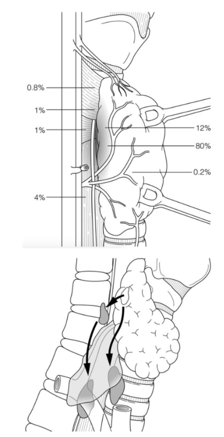
- In 85% of cases the above steps allow identification of Superior Parathyroid in its normal site
- Typical location - posteromedial surface of the superior pole
- At the level of the cricoid cartilage
- Posterior RLN
- 1cm superior to the junction of Inferior Thyroid Artery and Recurrent Laryngeal Nerve
- Typical location - posteromedial surface of the superior pole
- If abnormal/enlarged, P IV tends to migrate posteriorly and downwards
- Look beside/behind oesophagus
- May go very low (lower than Inferior Thyroid Artery)
- Lower it goes, the more posterior it becomes
- Can follow its vascular pedicle (originates middle/upper 1/3 of thyroid lobe)
- Can emerge with simple traction on the pedicle
- NB: Closely related to RLN, can be attached to their capsule
- Ectopic locations
- Tracheoesophageal groove
- Most common
- Retropharyngeal
- Retroesophageal
- Posterior mediastinal
- Intra- thyroidal
- Tracheoesophageal groove
Search for Inferior Parathyroid (P III)
- Higher variability (further to travel)
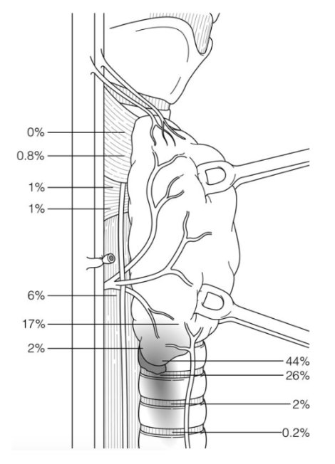
- Typical location - posteriorlateral surface of the inferior pole of the thyroid lobe
- Anterior to the RLN
- Along the posterolateral aspect of the inferior pole to the thyroid gland
- Approximately 1 cm caudal to the junction to the RLN and inferior thyroid artery
- Search from
- Inferior Thyroid Artery to Inferior Thyroid pole then down to thymus
- PIII’s are rarely posterior
- Become more anterior the lower they are
- Ectopic locations
- Thyro-thymic ligament and Thymus
- 25%
- Anterosuperior mediastinum
- Undescended in a submandibular location
- Within the thyroid gland
- Within the carotid sheath
- Thyro-thymic ligament and Thymus
- Typical location - posteriorlateral surface of the inferior pole of the thyroid lobe
If you can’t find the superior parathyroid
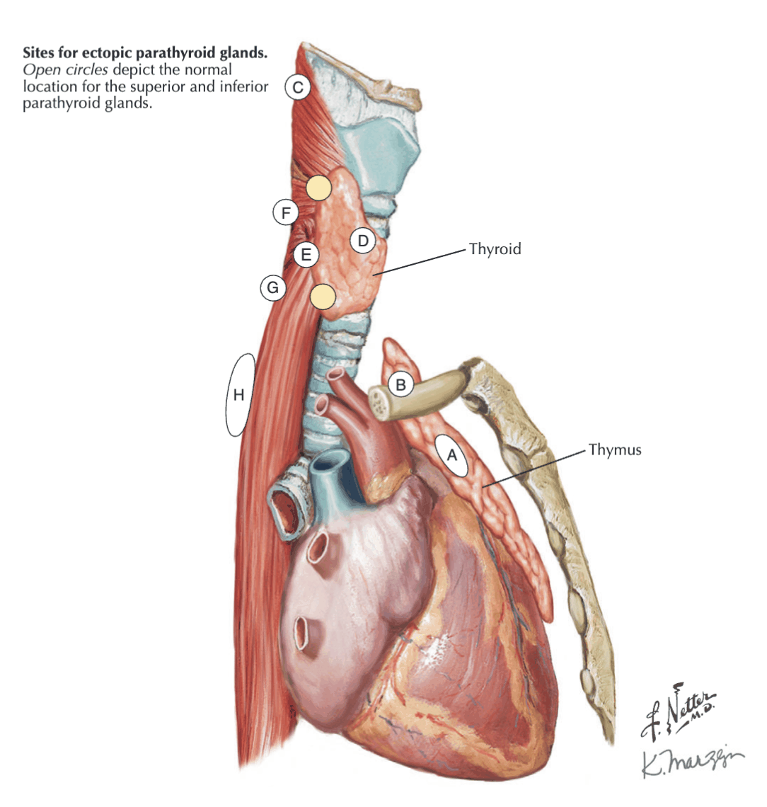
- Look in the retropharyngeal, tracheoesophageal groove, retro-oesophageal, and carotid sheath, and posterior superior mediastinum.
- This generally means you are dissecting posteriorly to its usual location.
If you can’t find the inferior parathyroid.
- Look in the thyro-thymic/ligament tract – runs from the inferior lobe of the thyroid down to the thymus.
- You should perform a Cervical thymectomy and examine its substance - this involves identifying the thymus at the lower border of the thyroid - gentle traction on the thymus superiorly out of the chest, and ligating the inferior thymic vein once visible.
- A hemithyroidectomy should also be considered as the gland can be within the substance of the thyroid.
- Other sites include within the anterior mediastinum, in the carotid sheath.
- You should also explore high - up near the submandibular gland.
Evaluation after Initial Bilateral Exploration
- Abandon/finish dissection if:
- All 4 glands discovered, and one or more abnormal
- Unless familial hyperparathyroidism
- One abnormal gland, other gland(s) normal, but fewer than four located
- All 4 glands discovered, and one or more abnormal
- Continue/peruse dissection if:
- No gland discovered
- Fewer than 4 glands discovered, but none are pathological
- Probable adenoma remains
- Fewer than 4 glands found, and ≥ 2 glands enlarged
- Multi-gland disease, must find remaining gland
- All 4 glands identified but none are pathological
- Likely ectopic supernumerary adenoma
Continuation of Exploration
- Congenital ectopias (neck or mediastinum) caused by abnormal embryological migration are related to P III’s (Inferior Parathyroids)
- Acquired ectopias in posterior mediastinum caused by migration associated with gravity, are related to P IV’s (Superior Parathyroids)
Minimally Invasive Options
- Limited incisions, target 1 specific gland
- Based on the fact most cases are single adenomas
- Contraindications
- Pre-op suspected multi-gland disease
- Familial hyperparathyroidism
- Suspected carcinoma
- Relative – previous surgery or large goiters
Open Minimally Invasive Parathyroidectomy
- Two approachs
- Lateral or medial approach (in relation to the strap muscles)
- Pre-op localisation prerequisite
- ioPTH also heavily used
- 2-4cm unilateral incision
- Can be day case, shorter op time
- Fewer overall complications
- Cure rates compatible with bilateral neck exploration
- Can also do minimally invasive radio-guided approach
Endoscopic Parathyroidectomy
- Can do like traditional endoscopic approach
- Use low pressure insufflation
- Can do MIVAP
- Minimally invasive video assisted parathyroidectomy
Reoperations
- Initial procedure have cure rates of > 95% and complication rates of < 2%
- Indications
- Persistent disease
- Failure of normalised Ca2+ post-op within 6 months of surgery
- Recurrent disease
- Over 6 month normocalcaemic interval prior to reappearance of hypercalcaemia
- Can be after several years
- Most seen in familial disease or multi-gland disease
- Over 6 month normocalcaemic interval prior to reappearance of hypercalcaemia
- Persistent disease
- Re-operative surgery can be technically challenging and associated with increased morbidity
- Threshold for surgical intervention should be higher
- Influenced by symptoms and signs as well as the ability to localize the diseased gland pre-op
- Attempt to localise using non-invasive methods
- May be more of a role for selective venous sampling if can’t otherwise locate
- Pre-operative imaging localisation mandatory
Total/Subtotal Parathyroidectomy
- Indications
- 4 Gland Hyperplasia
- 2° Hyperparathyroidism
- 3° Hyperparathyroidism
- Need to complete bilateral neck exploration initially
Subtotal Parathyroidectomy
- Remove nearly all of abnormal glands, leaving a well vascularised remnant of 1 gland in its anatomical location
- Low rate of post-op hypocalcaemia, but recurrence 5-30%
- First create remnant
- Means you can assess vascularity later in the case
- Aim to leave 50-100mg tissue (50-75% of gland)
- Try to leave an inferior gland
- Most readily accessible if need to re-operate, anterior to RLN
- Also leave titanium clip in the vicinity
- Remember blood supply from medial
- Take lateral portion
Total Parathyroidectomy with Reimplantation
- Removes all glands and implants morcellate pieces of tissue in non-anatomical sites
- Most common in non-dominant Brachioradialis or SCM, (can also be subcutaneous chest)
- Multiple small pockets created
- Most common in non-dominant Brachioradialis or SCM, (can also be subcutaneous chest)
- Recurrences easier to treat than for subtotal
- Don’t need further neck exploration
- Risk of profound hypocalcaemia if graft fails
- Not recommended for first time operations in 2° or 3° hyperparathyroidism due to risk of severe hypocalcaemia
Total Parathyroidectomy without Reimplantation
- Most effective surgical treatment for severe refractory 2° Hyperparathyroidism
- May need Ca2+ supplementation lifelong
Surgical Adjuncts
IoPTH
- Utilises short plasma circulating half-life of PTH - 4.5 mins
- “Miami” criteria
- Takes 4 measurements
- Pre-skin excision
- Pre-gland excision (Take higher of these two as ‘baseline’)
- 5 mins post-gland excision
- 10 mins post-gland excision
- 50% decrease in PTH levels from baseline
- 10 minutes after resection
- Indicates successful surgery in 97% cases
- If doesn’t drop by 50%
- Remeasure at 20 mins
- If still not, continue exploring
- Takes 4 measurements
- Dual Criteria
- Take 2 measurements
- Pre-gland excision
- 10 mins post-gland excision
- Take 2 measurements
- If drops by > 50%, and returns to normal range, then dual criteria met
- Has led to decreased operative failure rates
Gamma Probe Localisation
- Pre-op injection of 99mTc sestamibi
- Intra-op localization with a hand-held quantitative gamma counter
- Excised gland should be > 20% background
- Not done by most centers
RLN Monitoring
- A meta-analysis comparing
- RLN monitoring versus RLN visual identification
- Effect on true vocal cord palsy rates after thyroid surgery
- Found no statistically significant difference in RLN injury rates between the two groups
Complications
- Failure to cure
- Cure = Biochemical normalisation of calcium lasting > 6 months post-op
- Cure rates 99% for MIP, 97% for BNE in high vol centres
- ? Not as high in low volume centres
- RNL or ELN injury
- Post-op Hypocalcaemia
- Atrophy of remaining glands
- Damage to remaining glands in surgery
- Transient hypocalcaemia common
- Bleeding/Haematoma
- < 0.5% of patients
- Can be surgical emergency
- Hungry bone syndrome
- This can occur after surgery for secondary or tertiary hyperparathyroidism
- Occurs because after surgery, there is a sudden drop in PTH, which means bone Ca release stops, leading to severe hypocalcaemia, hypophosphatasemia and hypomagnesaemia.
- Managed with high dose IV calcium replacement