Section: Trauma Curriculum: Curriculum, page 78
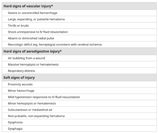
Zones of the Neck
- I - Sternal notch to lower border of Cricoid cartilage
- II - Cricoid cartilage to angle of mandible
- III - Angle of mandible to base of skull
Contents
- Zone 1 (6 structures)
- Vascular - Great vessels
- Aerodigestive - Trachea, Lung apices, Oesophagus
- Other - Thoracic duct, Upper mediastinum
- Zone 2 (7 structures)
- Vascular - Carotid and Vertebral A’s, Jugular V’s
- Aerodigestive - Pharynx, Larynx, Oesophagus, Trachea
- Zone 3 (4 structures)
- Vascular - Distal Extracranial Carotid A and Vertebral A, Jugular V segments
- Aerodigestive - Pharynx
When to explore
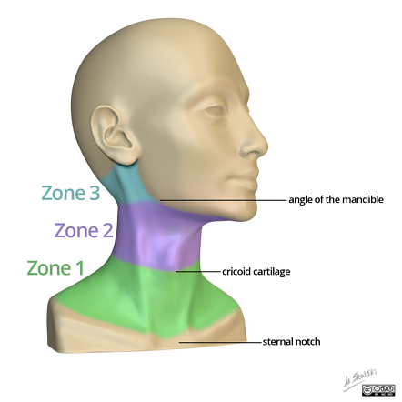
- Penetrating neck injury with hard signs of vascular or aerodigestive injury
- Modern day thinking is the “no zone approach” i.e if no indication for straight to theatre then all patients should be imaged
- Old thinking was mandatory zone 2 exploration
- Other Broad Principles
- Zone 1 - Angiography to aid operative planning due to potential vessel injury at the thoracic outlet
- Zone 2 - Angiography not as important but if suspicions of vertebral vessel injury then yes, often miss oesophageal injury with these
- Zone 3 - Angiography important to identify vascular injury near base of skull → Embolization
Access to Zones of the Neck
- Preparation
- Neck extended
- Rotate head away from site of injury
- Prep wide - including Chest
Zone 1 access
- Unstable patient
- For right
- Median sternotomy - exam answer as can get proximal control
-
- Supraclavicular extension
-
- Median sternotomy - exam answer as can get proximal control
- For Left
- DSCT say anterolateral thoracotomy as subclavian vessels are lower on the left
- But sternotomy and + Supraclavicular extension an option
- For right
- Stable
- Trans-clavicular
- Cut skin down on the clavicle
- Remove all muscle of clavicle
- Jiggly saw medial and then rotate lateral (can save) then wire it back
- This can be extended down the arm or sternotomy
- Infra-clavicular
- Supra-clavicular
- Trans-clavicular
Zone 2 access
- Incision
- Longitudinal at Anterior border of SCM
- Can extend all the way from the mastoid process down to the clavicle if needed
- Can be extended up and down or into a sternotomy
- Through subcutaneous tissue and platysma to reach muscle.
- Longitudinal at Anterior border of SCM
- Retract sternocleidomastoid laterally
- Omohyoid only strap to cross Carotid Sheath → CCA landmark
- Expose IJV
- Landmark for the carotid bifurcation - facial vein, which dives off the anterior border of the Internal jugular vein
- Divide fascial vein to take you on to the common carotid
- Lateral retractiopn of IVJ takes you to common carotid
- Beware of vagus nerve which is running in carotid sheath
- Lateral retraction of Carotid Sheath to access Trachea/Oesophagus/Thyroid
- Need to divide three structures
- Omohyoid
- Inferior Thyroid Vein
- Middle thyroid vein
- Need to divide three structures
- Medial retraction to access Prevertebral Fascia and Vertebral Artery
Zone 3 access
- Principles
- Nasotracheal tube if possible
- Neurosurgery
- Temporize by using Fogarty catheters via neck
- Incision
- Longitudinal at Anterior border of SCM
- Divide the sternocleidomastoid muscle at the mastoid if required
- Mindful of the Accessory Nerve, which is going to enter into the sternocleidomastoid about 3 centimetres distal to that
- Retractor on the mandible and pull that anteriorly to open up the space.
- Dislocate the mandible or divide the mandible in order to get you increased access if needed
- Same as zone 2
- Ligate the middle thyroid and the common facial veins, retract the sternocleidomastoid muscle and internal jugular laterally
- Careful of the vagus nerve in the carotid sheath.
- Find carotid artery proximal and follow superiorly
- Can divde posterior belly of diagastric
- Hypoglossal Nerve will run anterior to the artery at this point
Axillary injury
-
Can do supraclavicular incision to get proximal control
- Excise supraclavicular fat bad
- small blood vessels, lymph nodes and the Thoracic duct
- Get on to scalenus anterior
- preserve the Phrenic Nerve
- Divide Scalenus Anterior
- Then can access the subclavian artery which is sitting behind it
- Excise supraclavicular fat bad
-
What are the operative priorities in neck trauma
- #1 - AW control
- #2 - Stop bleeding - Pressure, Fogarty
Facial fractures
- Zygomatic Arch #
- Trismus and Infraorbital Nerve injury with paraesthesia
- Impinged Coronoid Process to cause trismus
- Requires maxfax repair within 10 days – Important for mastication
- Trismus and Infraorbital Nerve injury with paraesthesia
- Maxillary/Le Fort #
- Classification Le Fort 1 | 2 | 3 - progressively more involvement of the orbits
- Can cause dentition issues
- CT assessment requires thin slices - 1-2mm
- Repair by maxfax within 10 days
- Nasal # → ENT | Plastics | Maxfax
- Can get CSF leak
- Septal haematoma - drainage urgently to prevent septal cartilage necrosis
- Drain under LA/Topical anaesthetic
- Can be associated with orbital blow out #’s
- Can be reduced or complex #’s may need maxfax recon
- Orbital # (Assess all eye functions)
- Common to get diplopia with inferior rectus dysfunction (peri-muscular tissue entrapment)
- Irritation of infraorbital nerve
- Complications
- Injury to globe, optic nerve, infraorbital nerve
- Blindness, diplopia
- Lid malposition and poor function
- General mx principles
- Restore normal anatomy
- To prevent early and late complications including
- Vision loss, diplopia, cosmesis issues
- AW protection | Abx | ? Steroids | No nose blowing | No valsalva
C-Spine
- High risk of neck injury
- High speed MVA
- Death at the scene of MVA
- Fall from height > 3m
- Significant closed head injury or CT proven ICH
- Neurological sx/signs referred to the cervical spine (UL neurology)
- Pelvic or multiple extremity #’s
- Low risk factors - can clear c-spine clinically and without radiology
- No posterior midline cervical tenderness
- Normal level of alertness
- No evidence of intoxication
- No abnormal neurological findings
- No painful distracting injury
- Clearance
- If can’t clear clinically - Image and ortho
- Keep collar on
- Xray C-spine = 3 views
- AP | Lateral | Open mouth (Odontoid/peg view)
ABCs of Cervical Spine Imaging Evaluation
- A - Alignment & Anatomy
- B - Bony integrity
- C - Cartilage (Joint) spaces
- S - Soft Tissues
A - Alignment & Anatomy
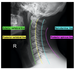
- Visually trace 4 lines
- Look for disrupted Anterior or Posterior Vertebral Body Lines
- Look for disruption of the Spinolaminar Line
- Look for Posterior Spinous Line
- Look for asymmetry of the facets
- Look for widening of the spaces between the individual vertebrae
- Look for widened the predental soft tissue space
B – Bony abnormalities
- Overt fracture lucencies/ deformities
- Once one fracture is seen, be sure to look for others
- Disruption of the ring of C1 (Odontoid Fx)
C – Cartilage (Joint) abnormalities
- Widened or narrowed disc space/facets
S – Soft tissue abnormalities
- Widened prevertebral space
-
7mm should prompt high suspicion for injury
Hard and Soft Signs
- Invx options in Neck Trauma
- CT - with IV and PO contrast (GG swallow on table) - arterial and portal venous phase
- Scopes - OGD and Bronchoscopy
- Interventional angiography in hybrid theatre - Stent/embolise
- Mx Options
- If unstable or hard signs should explore all
- If stable
- If stable all zones should be imaged
- Zone 1 - Prefer imaging (Great vessels, Trachea, Oesophagus)
- If imaging positive → Median Sternotomy and Neck exploration
- Zone 1 - Prefer imaging (Great vessels, Trachea, Oesophagus)
- Zone 2 - If no hard signs and stable → CT IV/PO 2 phase
- Zone 3 - Should image —> Difficult access
- Need help - Maxfax
- May need to disarticulate the jaw
- Start in zone 2 for proximal control
- Need help - Maxfax
- If stable all zones should be imaged
Laryngeal Trauma
- If need emergency airway likely need a surgical cricothyroidotomy
Oesophageal Injury
- Access dependent on location
- Need to dissect off trachea
- Repair in 2 layers - mucosal with 3.0 vicryl sutures in and 3.0 prolene or PDS muscular layer out
Blunt cerebrovascular injuries
- Definition
- Non-penetrating injury to the carotid or vertebral arteries.
- Pathophysiology
- Stretching or impingement of the vessel wall as the head and neck are forcefully moved in flexion extension or rotation.
- Causes
- Intimal tears and exposure of the subintimal layers to blood flow
- Leads
- Thrombus formation
- Dissections in the vessel wall
- Pseudo aneurysms
- Complete occlusion of the vessel
- Transection of the vessel.
- Strokes
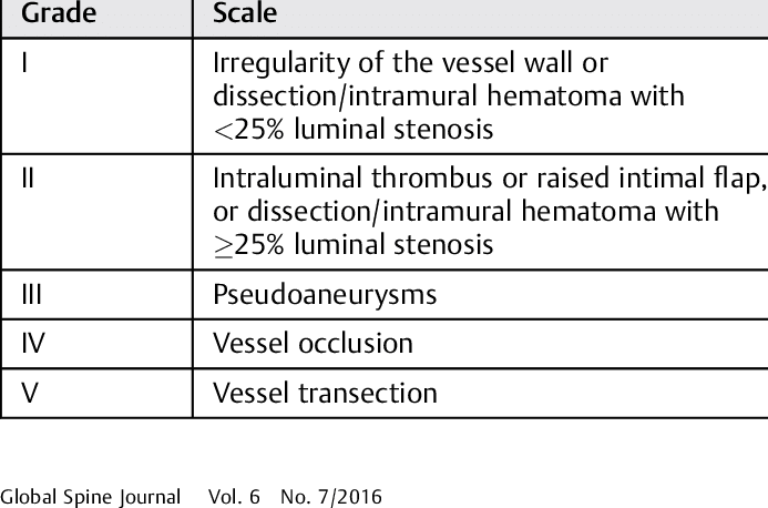
- Denver grading system
- Signs and symptoms
- Arterial hemorrhage from the neck, nose or mouth
- Expanding neck hematoma
- Cervical bruit in a patient less than 50 years old
- Focal neurological deficit that may indicate an ischemic injury to the brain
- Horner’s syndrome
- Stroke seen on imaging
- If any of the above
- CT angiogram
- Low threshold
- High energy neck trauma
- Displaced facial fractures, mandibular fractures, complex base of skull fractures, a severe traumatic brain injury, cervical spine fractures, a near hanging with hypoxic brain injury, a clothesline type injury or a seatbelt sign on the neck, significant scalp degloving, significant chest, upper rib or abdominal injuries.
- Low threshold
- CT angiogram
- Treatment
- Medical
- Antiplatelets or heparin
- Grade 1, 2 and 3
- If survive in grade 4 then for life long antiplatelet therapy
- Antiplatelets or heparin
- Endovascular
- Grade 1 - No role, self resolve
- Grade 2 - Consider in 2 if progresses on repeat imaging at 7-10 days
- Grade 3 - Consider in 3, often based on size (>1cm)
- Grade 4 - No role in grade 4
- Grade 5 - Can attempt in 5 but high mortality rate/stroke rate regardless.
- Surgery
- If hard signs
- Medical
- Follow up
- In medical management at 2 weeks and 3 months re-image
Carotid Artery Repair
- IR option available for stenting but not always amenable
- Contraindications to repair
- Ischaemic Infarct > 4hrs old
-
4hrs of interrupted flow with coma
- No back flow from carotid stump
- Repair options
- Shunt if needing to temporise
- Repair - Suture +/- Patch
- Replace - Graft - Interposition (reversed GSV)
- Bypass - Graft
- Transposition Graft - for ICA → replumb onto ECA
- Ligation - for high injuries, if no neurology or > 48 hrs neurology
- Access
- As above - depends on zone