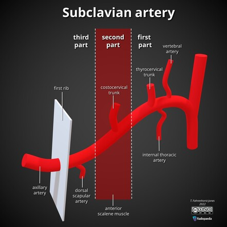
- Divided into 3 parts by the Anterior Scalene
First part
- Vertebral Artery
- Thyrocervical Trunk
- Internal thoracic artery Second part
- Costocervical Trunk
- Supreme Intercostal Artery Third part
- Dorsal Scapular Artery
The subclavian artery is a paired arterial vessel of the thorax. The right and left arteries have different origins; the left subclavian artery originates directly from the aortic arch, while the right subclavian artery originates from the brachiocephalic trunk.
From their origin, the left and right subclavian arteries arch superolaterally and course towards the axillary region. Along their course, they pass posterior to the anterior scalene muscles and anterior to the middle scalene muscles.
It passes between the clavicle and the first rib to enter the axilla. It becomes the Axillary artery at the lateral border of the first rib.
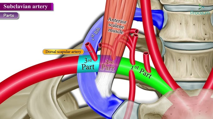
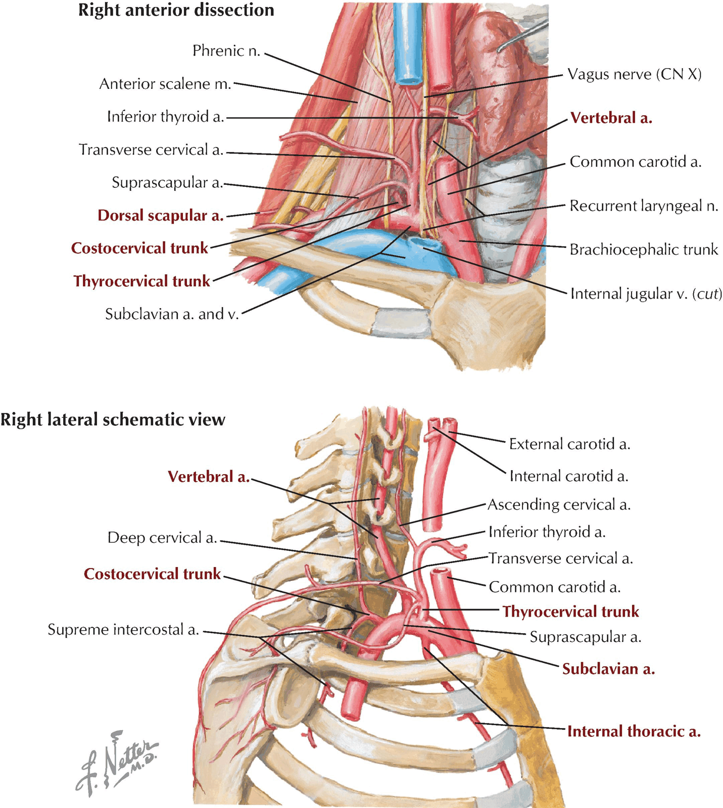
V - vertabrel I - internal thoracic T - thyrocececical
C - costocervical
D - doralscapula
- Divided into 3 parts by the Anterior Scalene
- 1st Part:
- Medial to Anterior Scalene
- Arches over the Suprapleural Membrane
- Impresses a groove on the Lung Apex
- Three Branches:
- Vertebral Artery
- Thyrocervical Trunk
- Inferior Thyroid Artery
- Ascending cervical artery
- Suprascapular artery
- Transverse cervical artery
- Internal thoracic artery
- Connecting loop between Middle and Inferior Cervical Ganglia passes in front of the Subclavian Artery
- Turns up posterior to it, forming the Ansa Cervicalis
- 2nd Part:
- Posterior to Anterior Scalene
- One Branch: Costocervical Trunk
- Supreme Intercostal Artery
- Deep Cervical Artery
- 3rd Part:
- Lateral to the Anterior Scalene
- One Branch: Dorsal Scapular Artery
Continue as Axillary artery after passing above the first rib
Vertebral Artery
- Aries from the Upper Convexity of the Artery
- Passes up to disappear at the Apex of the Pyramidal Space into the Foramen of the Transverse Process of C6 Vertebra
- Rarely: first part of Vertebral artery may initially enter the foramen of the transverse process of a higher vertebra than C6
- Accompany sympathetic nerve runs up posterior to artery
- Vertebral Vein:
- Emerges from the foramen in the Transverse process of C6 Vertebra
- Runs forwards anterior to the Vertebral Artery and Subclavian Artery
- Empties into the Brachiocephalic Vein
- May be accompanied by a companion vein that passes through the foramen of the Transverse process of C7 Vertebra
- Passes behind the Subclavian Artery to the same destination
Thyrocervical Trunk
- Arises lateral to the Vertebral Artery from the upper surface of the Subclavian
- Divides into:
- Inferior thyroid artery
- Ascending cervical artery
- Suprascapular artery
- Transverse cervical artery
Internal thoracic artery
- 2cm lateral to the sternal margin
- In neurovascular plane between the internal intercostal muscles and costal cartilages anteriorly, and the transversus thoracis muscle posteriorly
- At the level of the 1st rib, it is crossed anteriorly by the Phrenic Nerve
- Branches
- Anterior Intercostal Arteries
- Pericardiophrenic artery
- Perforating cutaneous branches
- Bifuraces at 6th -7th costal cartilage
Costocervical Trunk
- Passes back across the Suprapleural Membrane towards the neck of the 1st Rib
- There, it divides into a Descending Branch (Supreme Intercostal Artery) and an Ascending Branch (Deep Cervical Artery)
- Supreme Intercostal Artery:
- Enters the Thorax across the neck of the 1st Rib
- Deep Cervical Artery:
- Passes backwards between the Transverse Process of C7 Vertebra and the neck of the 1st Rib to run upwards behind the Cervical Transverse Processes
Dorsal Scapular Artery
-
Runs laterally through the Brachial Plexus anterior to Scalenus Medius
-
Runs deep to Levator Scapulae
-
Involved in the Scapular anastomosis
-
Frequently replaced by the deep branch of the Transverse Cervical Artery
- This branch then takes the name of Dorsal Scapular
-
Surface marking in the neck:
- Along a line arching upwards from the Sternoclavicular Joint to the middle of the Clavicle and about 2cm above it
-
Surgical Approach:
- Exposed by dividing clavicular head of Sternocleidomastoid from the Clavicle
- Detaching Scalenus Anterior from the 1st Rib
- Taking care not to damage the Phrenic Nerve