Arterial
Common Carotid Artery
- Arises on Left side from Arch of Aorta
- Lies anterior to Subclavian Artery up to the Sternoclavicular Joint
- On Right side, Brachiocephalic Trunk bifurcates behind the Sternoclavicular Joint into Common Carotid and Subclavian Arteries
- Gives off no branches proximal to its bifurcation
- Lies within medial part of Carotid Sheath
- Internal Jugular Vein lateral to it and Vagus nerve deeply placed between
- Sympathetic trunk behind the artery and outside sheath
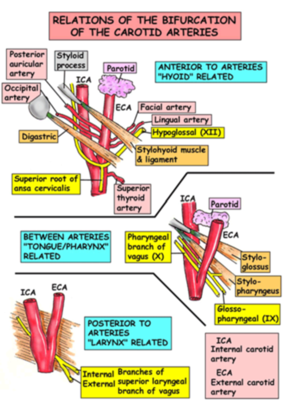
- Bifurcates at level of upper lamina of Thyroid Cartilage (upper border of C4)
- Terminal portion of artery often shows a slight dilatation (Carotid Sinus) – includes the commencement of the Internal Carotid Artery
- Common Carotid Relations
- Posterior (outside sheath) = sympathetic trunk, pharyngeal veins and superior laryngeal branch of vagus.
- Medial = ascending pharyngeal artery
- Lateral = IJV, and vagus nerve deep b/n artery and vein
- Superficial, near origin, it is crossed by: lingual vein, facial vein, XII, and superior root of ansa cervicalis (embedded in the sheath).
- Overlapped higher by SCM, and posterior border of digastric, stylohyoid and posterior auricular artery
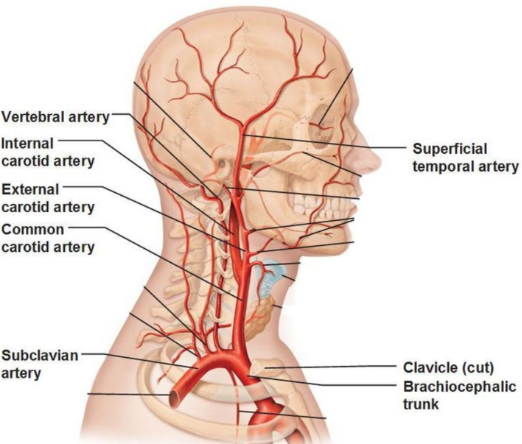
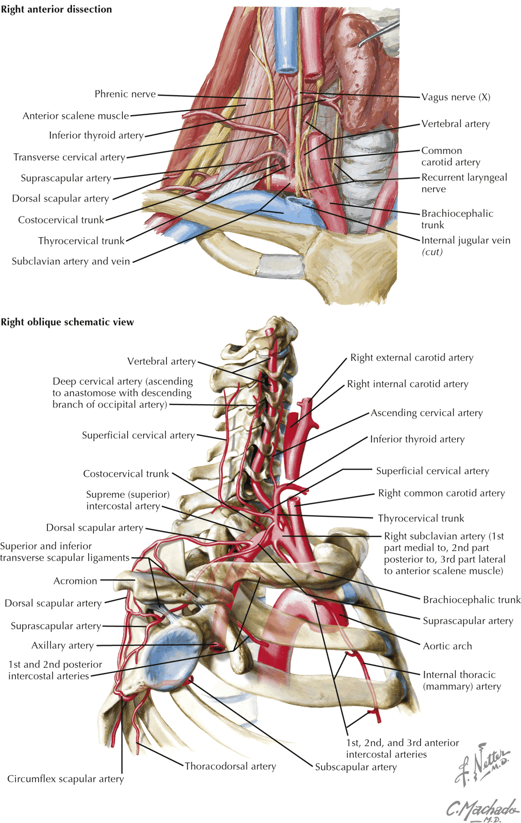
External Carotid Artery
- At its commencement, lies against side wall of Pharynx anteromedial to Internal Carotid Artery
- Ascends in front of Internal Carotid deep to the Posterior Belly of Digastric and Stylohyoid
- Pierces the Parotid Fascia to enter the Parotid Gland
- Divides within the gland behind the neck of the Mandible into Maxillary and Superficial Temporal Arteries
- As it lies within the Parotid Gland, it is separated from the Internal Carotid by the deep part of the Gland, its fascia, Styloid Process and it’s continuation the Stylohyoid Ligament, Styloglossus and the ‘Pharyngeal Structures’
- Stylopharyngeus muscle
- Glossopharyngeal Nerve
- Pharyngeal Branch of the Vagus Nerve
- Facial Vein crosses artery with Hypoglossal Nerve lying between
- Lies mostly in the anterior border of SCM
Branches of External Carotid Artery
- Before dividing into 2 terminal branches (Superficial Temporal Artery and Maxillary Artery), it gives off 6 branches – 3 from in front, 2 from behind, and one from its deep (medial) aspect
- Three from in Front:
- Two from Behind:
- One from deep:
- Some Anatomists Like Freaking Out Poor Medical Students
Internal Carotid Artery
- Arises at Bifurcation of Common Carotid
- Continues upwards within Carotid Sheath
- At its commencement, Carotid Sinus – arterial wall is thin and contains Baroreceptors
- Supplied by the Glossopharyngeal Nerve and Vagus Nerves
- Carotid Body – small structure lying behind the bifurcation – receives 2 or 3 small glomic arteries
- Chemoreceptors supplied by Glossopharyngeal and Vagus Nerves
- Lateral to External Carotid at its origin but soon passes posteriorly to a medial and deeper level
- Has no branches and passes to the Carotid Canal in base of the skull
Relations
- Posterior:
- Sympathetic Trunk (Outside Carotid Sheath)
- Pharyngeal Veins
- Superior Laryngeal Branch of the Vagus Nerve
- Medial:
- Ascending Pharyngeal Artery
- Lateral:
- Internal Jugular Vein
- Deep:
- Vagus Nerve between Artery and IJV
- Anterior:
- Crossed by Lingual Vein
- Facial Vein
- Occipital Artery
- Hypoglossal Nerve
- At a higher level, crossed by Posterior Belly of Digastric and Stylohyoid and Posterior Auricular Artery and the structures that separate it from the External Carotid
Subclavian Artery
Surgical Approach
- Incision along anterior border of SCM
- Muscle retracted backwards
- Facial and Lingual Veins divided
- Carotid Sheath incised
- Hypoglossal Nerve must be safeguarded and this may require division of the lower SCM branch of the Occipital Artery
- Emergence of branches from the External Carotid ensures its differentiation from the Internal Carotid
Venous
Internal Jugular Vein
- Emerges from the Jugular Bulb at the posterior compartment of the Jugular Foramen
- Continuation of the Sigmoid Sinus
- At first behind the Internal Carotid Artery
- Lies on transverse process of Atlas
- Crossed by the Accessory Nerve
- Receives Inferior Petrosal Sinus as its first tributary just below base of skull
- Sinus passes back lateral or medial to the Glossopharyngeal, Vagus and Accessory Nerves
- Vein passes down to gain the lateral side of first the Internal Carotid and then the Common Carotid, within the loose lateral part of Carotid Sheath
- Vagus Nerve placed deeply between the vein and arteries
- Overlapped in its lower aspect by SCM
- Joins the Subclavian Vein to form the Brachiocephalic Vein behind the Sternal end of the Clavicle
Relations of IJV
- Deep Cervical Lymph Nodes within the Carotid Sheath are closely adjacent to IJV throughout
- Posterior:
- Cervical Plexus lying on Levator Scapulae and Scalenus Medius
- Phrenic Nerve on Scalenus Anterior
- Thoracic Duct – crosses behind the Left IJV at the level of C7
- Anterior:
- Union of Inferior and Superior Roots of the Ansa Cervicalis
- Low down, the tendon of Omohyoid – useful guide
- Lateral:
- Inferior Root of Ansa Cervicalis
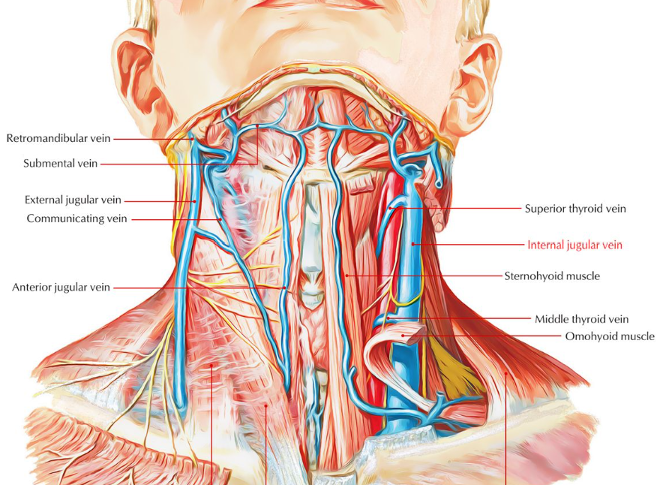
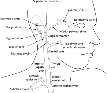
Tributaries
- Inferior Petrosal Sinus
- Pharyngeal Vein
- Lingual Vein
- Facial Vein
- Superior Thyroid Vein
- Middle Thyroid Vein