Intercostal Arteries
- Arteries enter intercostal spaces at the back and front
- Posterior
- Back upper 2 spaces: Supplied by the Superior intercostal artery, i.e. the descending branch of the Costocervical Trunk (from the 2nd part of the Subclavian Artery behind Scalenus Anterior)
- Enters thorax by passing across the front of the neck of 1st rib
- Relations here: Medial side: Sympathetic Trunk; Lateral side: 1st Thoracic nerve passes across 1st rib to join Brachial plexus
- Small 1st Posterior Intercostal Vein lies between artery and Sympathetic Trunk
- Relations here: Medial side: Sympathetic Trunk; Lateral side: 1st Thoracic nerve passes across 1st rib to join Brachial plexus
- Here: 1st Thoracic Sympathetic Ganglion frequently fused with Inferior Cervical Ganglion to form the Cervicothoracic (Stellate) Ganglion
- Enters thorax by passing across the front of the neck of 1st rib
- Remaining 9 spaces: Supplied by separate branch of Descending Thoracic Aorta
- Branches cross the front of the vertebrae and Anterior Longitudinal Ligaments, behind the Hemiazygous Veins and Thoracic Duct
- All 11 arteries = Posterior Intercostal Arteries
- Each artery gives off small collateral branch – passes around neurovascular plane at a lower level than the main trunk
- Back upper 2 spaces: Supplied by the Superior intercostal artery, i.e. the descending branch of the Costocervical Trunk (from the 2nd part of the Subclavian Artery behind Scalenus Anterior)
- Anterior
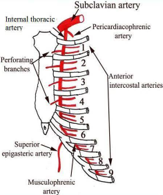
- At the front: Internal thoracic artery in the upper 6 spaces and the Musculophrenic Artery in the 7-9th spaces give off 2 Anterior Intercostal Arteries ⇒ Pass backwards and anastomose with Posterior vessels
- No anterior intercostal spaces in the last 2 spaces
- At the front: Internal thoracic artery in the upper 6 spaces and the Musculophrenic Artery in the 7-9th spaces give off 2 Anterior Intercostal Arteries ⇒ Pass backwards and anastomose with Posterior vessels
Intercostal Veins
- In each space: 1 Posterior and 2 Anterior Intercostal Veins
- Anterior Intercostal Veins:
- Drain into Musculophrenic and Internal Thoracic Veins
- Posterior Intercostal Veins:
- Supreme Intercostal Vein - 1st Posterior Intercostal Vein ⇒ Drains either into the Vertebral Vein or the Brachiocephalic Vein on its own side
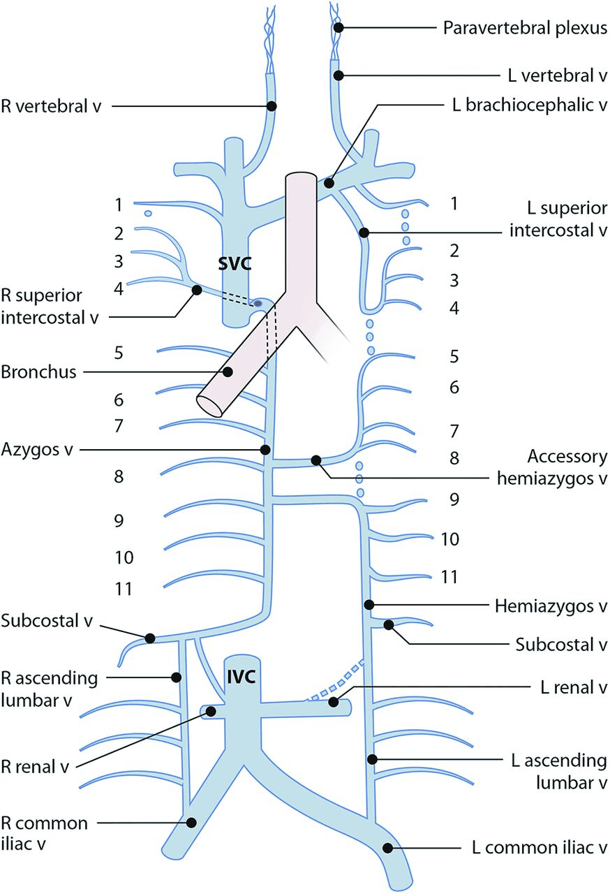
- Superior intercostal vein - 2nd and 3rd (and sometimes 4th) Posterior Intercostal Vein ⇒ Form a single trunk on each side (Superior Intercostal Vein) ⇒ On the right, drains into the Azygos vein; On the left, runs forward over Arch of Aorta, Lateral to Vagus Nerve and medial to Phrenic Nerve and empties into Left Brachiocephalic Vein
- Azygos Vein and Hemiazygos Veins
- In lower 8 spaces ⇒ Drain into Azygous system: Azygos Vein on the right and the Hemiazygos and Accessory Hemiazygos Veins on the left
Internal Thoracic Artery
-
From 1st part of the Subclavian Artery
-
Passes vertically downwards about 1cm lateral to sternal border
-
Gives off 2 Anterior Intercostal Arteries in each intercostal space
-
At costal margin, it divides into the Superior Epigastric Artery and the Musculophrenic Arteries
- Superior Epigastric Artery:
- Passes between xiphisternal and highest costal fibres of the diaphragm to enter the Rectus Sheath behind the muscle
- Musculophrenic Artery:
- Passes along the costodiaphragmatic gutter and gives off 2 Anterior Intercostal Arteries in each space until it ends by piercing the diaphragm in the 9th space to ramify on its abdominal surface
- Superior Epigastric Artery:
-
Internal Thoracic Artery – accompanied by 2 venae comitantes that empty into the Brachiocephalic Vein
Azygos Vein
- Origin: Formed by the union of the Right Ascending Lumbar Vein and Right Subcostal Vein
- Travels through the Aortic Opening under shelter of Right Crus
- Lateral to the Thoracic Duct
- Passes upwards lying on the sides of the Vertebral bodies on a plane posterior to that of the Oesophagus
- Termination: Superior Vena Cava
- At level of T4
- Azygos Vein arches forwards over the Hilum of the Right lung
- Receives blood from:
- Lower 8 Posterior Intercostal Veins of the Right Side
- Superior Intercostal Vein
- Bronchial veins from the Right Lung
- Pericardial Veins
- Veins from the Middle third of the Oesophagus
- Two Hemiazygos Veins usually join it at the levels of T7 and T8
Hemiazygos Veins
- Two veins
- Lie longitudinally on the left side of the bodies of the Thoracic Vertebrae
- Communicate with each other
- BUT characteristically drain separately from their adjoining ends behind the Oesophagus into the Azygos Vein
- Receive blood from:
- Lower 8 Posterior Intercostal Vein of the Left Side (4 each)
- Inferior vein, a.k.a. Hemiazygos Vein, formed in the abdomen by the union of the Left Ascending Lumbar Vein and the Left Subcostal Vein
- Often communicates with the Left Renal Vein
- Passes up through the Left Crus of the Diaphragm
- Receives blood from the lower Oesophagus
- Superior vein, a.k.a. Accessory Hemiazygos Vein, receives Bronchial veins from the Left Lung
Thoracic Aorta
- Commences at lower border of T4 where Arch of Aorta ends
- Initially to the left of midline
- Slants gradually to the midline
- Leaves Posterior Mediastinum at the level of T12 by passing behind the Diaphragm between the Crura, i.e. behind the Median Arcuate Ligament
- Branches:
- 9 x pairs of Posterior Intercostal Arteries
- 1 x pair of Subcostal Arteries
- Bronchial Arteries
- Oesophageal Arteries
- Few small Pericardial and Phrenic Branches
Brachiocephalic Veins
Overview
- Formed behind the Sternoclavicular Joints by confluence of Internal Jugular and Subclavian Veins
- Medial to the Anterior Scalene, located anterior to the Subclavian Artery (1st part) and receives tributaries corresponding to the branches of the 1st part of Subclavian – i.e. Vertebral, Inferior Thyroid, Internal Thoracic, Supreme Intercostal Vein and Superior intercostal vein (on left hand side only)
Right Brachiocephalic Vein
- Commences behind the right Sternoclavicular Joint
- Receives at its commencement: Right Jugular, Subclavian, Bronchomediastinal lymph trunks separately or via their confluent channel, the Right Lymphatic Duct
Left Brachiocephalic Vein
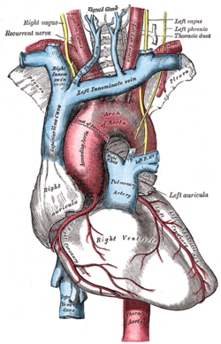
- Passes to the right with a downward inclination across the Superior Mediastinum
- Passes above the Arch of the Aorta
- Passes posterior to the Thymus and the Upper ½ of the Manubrium
- At its commencement, receives the Thoracic Duct
- Receives most of the Inferior Thyroid Veins, Left Superior Intercostal Vein, and the large Thymic Vein
Superior Vena Cava
- Commences at lower border of the 1st Right Costal Cartilage by confluence of the two Brachiocephalic Veins
- Passes vertically downwards behind the right border of the Sternum
- Pierces the Pericardium at the level of the 2nd Costal Cartilage
- Enters the Upper border of the Right Atrium at the lower border of the 3rd Right Costal Cartilage
- Receives the Azygos Vein behind the Sternal Angle and the right Superior intercostal vein
- No valves in the SVC, Brachiocephalic Vein, or Azygos Vein systems