Thoracic Wall Muscles
- Lie in the same three morphological layers as the abdo wall but divided by presence of ribs
- Innervated segmentally by anterior rami
- Outer layer = External Intercostal muscles
- Middle layer = Internal Intercostal muscles
- Inner layer = Subcostals + Innermost Intercostals + Transversus Thoracis
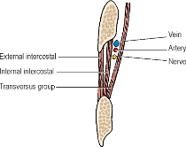
- Neurovascular plane = Between Inner and Middle layers
Outer Layer
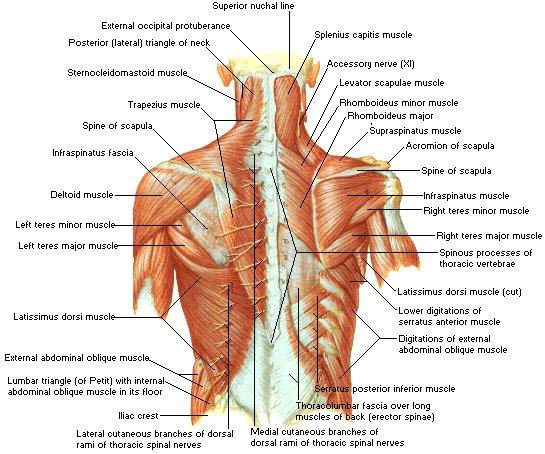
- Posterior Serratus muscles
- Two small muscles that migrated posteriorly and lie on surface of Erector Spinae mass
- Supplied by anterior rami
- Each arises from 4 spinous processes – 2 in the thorax and 2 beyond it
- Each insert into 4 ribs
- Serratus Posterior Superior
- Origin: Spinous processes of lowest 2 cervical and upper 2 thoracic vertebrae
- Insertion: Just lateral to angles of Ribs 2-5
- Dorsal scapular nerve and vessels run down its posterior surface – between it and the Rhomboids
- Elevates upper ribs (inspiration)
- Serratus Posterior Inferior
- Origin: Spinous processes of lowest 2 thoracic and upper 2 lumbar vertebrae
- Insertion: Just lateral to angles of Ribs 9-12
- Depresses lower ribs (expiration)
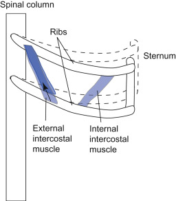
- External Intercostal muscles
- Fibres pass obliquely downwards and forwards from the sharp lower border of the rib above to the smooth upper border of the rib below
- Each muscle extends from the superior costotransverse ligament at the back of the intercostal space as far forwards as the costochondral junction
- Here it is replaced by the Anterior Intercostal Membrane – extends to side of the sternum
- Thus, between the bony ribs is muscle; between the costal cartilages is membrane
Middle Layer
- Internal Intercostal muscles
- Fibres run downwards and backwards from the costal groove to the upper border of the rib below
- Each muscle, unlike the External Intercostals, extends as far forwards as the side of the sternum
- Replaced posteriorly by the Posterior Intercostal Membrane – which extends from the angle of the rib to the superior costotransverse ligament at the posterior limit of the space
- Muscles are NOT purely inspiratory
Inner Layer
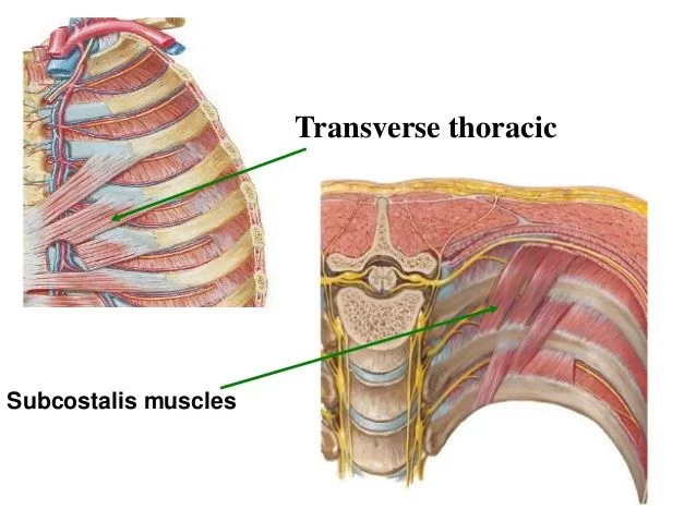
- Innermost Intercostals line the rib cage at the side; Subcostals at the back; Transversus Thoracis at the front
- They cross more than 1 intercostal space
- Subcostal Muscles
- Attached to inner surface of ribs
- Separated from posterior border of the Innermost Intercostals by a space
- Intercostal nerves and vessels are in contact with parietal pleural across this space
- Innermost Intercostal muscles
- Attached to the inner surfaces of ribs on lateral part of thoracic wall
- Transversus Thoracis
- Origin: Posterior surface of the lower part of the sternum
- Insertion: Digitations diverge on each side to the 2-6th costal cartilages
Intercostal Spaces
- Vein, Artery and Nerve lie in that order from above downwards under cover of the downward projection of the lower border of the rib
- Thus, needle or trocar for pleural drainage is inserted just above the rib that forms the lower boundary of the space
- Collateral branches of nerve and vessels that run along the upper border of the ribs are small and can be ignored
Thoracic vascular anatomy
- See notes
Intercostal Nerves
- Mixed spinal nerve
- ⇒ Emerges from Intervertebral Foramen and gives off its posterior ramus (NB: Thus, it is the anterior rami that becomes the remainder of the intercostal nerve)
- ⇒ Passes around neurovascular plane (between Internal Intercostals and Transversus Thoracis group)
- ⇒ Gives off a Collateral Branch and a Lateral Cutaneous Branch before it reaches costal angle
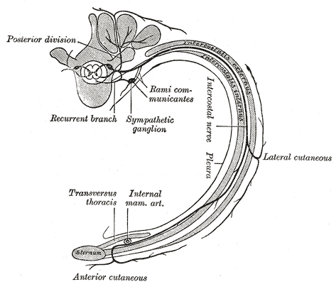
- Anterior rami
- Lateral Cutaneous Branch
- Pierces intercostal muscles and overlying muscles of body wall along the Midaxillary line and divides into Anterior and Posterior Branches to supply skin over the space
- Lateral Cutaneous Branch of the 2nd Intercostal Nerve: Crosses axilla to the medial side of the arm as the Intercostobrachial Nerve
- NB: Anterior rami have some fibres of the first 3 thoracic nerves that goes to the upper limb
- Continuation of Anterior Rami
- Muscles of thoracic wall innervated segmentally by the anterior rami
- Intercostal nerve ends as an Anterior Cutaneous Nerve
- Lower 5 Intercostal Nerves (T8 – T12) (and their Collateral Branches) slope downwards behind costal margin into the neurovascular plane of the abdominal wall which they supply
- NB: These nerves do NOT supply the parietal and visceral peritoneum
- Anterior ramus of T1 ascends across the neck of the 1st Rib to enter the Brachial Plexus
- Gives off small 1st Intercostal nerve before entering Brachial Plexus
- Subcostal Nerve (12th Thoracic)
- Although arises in thorax, quickly leaves by passing behind Lateral Arcuate Ligament of the Diaphragm into the abdomen, below the Subcostal Artery and Vein
- Lateral Cutaneous Branch
- Posterior Rami of the trunk:
- Supplies all muscles of the Erector Spinae and Transversospinalis groups that lie deep to the thoracolumbar fascia
- Supplies skin overlying
Lymph Drainage
- Lymph vessels of intercostal spaces follow arteries
- From the front: Vessels pass to Anterior Intercostal (Parasternal) Nodes that lie along the Internal Thoracic Artery
- From the back: Drain to Posterior Intercostal Nodes
Sternal Angle of Louis Anatomy
- At level of 2nd Costal Cartilages
- Level of lower border of T4
- Above this level up to the Thoracic Inlet = Superior Mediastinum
- Below this level = Inferior Mediastinum – divided into 3 parts by the Fibrous Pericardium – Anterior, Middle and Posterior
- Landmarks at this level:
- Bifurcation of the Trachea
- Concavity of the Arch of the Aorta
- Just above the bifurcation of the Pulmonary Trunk
- Azygos Vein enters the Superior Vena Cava
- Thoracic Duct reaches the Left Side of the Oesophagus
- Ligamentum Arteriosum with the Left Recurrent Laryngeal Nerve recurving below it
- Tracheobronchial Lymph Nodes
- Superficial and Deep Parts of the Cardiac Plexus
Thoracotomy Surgical Anatomy
Thoracotomy incision: postero-laterally extending from medial to the scapula posteriorly and curving around the lower edge of the scapula. Muscle layers divided/dissected during this approach.
- Latissimus dorsi
- Serratus anterior
- Rhomboids
- Intercostals (External, Internal, Innermost Intercostals)