Overview of Breast Anatomy
Location:
- Subcutaneous tissue (superficial fascia) of anterior thoracic wall
Base of Breast Landmarks
- From sternal edge to near the midaxillary line
- From the 2nd to 6th ribs
- Overlies Pectoralis Major overlapping onto Serratus Anterior and onto small part of rectus sheath and external oblique muscle
Axillary Tail
- Extension of upper outer quadrant towards axilla
- Usually lies in subcutaneous fat
- Rarely penetrates deep fascia of axillary floor and lie adjacent to axillary lymph nodes
Behind Breast
- Superficial fascia (upward continuation of membranous layer of Scarpa’s fascia of abdo wall) condensed to form posterior capsule
- Retromammary space – loose connective tissue between posterior capsule and fascia over pectoralis major
Overlying Skin
- Suspensory ligaments of Cooper – fibrous tissue connecting dermis of overlying skin to ducts of the breast and to posterior capsule
- Atrophy with age – breast becomes more pendulous
- Contracts via fibrosis associated with certain breast ca. causing dimpling of overlying skin
- Cause pitting of oedematous skin resulting from malignant involvement of dermal lymphatics (peau d’orange)
Male Breast
- No lobules or alveoli
- Small nipple and areola lie over 4th intercostal space
Ducts
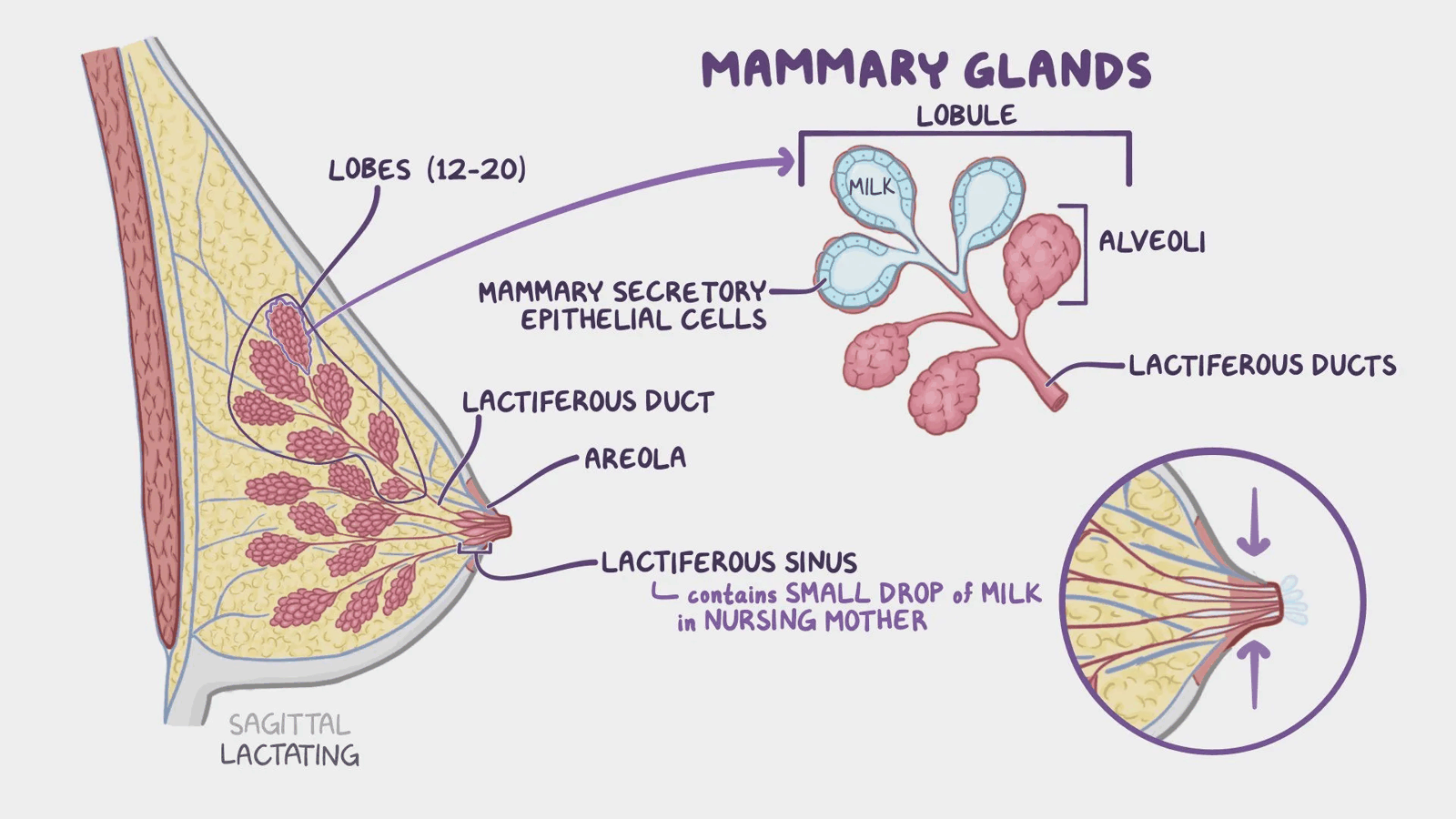
- Lactiferous ducts
- 15-20 in number
- Each drains a lobe of breast
- Converge in radial direction to open individually on tip of nipple
- Each duct has a dilated sinus at its terminal portion
Nipple
- Projection below centre of breast surrounded by area of pigmented skin, the areola
- Smooth muscle cells present – contraction ⇒ erection of nipple
- Sebaceous glands, sweat glands, and other areolar glands present in areola skin
- Areolar glands form small elevations (tubercles of Montgomery), esp. when they enlarge during pregnancy
Blood Supply
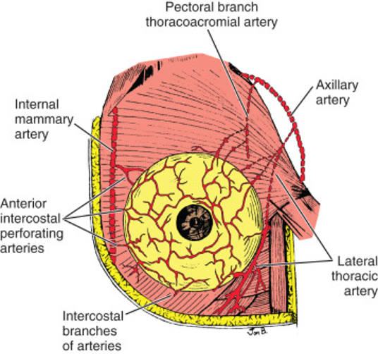
Arteries
-
- Origin: Medial surface of 2nd part of the Axillary artery (second part)
-
Branches curl around border of pectoralis major and other branches that pierce the muscle
-
At the level of the fifth rib, along the inferior bundle of the serratus anterior muscles, the long thoracic nerve runs beside the main branch of the lateral thoracic artery
-
Internal Thoracic Artery
- Origin: First part of Subclavian Artery
- Branches through the intercostal spaces behind sternum
- Largest branches: through the 2nd and 3rd intercostal spaces
-
Posterior Intercostal Arteries
- Origin: 1st and 2nd from Supreme Intercostal Artery (a branch of the Costocervical Trunk of the Subclavian Artery), lower 9 from back of the thoracic aorta
- Similar perforating branches to internal thoracic artery but smaller
- Origin: 1st and 2nd from Supreme Intercostal Artery (a branch of the Costocervical Trunk of the Subclavian Artery), lower 9 from back of the thoracic aorta
-
Pectoral branch of Thoracoacromial Artery
- Origin: 2nd part of the Axillary Artery
- Supplies upper part of the breast
Veins
- Circumareolar venous plexus + from glandular tissue ⇒ via deep veins running with main arteries to internal thoracic and axillary veins
- Posterior drainage via posterior intercostal veins
- Important link to the internal vertebral venous plexus veins and, thus, pathway for metastatic spread to bone
Lymph Drainage
- Subareolar plexus of lymphatics communicates with lymphatics within breast
- Axillary lymph nodes – 75% of lymphatic drainage
- Mainly anterior nodes
- Some to posterior nodes
- Direct drainage to central or apical nodes possible
- Parasternal nodes along internal thoracic artery – esp. medial part of breast
- Posterior intercostal nodes – a few lymphatics following intercostal arteries
- Infraclavicular nodes in deltopectoral groove – occasional
- Interpectoral nodes between pectoralis major and minor – occasional
- Lymph nodes of opposite breast – superficial lymphatics have connections with those of opposite breast and anterior abdominal wall
- Posterior mediastinal lymph nodes – via extraperitoneal tissues and drainage through the diaphragm
- Supraclavicular nodes (Inferior deep cervical nodes) – possible
- Minor pathways convey lymph when major channels obstructed by malignant disease
Development and Structure
- Development starts at 4th week gestation
- Downgrowth from a thickened mammary ridge of ectoderm along a line from the axilla to the inguinal region
- Supranumerary nipples or glands proper can form at lower levels on this line
- During puberty:
- Lobules form
- Each lactiferous duct is connected to a tree-like system of ducts and lobules intermingled and enclosed by connective tissue to form a lobe of the gland
- During pregnancy:
- Alveoli bud off from smaller ducts
- After menopause:
- Atrophy of lobes and ducts