Section: Breast Curriculum: Curriculum, page 13
Important points for the exam
Mastectomy
Can be classified as immediate vs delayed and autologous vs implant based
Immediate reconstruction
- Benefits – single surgery, some patients prefer this from a psychological standpoint of not loosing their breast, keep the skin envelope
- Disadvantage – complications of reconstruction may delay adjuvant treatment. Adjuvant treatment such as radiotherapy may be associated with complications and poor cosmetic result. Long surgical procedure
Delayed
- Advantages – Get cancer treatment done without any potential delay
- Disadvantage – multiple surgeries, patient left without breast during interval period (some patients may find this psychologically stressful), allows adjuvant treatment to be completed first, public healthcare system this is an issue due to delay, recruit or stretch skin
Autologous
- Advantage – Cosmetically more natural result, tissue changes with patient over years (with weight gain/loss
- Disadvantage – longer surgery, significant recovery period
Implant
- Advantage – faster surgery, shorter recovery
- Disadvantage – less natural cosmesis, risk (although low chance) of ALCL lymphoma related to implant
Breast conserving surgery
- Level 1 oncoplastic
- Up to 20% of the breast
- Techniques
- Skin incision
- Skin undermining
- Nipple-Areola Complex undermining
- Tissue excision
- Re-approximation of the glandular defect
- De-epithelialisation and NAC repositioning
- Level 2 oncoplastic
- Generally used for excisions of 20-50% of breast volume
- Replacement
- Local perforator flaps
- Displacement
- Reduction mamoplasty + need for contra-lateral symmetrisation
Definition
- Optimal tumour removal with tissue rearrangement to improve cosmesis and maintain contour of the breast
- Allows wider excision of breast cancers (C.f. BCS) without risking major deformity
- Margins important risk factor for local recurrence
- Can extend the scope of BCS to include larger tumours, without compromising adequacy of resection or cosmetic outcome
- Can be used after prior BCS and radiotherapy to correct unacceptable deformity
Indications
- Large tumour:breast ratio
-
5-10% superiorly, medially or inferiorly
-
10-20% laterally
-
Considerations
- Risk of margin involvement
- DCIS
- Invasive lobular cancer
- Young age
- Large tumour size
- Risk of poor outcome with mastectomy and reconstruction
- Need for radiotherapy after mastectomy
- Large BMI
- Need for AND
- No good or acceptable reconstructive option
Principles
- Maintain breast form & shape
- Simplest procedure
- Only perform techniques within skill set
- Do not compromise on wide local excision to facilitate better cosmesis
- Safety over cosmesis
- Aim for all necessary surgery to be completed at one operation
Volume Replacement or Reconstruction
- Imports volume from elsewhere to replace resected tissue
- Restores shape & size of breast
- No need for contralateral surgery
- Good for:
- Pts who want to avoid volume loss and contralateral surgery
- Good for immediate and delayed reconstruction
- Method of choice for correcting severe deformity after radiation
- Cons:
- Long theatre times, donor site morbidity, flap loss, extended convalescence
- Indications
- Consider esp. if resection of > 20% beast volume, central, inferior or medial locations, in patients wanting to avoid mastectomy and contralateral surgery
- Indications:
- Breast any size
- Resection of 10-50% breast volume
- Correcting deformity after previous BCS
- When contralateral surgery declined
- When radiotherapy planned after mastectomy
- Contraindications:
- Multicentric Tumours
- T4 Tumours
- Diffuse malignant microcalcification
- Comorbid
- Previous division of vascular pedicles (for LD flap)
- Previous ipsilateral thoracotomy (for LD flap)
Approaches
- Autologous Flap
- Pedicled
- Latissimus Dorsi (LD) myocutaneous
- LD myosubcutaneous
- Pedicled TRAM (Transverse Rectus Abdominis Myocutaneous)
- Free
- Free and msTRAM (muscle-sparing)
- DIEP (Deep Inferior Epigastric Perforator), SIEA (Superficial Inferior Epigastric Artery)
- Gluteal
- Inner thigh
- Local Perforator
- TDAP (Thoracodorsal Artery Perforator)
- LICAP (Lateral Intercostal Artery Perforator)
- Pedicled
- Lipomodelling
- Non-Autologous
Pedicled
Latissimus Dorsi Flap
-
LD Myocutaneous Flap
- Carries skin paddle to replace resected skin
- Can lead to ugly patch effect if skin different colour
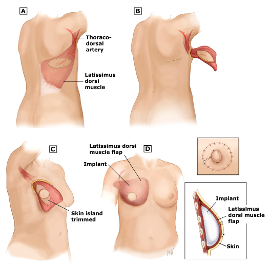 The latissimus dorsi (LD) flap is a reliable option for autologous tissue reconstruction. The flap is tunneled through the axilla, leaving its vascular pedicle, the thoracodorsal artery and vein, intact. Because the LD is not a bulky flap, it usually is used in combination with a saline or silicone implant to provide sufficient breast volume and projection.
-
Early Complications
- Infection
- Flap necrosis (rare)
- Haematoma
- Transient brachial plexopathy
- Breast oedema (settles 6-8/52)
- Donor site seroma (nearly always)
-
Late Complications
- Lateral flap retraction
- Hollowing of resection site
- Flap atrophy (start with slightly larger)
- Mammography not adversely affected
- No compromise to early detection of local recurrence
Pedicled transverse rectus abdominis myocutaneous (TRAM) Flap
- Run off Superior Epigastric vessels
- Inferior epigastrics need to be ligated
- Lower abdominal tissues similar consistency to breast
- Most commonly horizontal lower abdominal skin paddle
- Provides abdominoplasty also
- Limitations:
- Tissue has high metabolic demand, can get flap necrosis
- Longer recovery period, increased abdominal discomfort
- Can be limited by previous abdominal surgery
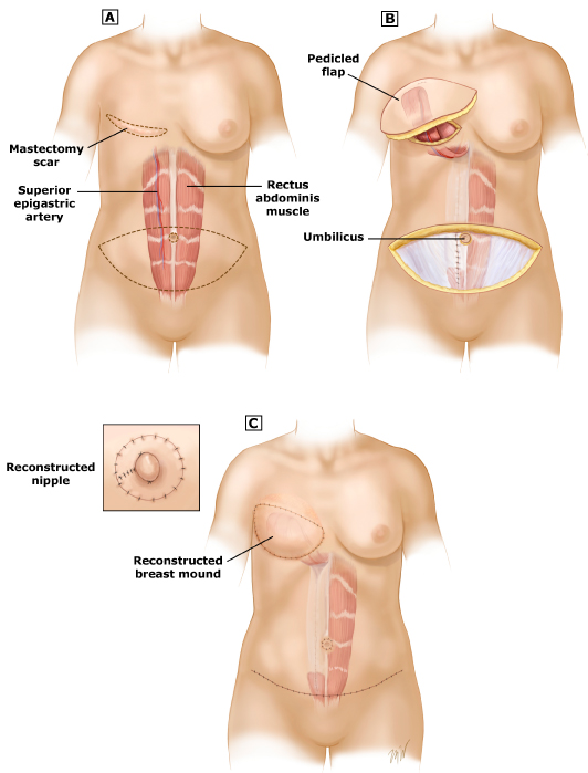 Either the contralateral or ipsilateral rectus abdominis muscle can be utilized. Incision placement and size will vary based on the patient’s body habitus. There are many ways to perform nipple reconstruction; one method is pictured here. The nipple is reconstructed after the flap heals and is then tattooed to the appropriate color so that there is symmetry with the contralateral breast.
Free
- Free TRAM Flaps
- Muscle sparing TRAM (msTRAM)
- Only muscle surrounding perforators taken
- Less morbidity
- Need adequate vessel size
- Deep Inferior Epigastric Perforator Flap (DIEP)
- No muscle taken
- Perforating vessels dissected out in a chain
- Less morbid than TRAM and msTRAM
DIEP
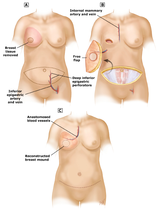 The most commonly used perforator flap for breast reconstruction is the deep inferior epigastric perforator (DIEP) flap. The DIEP flap uses the lower abdominal island of skin and fat and spares the rectus abdominis muscle. Although the underlying muscle must be split to dissect out the perforating vessels, the muscle itself is not included in the transfer. The deep inferior epigastric artery and vein, which provide the vascular supply for the free flap, are anastomosed to local recipient vessels, using either the internal mammary or thoracodorsal arteries and veins.
Free-TRAM
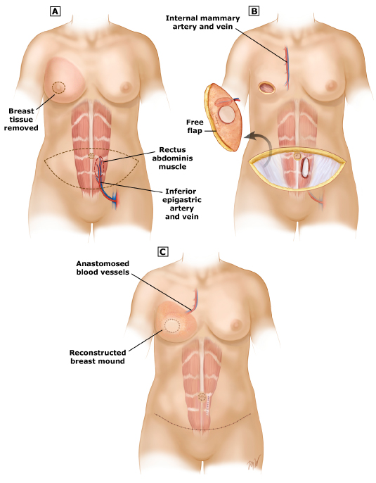 The deep inferior epigastric artery and vein, which provide the vascular supply for the free flap, are anastomosed to local recipient vessels, using either the internal mammary or thoracodorsal arteries and veins. This can be performed to provide immediate or delayed breast reconstruction. In this drawing, a skin sparing mastectomy is depicted. This drawing depicts the blood supply anastomosis to the thoracodorsal vessels; however, the internal mammary site is also used commonly for the site of anastomosis.
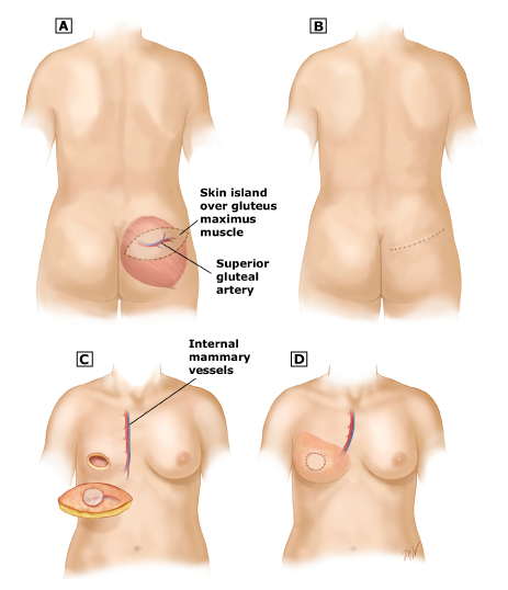
Gluteal
- Superior gluteal artery (SGAP flap) or the inferior gluteal artery (IGAP flap)
- Used if low abdominal fat so can’t do DIEP
- Perforator flap design (SGAP or IGAP)
- Limits donor site deformity and sciatica
- Cons
- Difficult dissection
- Short pedicle
- Size discrepancy of gluteal vein to internal mammary
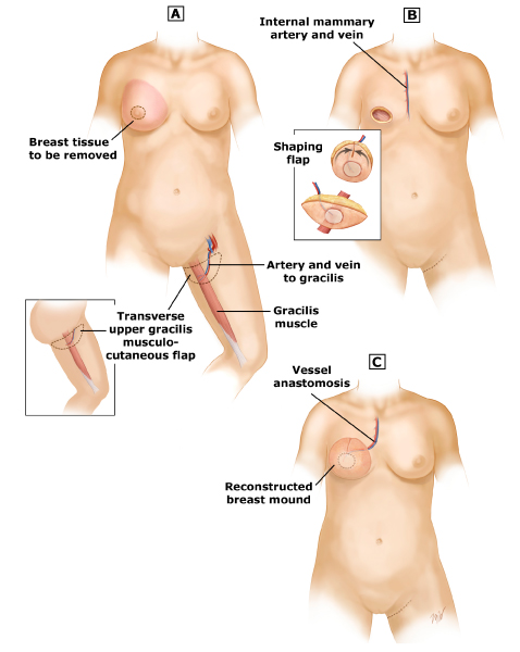
Transverse Upper Gracilis Flap (TUG)
- Uses upper inner thigh tissue
- Ideal for women without abdominal or gluteal tissue to use as donor site
- Can be good to use in salvage situation post-failed reconstruction
- Anatomy
- Based on branch of Medial Circumflex Femoral Artery
- Includes Gracilis and overlying horizontal paddle of skin/fat
- Scar camouflaged in groin/gluteal fold
- More straightforward dissection than perforators flaps (DIEP, GAP etc)
- Disadvantages
- Short pedicle length
- Smaller skin island
- Possible contour deformity of medial thigh
- Can get widened donor site scar
Profunda artery perforator (PAP) flap
- Fasciocutaneous flap based on perforators from the profunda femoris vessels that pass through adductor magnus muscle posterior to the gracilis
Local Perforator
- Pros
- Expendable – do not jeopardise total recon options if reoperation required.
- Available in all patients.
- Can be rotated on a single pedicle or as turnover like a book on multiple pedicles.
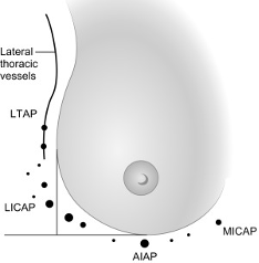
- Intercostal perforators usually in 4th to 6th intercostal spaces
- Types
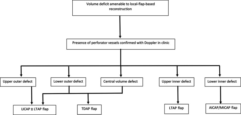
- LTAP flap
- Lateral thoracic artery perforator
- Intercostal perforators
- LICAP
- Lateral intercostal artery perforator
- AICAP
- Anterior intercostal perforator
- MICAP
- Medial intercostal perforators
- LICAP
- TDAP
- Thoracodorsal artery perforator flap
- LTAP flap
Lipomodelling
- Usually used to correct rather than prevent deformities
- Stem cells harvested from fat injected around defect and into Pec muscle and subcut fat
- Avoid breast parenchyma
- Avoids confusing mammographic findings from calcified fat necrosis
- Theoretical risk of tumour induction from injected stem cells!
Non-Autologous Volume Replacement
- Saline or silicone implants
- Anterior or posterior to Pec Major
- Best for bilateral reconstructions
- Otherwise difficult to get ptosis symmetry
- Often expanders placed initially at time of mastectomy
- Capsules (esp. if post-radiotherapy) can interfere with clinical exam and mammography
- MRI may be required
Complications of Volume Replacement
- Chemo has little effect on outcomes of breast reconstruction
- As long as appropriate period post-neoadjuvant therapy for immunologic function to return
- Radiotherapy does impact
- Implant-based has high failure rates
- Complications important as may cause delay to adjuvant therapy!
- Partial/total flap loss
- Mastectomy flap loss
- Wound breakdown
- Infection
- Donor site seroma
- Most common complication of LD flaps
- Fat necrosis
- Most common complication of pedicled TRAMs
- Donor site hernia
More common in:
- Women who require adjuvant therapy
- Implant-based reconstruction
- 15% complications without radiation, 42% with radiation
- Most commonly capsular contraction
- Do capsulectomy, capsulotomy, implant removal
- Smokers
- Decreased microvascular distal circulation
Volume Displacement/ Oncoplasty
Indication
- Need to excise large lesions or significant percentage of the breast via BCS
- Consider also for
- Extensive DCIS
- ILC
- Multifocality
- Partial/poor response to neoadj rx
- Positive margins post-standard BCS
Selection Criteria
- Volume
- Most predictive factor for cosmetic outcome
- If 10-20% of breast removed, clear risk of deformity
- General rule 80g the most removed from medium sized breast before deformity
- Tumour location
- Upper outer quadrant favourable cosmetic results
- Lower pole, upper inner, or central tumours unfavourable
- Glandular characteristic of breast
- Fatty is more difficult to move with higher risk fat necrosis with level 1 procedures, may need level 2
- Dense glandular breasts more amenable to manipulation
Oncological Safety of Oncoplastic Surgery
- No long-term RCTs
- Prospective series show essentially equivalent survival outcomes
- OPS should not cause a change to neoadjuvant or adjuvant practices
General Considerations
- Important to counsel
- Stress outcomes good but do vary
- OPS may result in longer and multiple scars
- May have asymmetry
- Expected, but necessary to prevent distortion and deformity
- May require contralateral procedure
- Patient should be pre-marked sitting/standing
- Able to be sat up on the operating table
Decision on Level of Technique
- Level 1 techniques should be able to be performed by all breast surgeons
- Skin and glandular undermining
- NAC recentralisation
- Level 2 require more complex procedures
- Skin excision and glandular mobilisation
- Derived from breast reduction techniques
- Require additional training
- If < 20% of breast tissue, usually level 2 not required
- Unless fatty composition
-
10% of breast
-
Level 1 Oncoplastic Techniques
- All surgeons should be able to adopt the following steps
- Skin incision
- Skin undermining
- Nipple-Areola Complex undermining
- Tissue excision
- Re-approximation of the glandular defect
- De-epithelialisation and NAC repositioning
1. Skin Incision
- Oncoplastics not minimally invasive
- Short incisions limit mobilisation
- Unable to create adequate glandular flaps
- Effective mobilisation of the gland is key to achieving a natural shape
- Incisions should allow
- En-bloc resection of tumour without fragmentation
- Undermining of surrounding breast tissue to facilitate reshaping
- Generally should follow Kraissl’s lines of maximum resting skin tension
- Many cases may allow an incision away from the tumour itself (e.g. periareolar)
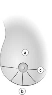 a) Periareolar for upper half.
b) Inframammary for low
c) Radial cause less deformity in lower half
a) Periareolar for upper half.
b) Inframammary for low
c) Radial cause less deformity in lower half
2. Skin Undermining
- Extensive subcutaneous undermining may be necessary
- Can be up to 1/2 – 2/3 of breast
- Needed to facilitate glandular redistribution
- Easier to undermine large skin area before excising the lesion
- Perform in plane between subcut fat and breast tissue
- Can facilitate by using hydrodissection (NaCl with adrenaline 1:500,000)
- Assess risk factors for vascular compromise before performing extensive undermining
- Smoking
- Diabetes
- Connective tissue diseases
- Final factor for determining how much to undermine is composition of the breast
- If fatty, Level 2 procedure may be better (less undermining, more direct glandular excision)
3. Nipple-Areola Complex Undermining
- Major NAC distortion is a common cause of breast deformity
- Fibrosis after surgery creates tension on adjacent tissue
- NAC deviates towards area of excision
- NAC repositioning can be done easily with simple undermining
- Key component to both L1 and L2 OPS
- First step is to completely transect terminal ducts under the nipple and separate NAC from breast tissue
- 0.5-1cm glandular tissue is generally left on NAC to ensure vascular integrity
- Prevents NAC necrosis and limits venous congestion
- NB: Even if ducts are divided immediately under the skin, > 90% of nipples will survive!
- Level of NAC undermining is related to loss of nipple sensitivity
- Need to warn patients
4. Tissue Excision
- Perform full thickness excision from subcutaneous fat underlying the skin, to pectoral fascia
- Ensures free anterior and posterior margins, leaving only lateral margins to worry about
- NB: Not always required
- If tumour superficial or deep, and breast > 4cm anterior to posterior dimensions
- NB: Not always required
- Parenchyma can be taken in fusiform fashion (tapering at both ends) to facilitate approximation
- Can potentially increase total volume of tissue excised
- Before closing, put clips at edges of tissue defects
5. Re-approximation of Glandular Defect
- Don’t leave cavity for seroma
- Non-predictable results in larger excisions
- Seroma reabsorption → Cavity contraction → Defect with distortion → NAC displacement
- Instead redistribute the remaining tissue to redistribute the loss
- Can mobilise tissue from lateral or central portions
- May require further undermining in superficial mastectomy plane or retro-mammary space
6. De-epithelialisation and NAC Repositioning
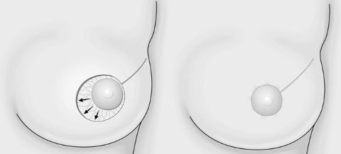
- Major source of dissatisfaction is NAC deviation
- Will deviate towards resection after any large resection
- NAC repositioning very difficult after radiotherapy
- Do it immediately
- NAC repositioned to adjust for
- Anticipated deviation of the nipple
- New shape of the breast
- Cresenteric area of periareolar skin opposite the excision defect is de-epithelialised
- NB: NAC blood supply based on dermal plexus, should not be affected if correct de-epithelialisation performed
Level 2 Oncoplastic Techniques
- Generally used for excisions of 20-50% of breast volume
- Can be less if fatty breast
- Generally results in smaller breast, that is rounder and higher on the chest wall then contralateral breast
- Need for contralateral symmetrisation
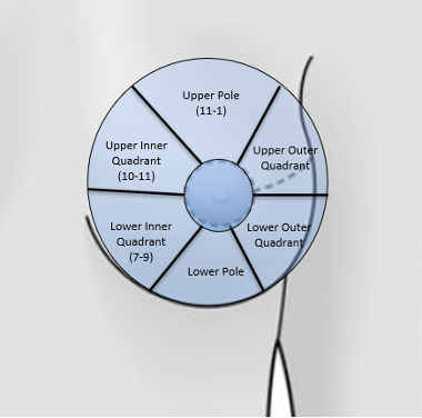
- Follows oncoplastic atlas
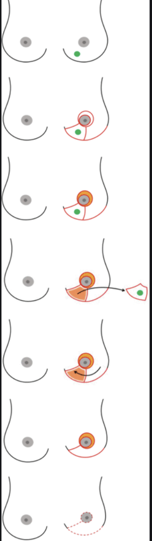
- Lower pole (4-7 o’clock)
- Lower inner quadrant (7-9 o’clock)
- Upper inner quadrant (10-11 o’clock)
- Upper pole (11-1 o’clock)
- Upper outer quadrant (1-3 o’clock
- Lower outer quadrant (4-5 o’clock)
- Retroareolar
Lower Pole
- First region to be recognised as at risk of deformity
- Downward retraction after resection = Bird’s beak deformity
Superior Pedicle Mammoplasty
- Inverted “T” and periareolar scars
- Permits large volume excision of lower pole without NAC deviation
- Added benefit of breast reshaping and elevation
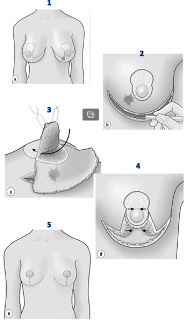
Procedure
- Begins with de-epithelialisation of area surrounding NAC
- Then NAC dissected away from surrounding breast tissue
- Superior pedicle preserved to maintain NAC blood supply
- Infra-mammary incision completed
- Followed by wide undermining of breast tissue of pec fascia (fascia preserved)
- Undermining starts inferiorly, goes superiorly beneath tumour (can palpate tumour)
- Encompasses medial and lateral aspects of breast and NAC
- Tumour then removed en-block with surrounding (1cm) breast tissue and overlying skin as per pre-op markings
- Remodelling
- Medial and lateral glandular columns re-approximated
- NAC recentralisation
- Weigh excised tissue (provides guide for contralateral side)
- Remove 10-20% less on cancer side as will shrink with radiotherapy
Lower Inner Quadrant (7-9)
- Standard superior pedicle mammoplasty can go to 7 o’clock only
- V Mammoplasty
- Excises a pyramidal section of skin and breast tissue
- Base sub-mammary fold
- Apex border of areola
- Skin and glandular tissue removed En-block
- Incision then made along inframammary fold towards anterior axillary line
- Lower pole mobilised off pec fascia and laterally
- Used as advancement flap to fill defect
- NAC then recentralised on de-epithelialised superior lateral pedicle
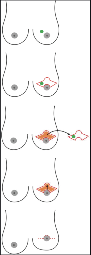
Upper Inner Quadrant (10-11)
- This area requires caution
- WLE can have significant impact on quality of breast shape
- Distorts visible breast line (decollete)
- Standard level 2 procedures limited for this area
- Omega (batwing) mammoplasty can be used
- Excess tissue needs to be excised
- Large scar
- Removes pectoral fascia and skin
- Smaller level 1 procedures and immediate lipofilling may be best approach
- Omega (batwing) mammoplasty can be used
Upper Pole (11-1)
- Superior tissue can be widely excised and defect filled with central tissue
- Gives good durable results
- Can do either
- Inferior Pedicle Mammoplasty
- Round Block Mammoplasty
- Danger when trying to preserve tissue superior to nipple
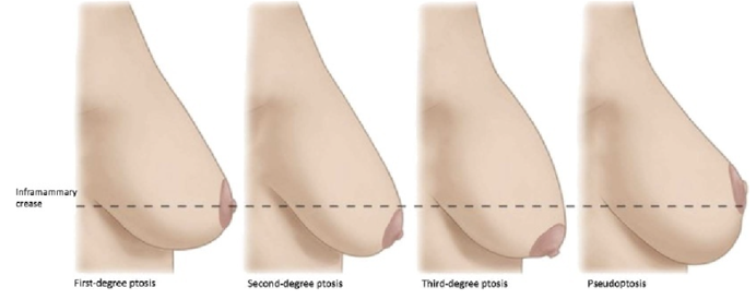
- get “bottoming out” (pseudoproptosis)
- Danger when trying to preserve tissue superior to nipple
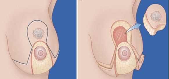
Inferior Pedicle Mammoplasty
- Inverted T scar, skin markings the same as superior pedicle mammoplasty
- Resection at upper pole
- Vascular supply of NAC based on inferior pedicle
- Excision of cancer is through incision in skin that will be removed
- Cancer removed and X-rayed
- Inferior pole de-epithelialised and advanced towards excision defect
- Resection of tissue in inner + outer lower quadrants to optimise shape
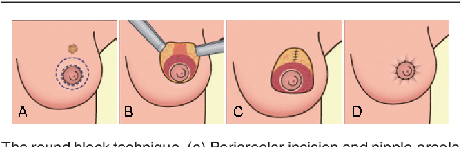
Round Block Mammoplasty
- Mainly used for upper pole tumours, but is versatile and can be adapted to tumours anywhere in the breast
- Not most surgeons favoured method of reduction due to some limitations
- Technique
- Periareolar incision
- 2 concentric periareolar incisions followed by de-epithelialisation of intervening skin
- Peripheral skin envelope developed to give access to tumour
- NAC remains vascularised through posterior glandular base
- Lesion resected
- Subcut tissue to pec fascia
- Forms external and internal glandular flap
- Flaps mobilised off pec fascia and moved towards each other
- Two incisions then approximated to give single periareolar scar
Factors Affecting Timing of Breast Reconstruction
- Likelihood of requiring radiotherapy
- Implant can still be used for immediate reconstruction
- Autologous usually delayed to allow for irradiated breast and chest wall tissue to be replaced with healthy tissue from elsewhere
- Inflammatory Breast Cancer or cancer with significant skin involvement
- More skin removal required – thus, immediate reconstruction more challenging
- Time to consider options
- More time to think about options when not worrying about cancer surgery
Factors Affecting Choice of Breast Reconstruction
- Patient factors:
- Size and shape of breasts
- Age and comorbidities
- Previous surgeries
- Smoking status
- Obesity
- Availability of autologous tissue
- Disease factors:
- Location of tumour in breast
- Specific consideration factors related to implants:
- Enough skin and muscle must remain after mastectomy to cover implant
- Shorter surgical procedure than autologous reconstruction
- Shorter recovery period
- Many follow-up visits may be required to inflate expander and insert implant
- May not be adequate for large breasts
- Will not last a lifetime
- Specific consideration factors related to autologous reconstruction:
- Longer surgical procedure
- Longer recovery period
- Provides a more natural breast shape
- Can be used to replace tissue damage by radiotherapy
Breast implant related issues
Capsular Contraction
- A chronic inflammatory infiltrate + a fibrous capsule usually forms around the implant
- 15-20% develop capsule contraction / scarring around the implant
- Classification - Baker Grading (I-V) for contracture of implants
- Grade I — the breast is normally soft and appears natural in size and shape
- Grade II — the breast is a little firm, but appears normal
- Grade III — the breast is firm and appears abnormal
- Grade IV — the breast is hard, painful to the touch, and appears abnormal
- Aetiology:
- Can occur with both saline and silicone implants
- RTx ↑s risk
- Clinical:
- Capsule contraction / scarring → firmness / distortion, may be painful
- Baker Grade III or IV contracture: implants are so hard they may impair mammography
- Mx
- Symptomatic women may require removal of implant + capsulotomy/capsulectomy
- Baker Grade III or IV contracture: implants often removed
- Natural Hx:
- Surgical Rx of the contracture → High recurrence rates if implant not removed
Implant Rupture
- Aetiology
- Degeneration caused by daily stresses & aging of the implant, trauma, iatrogenic cut during surgery / Bx
- Clinical:
- Ruptured saline implants usually deflate quickly → Change in size of breast
- ± Chest pain, breast rash
- Silicone leak
- May go unnoticed, even for a period of years
- ↓ Size of breast, uneven breasts, pain / tenderness, swelling, numbness / burning / change in sensation, lumps
- Ix:
- +/- USS may demonstrate a leak
- Mammogram is not accurate in detecting rupture / leak
- MRI = Best modality to assess implant integrity
- Mx:
- Remove implants ASAP, esp. if silicone implant
- Natural Hx:
- Most implants rupture by the time they are 25 years old
Other implant related issues
- Infection following implant placement
- Breast implant illness
- Systemic response following placement – currently not proven; just reports
- Non-specific symptoms
- Fatigue
- Malaise
- Fibromyalgia Sx
- Joint pains / stiffness
- Autoimmune disease
- SLE, scleroderma, RA, Sjögren’s disease
- Non-specific symptoms
- Systemic response following placement – currently not proven; just reports
- NB: If woman develops Ca → Treat the same as general population; but beware that RTx may result in marked capsular contracture
[Breast Implant Associated Anaplastic Large Cell Lymphoma ]
- Definition
- Peripheral T-Cell lymphoma associated with textured surfaces of breast implants
- Incidence
- Rare (1:10,000)
- Presentation
- Usually localised disease 10yrs post insertion
- Seroma 60%
- Often found incidentally during revision surgery for late-onset seroma
- Mass 17%
- Aetiology unknown
- ? Implant induced chronic inflammation, ? Biofilm
- Association with Allergan BIOCELL textured implants (91% of cases)
- Pathology
- Large anaplastic pleomorphic cells with abundant cytoplasm, horseshoe nuclei
- IHC shows strong uniform CD30 staining, ALK negative
- NB: This can also be seen in inflammation, must have anaplastic morphology and single T-Cell clone also
- Investigations
- USS guided aspirate of seroma and send for cytology, cultures
- Management
- Localised disease
- Surgical removal of implant, associated masses/disease, and total capsulectomy
- Treatment is usually curative
- Disseminated disease
- Systemic chemotherapy
- SLNB not required, but excision biopsy of suspiciously enlarged nodes performed
- Regular F/U
- Localised disease
- Prognosis
- Biologically indolent disease with a good prognosis with complete surgical resection provided there is no extension beyond the implant capsule