Thoracic Aortic Dissection
- Dissection of the thoracic aorta
Incidence
- Aortic dissection = Most common catastrophe of Aorta (more common than AAA)
- M > F (3:1)
- Peak age: 50-65; (75% occur in pts aged 40-70yrs)
Classification
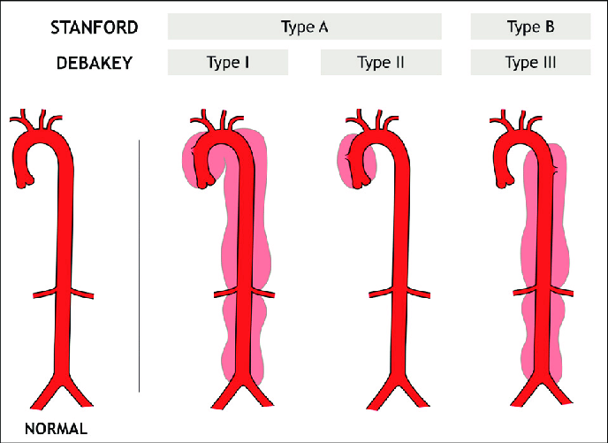
Aetiology
- Marfans, Ehlers-Danlos & other connective tissue diseases
- HTN can propagate dissection
- Atherosclerosis
- Iatrogenic injury (e.g. Catheter, Open heart procedures)
- Blunt chest trauma
- Can be associated with Pregnancy
Clinical
- Chest pain
- Ripping / tearing/ sudden onset
- Neck, jaw, interscapular pain
- Pain tends to be felt anteriorly in ascending dissection & posterior interscapular in descending dissection
- Neurological Sx / stroke Sx / limb paraesthesia, pain or weakness – in 20%
- BP differential of > 20mmHg
- Renal failure, acute mesenteric occlusion & lower extremity arterial occlusion may occur
- If the dissection involves the pericardial space, tamponade may result
- Occlusion of coronary arteries may also occur
- Aortic root involvement is also associated with prolapse of the Aortic valve
Pathology
- 90% occur within 10cm of the Aortic valve
- Tear in the intimal layer → Formation & propagation of subintimal haematoma
- Cystic Medial Necrosis
- Hallmark change associated with dissection, esp. in Marfan’s
- Degenerative changes in the media leading to breakdown of the collagen, elastin and smooth muscle
- Medial degeneration predisposes to rupture of Vasa Vasorum → Intramural haematoma formation → Dissection
Investigations
- ECG
- 8% of pts with type A dissections have ST elevation (none with type B)
- CXR
- Normal in up to 50%
- Widened mediastinum (> 8cm on PA CXR)
- Abnormal (blunted) Aortic knuckle
- Left Apical Cap
- Angiography
- Accurate Dx in 95% (historically the gold standard)
- CT
- Helical / multiplanar CT = Replacing angiography as the test of choice
- Sensitivity 83-94%, specificity 87-100%
- Echo
- Transthoracic – Sensitivity 80%, specificity 90%
- Transoesophageal – Sensitivity 97-99%, specificity 97-100%
- MRI
- Sensitivity >90%, specificity >95%
Management
- Type A: Surgery → Dacron graft
- Type B: Medical therapy → Control of BP (β-blockers)
- But ≈ 20% will require surgery for organ ischaemia
Prognosis / Natural Hx:
- If untreated, Acute Type A → 33% die within 24/24, 50% die in 48/24; 70% by 1/52
- 2-week mortality ≈ 75% in pts with undiagnosed Ascending Aortic dissection
- After surgery
- Operative mortality: 5-15%
- 5YS Open surgery 50%
- Type B risks: 3% Paraplegia, 34% Renal failure, 7% Bowel ischaemia, 4.7% CVA
Follow-up
- For Type B dissection, 20% develop aneurysmal dilation of the aorta, therefore yearly imaging surveillance advised
Buerger’s disease
- Definition
- A non-atherosclerotic, segmental, inflammatory vasculitis primarily affecting small- and medium-sized arteries and veins. It is strongly associated with tobacco use.
- Epidemiology
- Occurs mostly in young males aged 20–45 years and has a higher prevalence in smokers and regions with common tobacco use.
- Pathophysiology
- Chronic inflammation of vessel walls leads to thrombosis, ischemia, and tissue necrosis, predominantly involving distal extremities and leading to gangrene in advanced cases.
- Risk factors
- Tobacco use, including smoking or chewing, is the major trigger, and genetic predisposition may play a role in some populations.
- Clinical
- Early symptoms include pain in affected limbs, claudication, or Raynaud’s phenomenon, while advanced symptoms include rest pain, ulceration, and gangrene in fingers or toes.
- Superficial thrombophlebitis may also occur.
- Diagnosis
- Involves Shionoya’s clinical criteria: onset before age 50, tobacco use history, involvement of distal vessels, exclusion of other diseases like atherosclerosis or autoimmune conditions, and positive angiographic findings showing “corkscrew” collateral vessels in distal extremities.
- Investigations
- Angiography to show segmental occlusion and collateral formation and blood tests to exclude autoimmune or hypercoagulable conditions.
- Management
- Includes complete cessation of tobacco, which is the cornerstone of treatment and halts disease progression.
- Smoking cessation programs or nicotine replacement therapy may be required.
- Vasodilators like calcium channel blockers are used for ischemic symptoms, and antiplatelets reduce thrombus formation. Pain is controlled with analgesics.
- Sympathectomy may improve symptoms in severe ischemia, while amputation is reserved for nonviable tissue or uncontrolled infection.
- Includes complete cessation of tobacco, which is the cornerstone of treatment and halts disease progression.
- Prognosis
- Depends on tobacco cessation, as continued smoking leads to recurrent thrombosis, severe ischemia, and limb loss.
Raynaud’s Syndrome
- Episodic digital asphyxia caused by arterial insufficiency
- Primary – Exaggerated vasospastic response in normal vessel – hypersensitivity of receptors in vessel wall
- Bilateral, gangrene not a feature
- Secondary – normal vasospastic response in abnormal vessel
- Unilateral, gangrene can be a feature
- Look for thoracic outlet obstruction, angiography
- Young women in cool damp climates
Clinical
- Sequential skin changes of pallor, cyanosis, rubor of fingers or toes after exposure to cold = Raynaud’s phenomenon.
- Hands > feet, also affects nose, ears, nipples
- White-blue-red color changes produced by excessive vasoconstriction, sluggish flow & reflex vasodilation
- Sudden onset / progression suggest underlying arterial lesions → More aggressive
- Associated CREST, SLE, Polymyositis, Thoracic outlet syndrome & occasionally others (cold agglutinins, CRF, neoplasia, vibration injury, drugs (OCP, ergotamine))
- Acrocyanosis is a related chronic benign condition seen in young women, characterized by cold-induced cyanosis of the hands and feet ± with skin of calf/thigh/forearm displaying livedo reticularis
Investigations
- Autoantibodies, Cryoglobulins, RhF, ESR, CRP, Electrophoresis
Management
- Avoid cold exposure, tobacco, OCP, β-blockers & Ergotamine
- Ca2+ Channel blockers (e.g. Nifedipine), ACEI, Prazosin
- Iloprost – seasonal treatment
- Sympathectomy in severe disease
Arterio-venous fistula
- Abnormal communications between arteries & veins, effect depend on size & location
Aetiology
- Congenital
- Acquired
- Trauma
- Iatrogenic
- Surgical – haemodialysis access
Clinical
- Tachycardia occurs in some pts due to increased cardiac output
- When fistula occluded by compression, the pulse rate slows (Branham’s sign)
- High-output heart failure can occur
- May cause bleeding
- Bleeding AV fistula in GI tract
- Osler-Weber-Rendu Syndrome (hereditary hemorrhagic telangiectasia) - large AV malformations in GIT & lungs, AD, GI bleeding & epistaxis
- Pulmonary lesions cause recirculation with lower PO2, polycythaemia, clubbing and cyanosis
Investigations
- MRI
- CTA
- Selective angiograms
Management
- Many can be treated conservatively
- Indications for intervention: haemorrhage, local expansion, severe venous/arterial insufficiency, cosmetic deformity, heart failure
- Most are managed by embolization under radiographic control
- Surgery for large, acquired fistulas
- Avoid surgery for congenital fistulas as cure requires en bloc resection of all tissue involved
- Local ligation is a temporary measure that invariably results in recurrence
Prognosis / Natural Hx
- Traumatic fistulas have the most favourable prognosis
- Congenital fistulas are difficult to eradicate, due to numerous AV connections
Subclavian Steal Syndrome
- Signs & symptoms that arise from retrograde vertebral artery flow or retrograde internal thoracic artery flow due to proximal Subclavian Artery stenosis or occlusion
Classification
- Vertebral-Subclavian Steal syndrome:
- Vertebral Artery serves as a collateral to supply blood to the arm (the blood from the brain to the upper limb is considered to be “stolen” as it is blood flow that the brain must do without) → Possible cerebral ischemia
- Coronary-Subclavian Steal syndrome:
- Post-op CABG using Internal Thoracic Artery = Distal Internal Thoracic Artery diverted to the LAD → If proximal Subclavian Artery Stenosis, blood may flow backward from the heart causing myocardial ischaemia
Aetiology
- Atherosclerosis of Subclavian Artery
- Cervical rib → Causing compression of subclavian artery
- Takayasu’s arteritis
- Tumour encasement
- Iatrogenic: Post-Aortic Stent graft placement for Thoracic Aneurysm
Clinical
- Symptoms of effort fatigue in involved extremity
- Presyncope / syncope
- Blood pressure differential between arms
- Look for other stigmata of PVD
Investigations
- USS, CTA, Angiogram
Management
- Stenting / angioplasty of subclavian stenosis (but this is not useful in the setting of a thoracic outlet syndrome / cervical rib, as the stent will itself become compressed
Surgery
- Endarterectomy
- Bypass grafting from the common carotid to the subclavian artery distal to the lesion or transposition of the subclavian a beyond the lesion
Takayasu’s Arteritis
- Obliterative arteriopathy principally involving aortic arch vessels that often affects young women – Large Artery Vasculitis
- Cause’s Aortic Arch Syndrome
Incidence
- Mainly young women M:F 1:3
- Eastern European / Asian
Clinical
-
Upper extremity claudication
-
Global cerebral perfusion
-
Pathology
-
Aortic arch & origin of great vessels most often affected
-
Giant cell arteritis, massive intimal fibrosis – vascular narrowing
-
Pararenal Aorta & pulmonary arteries may also be affected
Management
- High dose corticosteroids and cyclophosphamide arrest ± reverse
- Operative Rx avoided when arteritis is active, but may be successful in quiescent
Giant Cell Arteritis
- Temporal Arteritis
- Chronic systemic condition involving vasculitis of large and medium-sized arteries – Large Artery Vasculitis
Incidence
- F > M 2:1, average age at presentation 75yrs
Clinical
- Unilateral headache
- Tender temporal artery + nodular change
- Jaw claudication
- 50% have PMR
- Visual change > 50%
- Retinal artery occlusion
- Ischaemic optic neuritis
Pathology
- Inflammation of temporal, vertebral, ophthalmic, aortic arch (90% = branches of ECA)
- Histology: fragmentation of internal elastic lamina with mononuclear cell infiltration and accumulation of giant cells; skip lesions
Investigations
- ESR
- Temporal Artery biopsy
- At least 20mm in length
- Performed within 2/52 of starting steroids (Inflammatory changes probably persist 2-4/52)
Management
- High dose steroids: start at 60mmHg → Then taper
Aortic Coarctation
Aetiology
- Congenital
- May result from an inflammatory large vessel arteritis – e.g. Kawasaki’s or Takayasu’s
Clinical
- Sx of lower leg / mesenteric / renal ischemia
Management
- Congenital → Surgical Mx
- If on-going inflammation → Repair not recommended as these pts do poorly
- But if disease is quiescent with a normal ESR → surgical repair OK
Reflex Sympathetic Dystrophy
- Post-traumatic pain syndrome characterized by pain with ↑SNS activity
- a.k.a. causalgia, posttraumatic sympathetic dystrophy, Sudeck’s atrophy, shoulder-hand syndrome, traumatic neuralgia, Mitchell’s causalgia
Incidence
- M = F
- UL = LL
- The type or severity of initial injury dose not predict development of this syndrome
Clinical
- Severe pain – Burning, involving entire hand/foot
- Small stimulus → Sudden ↑ in severity of pain
- Marked vasoconstriction → Cold, cyanotic & moist extremity
- If left untreated → Limb shows dystrophy & atrophy
Management
- Sympathetic block with LA (short-term) or surgical/chemical sympathectomy (long-term)
Klippel-Trenaunay-Weber Syndrome
- Triad (Most have all three)
- Varicose veins
- Bony and soft tissue hypertrophy involving an extremity (bone lengthening)
- Port-wine stain (Haemangioma)
- ± addition of AV malformation as Parkes Weber syndrome
Incidence
- ~ 2 per 100,000
Aetiology
- Most cases are sporadic, although a few cases reported as AD
- Deep vein hypoplasia & absence of valves, with resultant obstruction of venous flow, lead to venous hypertension, development of varices, and limb hypertrophy
- Mesodermal defect during fetal development causes maintenance of microscopic arteriovenous communications
Clinical
- Generally affects a single extremity, although cases of multiple affected limbs reported
- Leg most common site (then arms > trunk, rarely head/ neck)
- 90% limbs
- 10% head & neck involvement
- Haemangioma is often noted on the lateral aspect of the limb
- May be limited to skin or extend deeper to subcutaneous tissue, including muscle & bone
- Visceral organs, such as pleura, spleen, liver, bladder, and colon may also be affected.
- Visceral organ involvement portends greater morbidity secondary to internal haemorrhage
- The Klippel-Trenaunay vein is a large lateral (i.e. atypical for a varicosity), superficial vein sometimes seen at birth
- Vein begins in the foot or the lower leg and travels proximally until it enters thigh or gluteal area
- Varicosities may be extensive, though often spare saphenous distribution
- Varicosities may affect superficial, deep, and perforating veins
- Varicosities may remain stable or gradually expand
- Pain and lymphoedema common.
- Symptoms may worsen during pregnancy
Pathology
- Deep veins often anomalous or absent
- So saphenous vein stripping = Hazardous!
Investigations
- Evaluation of the deep venous system can be completed with duplex scanning contrast venography, USS, contrast venography and arteriography and nuclear MRI studies
- Arteriography is especially helpful in the diagnosis of AV fistula
Management
- Conservative if asymptomatic
- Compression garments (usually class II) for chronic venous insufficiency, lymphoedema, recurrent cellulitis, and recurrent bleeding from capillary or venous malformations
- However, if absent or hypoplastic deep venous system, elastic compression may increase venous stasis and cause discomfort
- Referral to a pain clinic
- Cellulitis and thrombophlebitis - analgesics, elevation, antibiotics & corticosteroids
- Anticoagulant therapy - acute thrombosis/ prophylactically prior to surgical procedures
- Increased risk of thrombotic events, avoid using OCP
- Laser treatment for haemangioma
- Can be effective in lightening colour of port-wine stain
- Indicated in case of ulceration
- Compression garments (usually class II) for chronic venous insufficiency, lymphoedema, recurrent cellulitis, and recurrent bleeding from capillary or venous malformations
- Surgical intervention for varicosities & venous malformations controversial
- Venous stripping, ligation, excision or sclerotherapy are contraindicated unless the surgery involves the superficial system and the underlying deep system is normal or demonstrates only mild-to-moderate reflux
- Inadequate evaluation prior to excision increases surgical complications
- Radiotherapy has been reported to be of help
- Induce regression of haemangiomas; however results can be slow to develop
- Endovenous laser therapy of the greater saphenous vein gaining support
Carotid Artery Stenosis
- Narrowing of the lumen of the carotid arteries, usually caused by atherosclerosis
Incidence:
- Prevalence of asymptomatic carotid stenosis = 2-18%
- Up to 20% of pts undergoing CABG may have Carotid Stenosis of > 50%
Aetiology
- Risk Factors:
- ↑ Age
- Smoking
- HTN
- IHD
- TIA
- DM
- PVD
- Cholesterol
Clinical
- May be asymptomatic
- TIA (neurological deficit lasting < 24hrs)
- Amaurosis Fugax = Temporary monocular vision loss caused by a microembolus to the ophthalmic artery
- Pulsatile tinnitus
- Hollenhorst Plaques = Small bright flecks in field of vision, due to emboli lodged in arterial bifurcations in the retina
- Stroke
- Compared with Vertebrobasilar disease → Emboli in posterior circulation → Vertigo, diplopia, dysphagia & dysequilibirum
Pathology
- Origin of the ICA & Carotid Sinus is prone to atherosclerosis (change in flow, turbulence & dilatation at this point)
- Plaques can undergo acute disruption → Thrombus formation & 2° thromboembolism
- Trigger for acute change in plaque morphology is unknown, but may be related to increased expression of MMP
- High Risk Features:
- 90 - 94% Stenosis
- Irregular / ulcerated plaque
Investigations
- Duplex USS - 94% sensitive and specific for detecting severe stenoses
- Flow ratio (velocity) estimates stenosis
- MRA
- CTA
- Angiography = Gold standard – 1 - 4% risk of stroke
- Absolute indications
- Technically inadequate duplex
- Lesion beyond carotid bulb
- Anatomic variants
- Relative
- Unusually high bifurcation
- Recurrent stenosis
- Severe fixed neurological deficit
- Other complications
- Allergy to contrast < 2%
- Contrast-induced nephropathy
- Puncture site problem
- Absolute indications
Screening
- Asymptomatic pts > 65yrs with ≥ 3 cardiovascular risk factors but US Preventative Services Task Force (USPSTF) guidelines say that routine screening is not indicated as benefits do not outweigh risks
- Symptomatic PVD (but not AAA alone)
- Radiotherapy (≥ 45Gy) for head & neck Ca – screen 10 yrs post-Rx
- Post-Carotid Endarterectomy – can screen contralateral side, esp. if baseline stenosis ≥50%
- Renal ischaemic events
- Not necessarily if just renal artery stenosis
Management
- Carotid Endarterectomy
- Oblique anterior SCM incision
- CCA/ICA/ECA exposed
- Facial vein transected, lateral retraction of IJV
- Dissection up to hypoglossal nerve (may need to ligate muscular branches to SCM from ECA & ligate small branches to IJV)
- CCA/ICA/ECA dissected circumferentially & looped
- Systemic heparinisation (100U/kg)
- Sequential clamping ICA, CCA, ECA
- Monitor ICA stump pressures
- Anterolateral arteriotomy – common to ICA
- (Shunt – Javid/ Pruitt)
- Elevation of plaque with tacking sutures at distal end
- Primary or vein/gortex patch closure
- Flush arteriotomy
- Remove clamps (ECA/CCA/ICA)
- Skin closure
Indicated for:
- Symptomatic lesions > 70%
- Asymptomatic lesions > 60% if operative risk is < 3% & life expectancy is > 5%
- Symptomatic lesions
- Decreases stroke risk for symptomatic patients with > 70% stenosis from 26% → 9% at 2 years
- Some benefit in symptomatic patients with 50-69% stenosis: 22% → 15.7% risk of stroke at 5 yrs
- NNT= 6 to prevent one stroke / death
- Asymptomatic lesion
- Small risk reduction for asymptomatic pts with > 70% stenosis (12% → 6%)
- ACAS North America & the European ACST
- NNT=19 to prevent one stroke / death (*)
- Overall: ≈ 30% risk reduction for stroke over three years (Cochrane 2005)
- Semi-acute OT – after small TIA / stroke
- After small stroke in pts ASA I/II: surgery within 2-4 weeks → perioperative stroke risk = 2-7%;
- but if big stroke / unstable neurological deficit / ASA III or IV → ↑ perioperative stroke risk therefore best to delay surgery by ≈ 6 weeks
General Points
- Thromboendarterectomy cannot be performed when the ICA completely occluded
- Coexistent coronary disease treated first
- Most strokes during cardiac procedure are from atheroma in aortic arch, not low flow through carotids
- Stroke risk within 30 days = 3%; death rate < 1%
- May be higher if contralateral occlusion
- Possible with regional anaesthesia (superficial & deep cervical plexus block – at posterior aspect of SCM) + awake neurological monitoring with selective shunting (only use intra-op shunt in the people who start to develop neurological Sx after cross-clamping)
- Regional anaesthesia may ↓ strokes, deaths & AMIs
- Shunting everyone → ↑ complication rates
- Patch angioplasty (cf primary closure) may ↓ stroke & re-stenosis rates
- Post-op lose cerebral autoregulation – Keep BP 100-140 Systolic – nitroprusside/ GTN
- ± Post-op dextran (500mL dextran 40 over 4-6hrs) to ↓ platelet adhesiveness
- Dextran: Polysaccharide that decreases blood viscosity
Carotid Artery Stenting
- CAVATAS trial = Carotid artery & vertebral artery transluminal angioplasty & stenting: 500 symptomatic pts RCT
- Stroke rate ≈ 10% in both arms
- Trial also not useful because no protection device was used
- SAPPHIRE = Stenting and angioplasty with protection in pts at high risk for endarterectomy: >300pts; 2/3 were asymptomatic with >80% stenosis
- Similar results for high risk pts undergoing stenting vs endarterectomy – for death & stroke & for stroke rates at 3yrs
- Fewer immediate peri-op complications after stenting
- Results improving (esp. since the intro of cerebral protection devices)
- Useful for lesions that recur after surgical treatment
- CREST study: similar results as surgery; higher morbidity in pts >80yrs
Carotid Artery Bypass
- Rarely used for: long segment atherosclerosis, aneurysm disease, traumatic arterial disruption, re-stenosis after endarterectomy, as part of neck resections
Complications
- CVA/ Death
- Haemorrhage
- Nerve damage
- Hypoglossal, Ansa Cervicalis, Carotid Sinus nerve
- Marginal Mandibular
- Vagus, Recurrent Laryngeal, External Laryngeal
- Great Auricular, Transverse cervical, Spinal Accessory
- Glossopharyngeal, Sympathetic
Prognosis / Natural Hx:
- Stroke risk in symptomatic after 2 yrs → 25-30% (↓ to 2-10% with surgery)
- Stroke risk lower (≈ 20%) in pts with amaurosis fugax
- After Bypass:
- Peri-op stroke & mortality rate = 7.1%
- 5-yr graft patency rate: 74% for PTFE & 92% for autogenous vein grafts
- Risk factors for peri-op stroke (& likely to need intra-op shunting)
- Symptomatic pre-op, includes previous TIAs/AF
- Pre-op stroke = Highest risk
Carotid Artery Dissection
Incidence
- Spontaneous dissection: incidence ≈ 2 per 100,000, mostly seen in 5th decade
Aetiology
- Severe hypertension
- Neck trauma
- May develop spontaneously in young adults
- Genetic / hereditary disorders likely – e.g. Ehlers-Danlos
Clinical
- Sx due to cerebral ischaemia
- Horner’s syndrome
- Acute neck pain associated with localized cervical tenderness (at angle of mandible)
Pathology
- Primary lesion is an intimal tear at the distal end of carotid bulb
- Tear of tunica intima or between tunica media & adventitia (? originating from vasa vasorum)
Investigations
- Arteriography: characteristic pattern of tapered narrowing at / just beyond the distal portion of the carotid bulb ± intimal flap
Management
- Anticoagulation = Mainstay of Rx
- Heparin → Warfarin
- Antiplatelet agents: Aspirin
- Intervention reserved for pts with recurrent TIAs → Stenting
- If stenting unsuccessful & sx persist, ligation can be performed if the carotid back pressure is > 65mmHg
- Extracranial to intracranial bypass will be needed if the pressure is low
Prognosis / Natural Hx
- In most pts, intramural clot will be resorbed, restoring a normal lumen
- Healing usually takes 3 – 6 months
- Incidence of contralateral disease higher than the general population
- Mortality rate < 5%
Follow-up
- Imaging of both carotids
Fibromuscular Dysplasia
- Non-atherosclerotic angiopathy of unknown cause that affects specific arteries
Incidence
- Young women (< 45yo)
Clinical
- If carotid artery affected → Sx of cerebrovascular disease
- TIAs = Most common manifestation
-
20% have a stroke by the time of presentation
- HTN ≈ 1/3 of pts due to renal artery involvement
Pathology
- Overgrowth of media (smooth muscle & fibrous tissue) in a segmental distribution → Concentric rings of Hyperplasia that project into the arterial lumen
- Very rarely a problem of intima or adventitia
- Involves straight central vessels
- Involves primarily:
- Middle 1/3 of extracranial ICA
- Middle/ distal 1/3 of Renal Artery
- External Iliacs
- Usually bilateral if cerebral & bilateral in 50% of renal cases
- In renal artery FMD - renal artery aneurysms frequently coexist
Investigaitons
- Radiology: String of beads in a long Internal Carotid Artery
- Angiography, MRA
Management
- Carotid:
- Due to high risk of stroke, lesion should be corrected by angioplasty with distal protection when pts develop Sx
- Surgery with dilation of carotid artery with graduated dilators or balloon dilation an alternative
- Renal:
- Balloon dilatation & stenting or surgery
Vasculitides
-
Large
- GCA
- Takayasu’s
-
Medium
- Polyarteritis Nodosa
- Inflammation of muscular arteries
- M:F 2:1
- Splanchnic, cardiac, skin
- Kawasaki’s
- Saccular aneurysms of coronary arteries - Children
- Behçet’s
- Pan-arteritis of vein & artery
- Autoimmune
- Clinical – iritis, oral & genital ulceration, superficial thrombophlebitis & DVT, aneurysms after intervention
- Rx: Immunosuppressive
- Patients notoriously don’t heal
- Polyarteritis Nodosa
Small
- Collagen vascular diseases
- Wegener’s
- SLE
- Scleroderma
- Buerger’s (small to medium)
- Hypersensitivity angiitis group
- Raynaud’s
- Henoch Schonlein
- Immune complex deposition
- Rx – Steroids, operations for haemorrhagic & gangrenous complications
Aortoenteric fisula
Aortoenteric fistula (AEF) is a rare but potentially fatal condition defined as an abnormal connection between the aorta (or aortoiliac vessels) and the gastrointestinal tract. It most commonly arises from either direct erosion of an aortic aneurysm (primary AEF) or as a complication of an aortic prosthetic graft (secondary AEF).
- Cause
- Primary Aortoenteric Fistula
- Occurs de novo, typically due to an aneurysm, infection, malignancy, radiation, or foreign body.
- Over 75% involve the third and fourth parts of the duodenum
- Secondary Aortoenteric Fistula
- Occurs after aortic surgery, usually from mechanical graft erosion or infection. It most commonly affects the duodenum, though it can involve other bowel segments.
- Other causes
- Esophageal causes: Reflux esophagitis
- Peptic ulcer disease
- Duodenal pathology: Erosion of a distal duodenal diverticulum into the aorta
- Pancreatic pathology: Pancreatic pseudocyst
- Gastrointestinal malignancy
- Vascular intervention-related: Erosion of embolization coils
- Infectious causes: Septic, nonaneurysmal aortitis
- Surgical complications: Remote complications of Billroth II gastric resection
- Aortic pathology: Penetrating aortic ulcer
- Endoscopic intervention-related: Enteral stenting
- Foreign bodies
- Paraaortic radiation
- Rare infectious and inflammatory causes: Syphilis, tuberculosis, bacterial or fungal aortic infection, collagen vascular disease
- Primary Aortoenteric Fistula
- Presentation
- Massive GI hemorrhage from direct aortic-bowel communication.
- Occult bleeding, intermittent but progressive.
- Sepsis due to graft infection without bleeding.
- The “herald bleed” (a small self-limiting GI bleed before massive exsanguination) is a key warning sign
- Clinical triad: bleeding, abdominal pain, and a palpable abdominal mass
- Unreliable