Diaphragm embryology
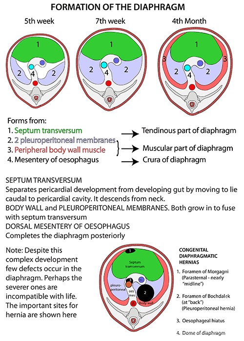
- Develops from 4 sources
- Septum Transversum – gives rise to Central Tendon
- Pleuroperitoneal Membranes (Mesodermal folds) – close the connection between the thoracic and abdominal parts of the Coelom
- Oesophageal Mesentery – also contributes to the development of the Diaphragm
- Cervical Myotomes
- Muscle cells derived from the 3rd, 4th and 5th Cervical Myotomes
- Muscle cells carry their own nerve supply with them – hence the motor supply from the Phrenic Nerves
- Failure of Pleuroperitoneal Membrane development – most common cause of Congenital Diaphragmatic hernia
- The defect (Bochdalek’s Foramen) is posteriorly placed
- Clinically manifests more often on left side probably due to the presence of the liver on the right
- Morgagni’s Foramen – possible small hernia site at the junction of the costal and xiphoid origins
Overview
- Derivative of the Inner (Transversus) layer of the muscles of the body wall
- Fibres arise in continuity with Transversus Abdominis from within costal margin
- Completed behind the costal margin by fibres arising from the Arcuate Ligaments and the Crura
- From circumference of oval origin, fibres arch upwards into pair of domes and then descend to a Central Tendon which lies at the level of the xiphisternal joint
- Right dome – ascends in full expiration as high as nipple line (4th space)
- Left dome – ascends as high as 5th rib
- Viewed from side: diaphragm resembles inverted J – long limb extending up from Crura (upper lumbar vertebrae) and the short limb attached to xiphisternum (T8 level)
- About 55% of muscle fibres (and 65% of intercostal muscle fibres) are slow twitch fatigue-resistant type
Attachments
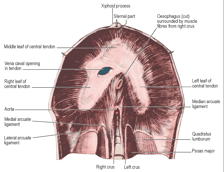
- Sternal part
- Arises from the posterior surface of the xiphoid process
- Costal part
- Arises from the inner surfaces of ribs 7–12 and their costal cartilages
- Lumbar part
- Right crus: from L1–L3 (or L4) vertebral bodies and intervertebral discs
- Left crus: from L1–L2 (or L3) vertebral bodies and discs
- Median arcuate ligament: spans over the aortic hiatus
- Medial arcuate ligaments: arch over the psoas major
- Lateral arcuate ligaments: arch over the quadratus lumborum
- Insertion
- All fibres converge into the central tendon of the diaphragm
Crura
-
Crura are the strong tendons attached to the anterolateral surfaces of the bodies of the upper lumbar vertebrae
-
Muscle fibres radiate from each crus, overlap, and pass vertically upwards before curving forwards into the central tendon
-
Median Arcuate Ligament
- Formed from the union of tendinous fibres from the medial edge of each crus in front of the Aorta at level of T12
-
Medial Arcuate Ligament
- Thickening of the Psoas Fascia
- Extends from the side of the L1 or L2 vertebral body to a ridge on the anterior surface of L1 vertebra transverse process at the lateral margin of Psoas
-
Lateral Arcuate Ligament
- Thickening of the anterior layer of the Lumbar Fascia on the front of Quadratus Lumborum
- Extends across to the middle of the lower margin of the 12th rib
-
Central Tendon
- Anteriorly situated middle leaf fused on each side to lateral leaves that extend back towards the paravertebral gutters
- Inseparable from the fibrous pericardium – same embryological origin
- Attached to the Falciform Ligament
- NOT attached to the Phreno-Oesophageal Ligament
-
Right Crus
- Fixed to upper 3 Lumbar vertebrae and intervening discs
- Some fibres pass up on the abdominal surface of fibres from Left Crus and surround oesophageal orifice in a sling-like loop
-
Left Crus
- Fixed to upper 2 Lumbar vertebrae
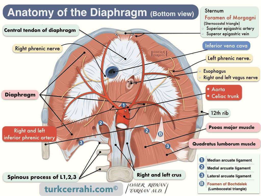
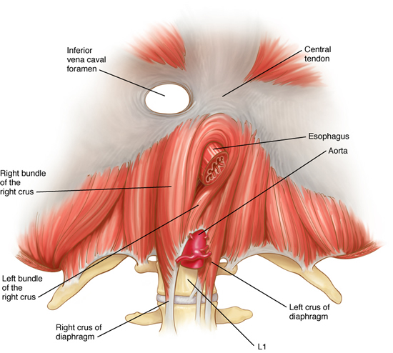
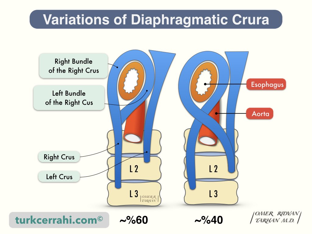
Openings in the Diaphragm
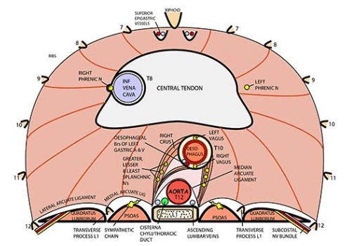
Aortic Opening
- Opposite T12 vertebra
- Midline
- Behind Median Arcuate Ligament
- Transmits:
- Aorta
- Azygos Vein
- Thoracic duct leading up from the Cisterna Chyli
- From right to left: Azygos Vein, Thoracic Duct, Aorta
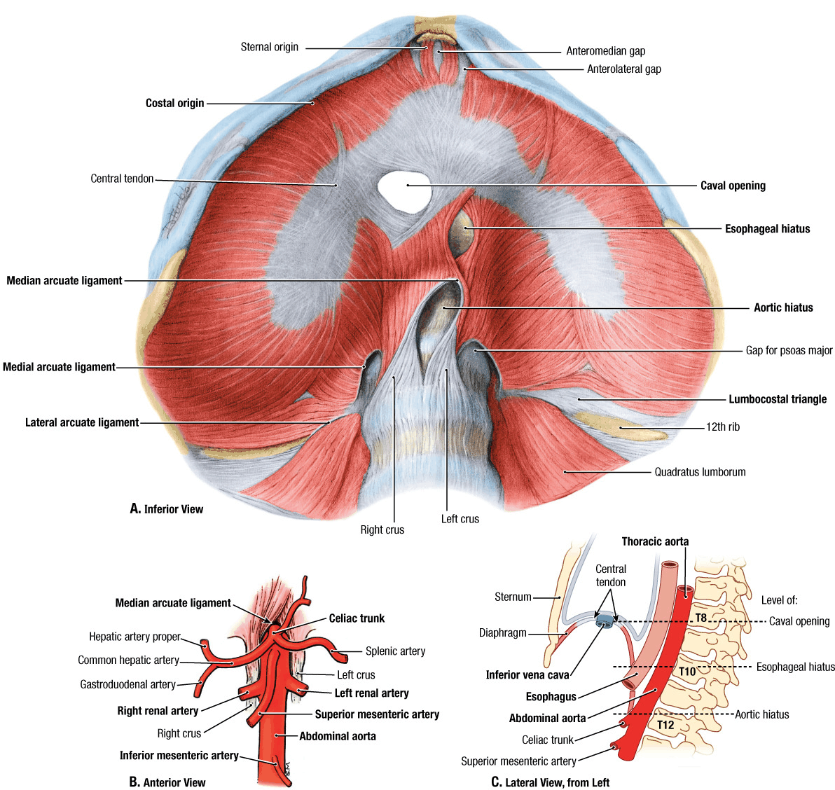
Oesophageal Opening
- Opposite T10 vertebra
- 2.5cm to the left of midline
- Behind the 7th Left Costal Cartilage
- Lies in the fibres of the Left Crus
- Sling of fibres from the Right Crus loop around it
- Transversalis fascia on the under-surface of the diaphragm extends up through the opening and blends with the endothoracic fascia above the diaphragm
- Attached to the diaphragm 2-3cm above the GOJ – i.e. the Phreno- oesophageal Ligament
- Phreno-oesophageal Ligament– stretched in the sliding hiatus hernia
- Transmits:
- Oesophagus
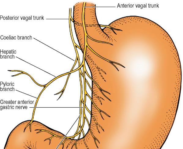
- Vagal Trunks
- Anterior vagal trunk
- Comprised mainly of left vagal fibres from the oesophageal plexus in the posterior mediastinum
- Lies in contact with the anterior oesophageal wall, usually nearer to its right margin
- Comprised mainly of left vagal fibres from the oesophageal plexus in the posterior mediastinum
- Posterior vagal trunk
- Comprised mainly of right vagal fibres
- Lies in loose tissue a little behind and to the right, NOT in contact with the posterior surface of the oesophagus
- Comprised mainly of right vagal fibres
- Anterior vagal trunk
- Left Gastric Artery (Oesophageal branches)
- Veins
- Venous drainage – passes caudally to the Portal venous system and cranially to the Azygos venous system – major site of portal-systemic anastomosis
- Lymphatics
Vena Caval Foramen
- Opposite T8 vertebra
- Right of the midline
- Behind the 6th Right Costal Cartilage
- Lies between the middle and right leaves of the Central Tendon; fibres of which fuse firmly with the adventitial wall of the IVC
- Transmits:
- Inferior Vena Cava
- Right Phrenic Nerve passes through Central Tendon alongside IVC at its opening
Other Structures and Openings
- Hemiazygos Vein – passes through Left Crus
- Greater, Lesser and Least Splanchnic Nerves – pierce each Crus
- Sympathetic Chain– behind Medial Arcuate Ligament
- Subcostal Nerve and Vessels – behind Lateral Arcuate Ligament
- Left Phrenic Nerve – pierces muscle of Left Dome
- Neurovascular bundles of 7th to 11th Intercostal Spaces – pass between digitations of Diaphragm and Transversus Abdominis into the neurovascular plane of the abdominal wall
- Superior Epigastric Vessels – pass between xiphisternal and costal (7th) fibres of the Diaphragm
- Extraperitoneal Lymph vessels on the abdominal surface pass through Diaphragm to lymph nodes lying on its thoracic surface – mainly in posterior mediastinum
- At posterior part of Diaphragm – may be a gap between lowest costal fibres and those arising from Lateral Arcuate Ligament
- Posterior surface of kidney and its peri-renal fascia is separated from the pleural only by areolar tissue of the Endothoracic Fascia
Blood Supply
- Lower 5 Intercostal and Subcostal arteries: Supplies costal margin
- Right and Left Inferior Phrenic arteries (from the Abdominal Aorta): Supplies main mass of fibres from the Crura
- Pericardiacophrenic and Musculophrenic branches of the Internal Thoracic Artery and Superior Phrenic branches of the Thoracic Aorta: Small contributions
Nerve Supply
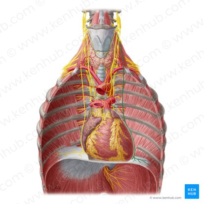
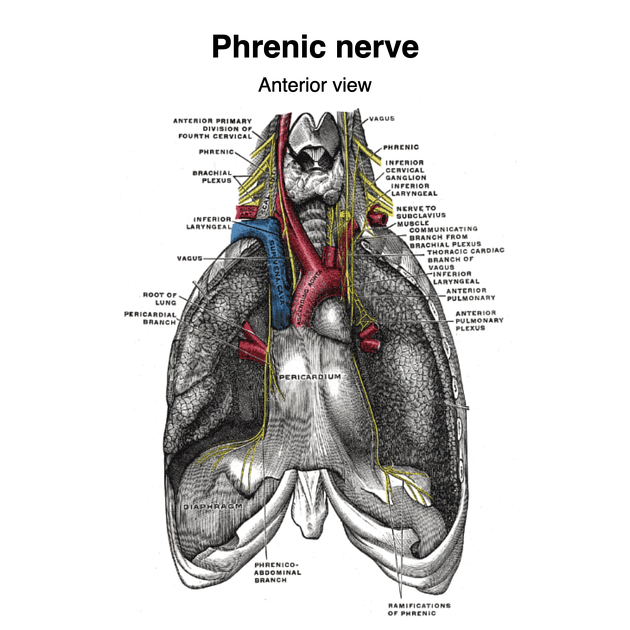
- Arrises from the Cervical Plexus
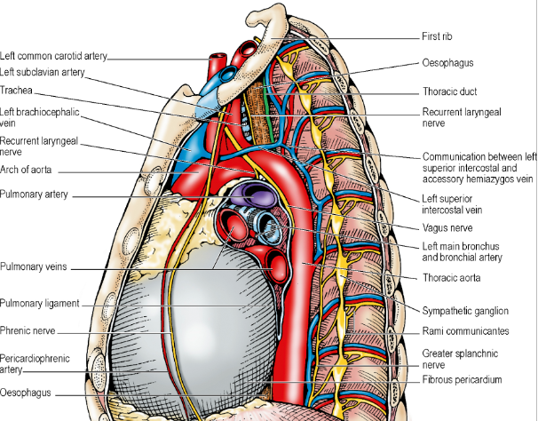
- Left and Right Phrenic Nerves (C3, 4, 5 but predominantly C4) – sole motor nerve supply
- Course of Phrenic Nerves:
- Arise principally from C4 in the neck
- Passes down over the Anterior Scalene muscle
- Deep to the Prevertebral Fascia
- Passes from lateral to medial over the Anterior Scalene surface
- Crosses in front of the Subclavian Artery and its Internal Thoracic Artery branch
- Across dome of the pleura behind the Subclavian Vein
- Can sometimes pass anterior or even through the Subclavian Vein
- May contain C5 fibres that pass anterior to the Subclavian Vein
- Can sometimes pass posterior to the Internal Thoracic Artery
- Lies lateral to the Ascending Cervical Artery (branch of the Inferior Thyroid Artery or Thyrocervical Trunk)
- Ascending Cervical Artery runs up on the Prevertebral Fascia
- Crosses anterior to the Vagus
- Runs through the mediastinum in front of the lung root
- Each nerve is in contact laterally with the mediastinal pleura throughout the entire course
- Right Phrenic Nerve
- Related medially with venous structures throughout its thoracic course
- Right Brachiocephalic Vein, Superior Vena Cava, Pericardium over the Right Atrium, and the Inferior Vena Cava
- i.e. The Right Phrenic Nerve is lateral to the above structures
- Reaches under-surface of Diaphragm by piercing the Central Tendon adjacent to the Caval Opening
- Related medially with venous structures throughout its thoracic course
- Course of Phrenic Nerves:
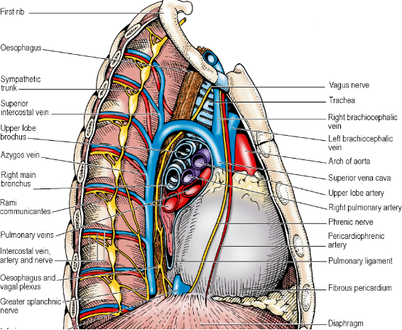
- Left Phrenic Nerve
- Related medially to arterial structures throughout its thoracic course
- Left Common Carotid Artery, Left Subclavian Artery
- Crosses the arch lateral to the Superior Intercostal Vein; Anterior to the Vagus Nerve
- Runs laterally down the pericardium over the Left Ventricle towards the Apex of the heart
- Reaches under-surface of Diaphragm by piercing the muscular part just to the left of the pericardium
- Related medially to arterial structures throughout its thoracic course
- Both
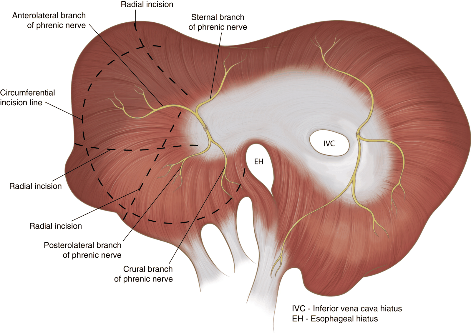
- On reaching abdominal surface of Diaphragm, divides into Anterior, Lateral and Posterior branches which run radially giving off branches that enter the muscle from below
- Fibres of the Right Crus that loop to the left around the Oesophageal Opening: supplied by the Left Phrenic Nerve
- Lower intercostal nerves give proprioceptive fibres to Diaphragm periphery
- Phrenic Nerves – proprioception to central part
- About 2/3rd of the Phrenic Nerve fibres are motor to the Diaphragm
- Rest are sensory to the:
- Diaphragm (except the most peripheral parts, which receive intercostal afferent fibres)
- Mediastinal pleura
- Fibrous pericardium
- Parietal layer of the serous pericardium
- Central parts of the diaphragmatic pleura and peritoneum
- Pain from diaphragmatic part of peritoneum classically felt in shoulder tip (C4)
- Pain from thoracic surfaces supplied by the Phrenic Nerve (pleura and pericardium) is usually only vaguely located there
- Rest are sensory to the:
- Diaphragm moves in respiration under central control of cell groups in the region of the Nucleus of the Tractus Solitarius