Section: Trauma Curriculum: Curriculum, page 79
- Liver - Curriculum, page 87
- Pancreatic-duodenal trauma - Curriculum, page 91
- Splenic trauma - Curriculum, page 92
- Small bowel trauma - Curriculum, page 62
Resus vs Definitive surgery
- Resus - Damage control resuscitation and damage control surgery
- Only lifesaving surgery - stopping bleeding and more contamination
- Physiologically unstable
- Complex Abdominal Injury requiring damage control approaches
- Major liver injuries
- Mobilization and packing +/- angioembolization
- Pancreaticoduodenal injuries
- Aortic and vena caval injuries
- Complex pelvic #’s
- Major liver injuries
- Complex Abdominal Injury requiring damage control approaches
- Definitive surgical treatment
- Physiological stable enough for definitive procedure
Trauma Laparotomy
Overview
- Rapid entry
- Large and adequate incision from xiphisternum to pubis
- Control of massive haemorrhage by:
- ID of all injuries
- Packing
- Direct control
- Proximal control
- ID of injuries
- Control of contamination
- Reconstruction if possible
- Give abx prophylaxis – Cef + Met or Augmentin
- Give longer duration abx if enteric or colonic soiling
- Temperature
- Should be higher than usual
- Bair hugger/warm air blankets during surgery
- Warmed anaesthetic agents, IVF and wash
- Blood collection and autotransfusion
- With cell saver if available
- Draping
- Expose from jugular notch to knees
- Allows access for Median sternotomy, thoracotomy and groin access
Steps
- Incision
- 2 swipes of blade - 1 to fascia and into abdomen
- Mayo scissors for fascia and parietal peritoneum
- If hostile abdomen anticipated - do thoracotomy for proximal control first
- Aortic cross clamping OR
- REBOA - Retrograde Endovascular Balloon Occlusion of Aorta
- Procedure
- Scoop out clot/contaminants and use Poole sucker
- Eviscerate SB, assess for major vessel injury, control bleeding
- Packing with dry swabs
- Paracolic gutters
- Pelvis
- Under left diaphragm and above/right of liver
- Subhepatic pouch and other areas of bleeding
- Identify where major source of bleeding is and pack
- Let anaesthetics catch up physiologically
- Work from top down/systematically after major things have been dealt with
- To assess if bleeding is mesenteric or retroperitoneal
- Need to mobilise the whole root of mesentery up from Caecum to DJF
- Deal with lesions in order of lethality
- Haemorrhage
- Major blood vessels
- Solid organs
- Mesenteric and hollow organ
- Retroperitoneal
- Contamination
- Haemorrhage
- Definitive Packing
- Facts
- Dry folded packs placed flat
- Effective for venous bleeding
- Be efficient with pack use
- Liver
- Dissect gastrohepatic ligament and vessel loop portal triad (Pringle’s maneuver) in preparation for liver bleeding if it is suspected
- Pack liver - If controlled → venous and should settle
- If not controlled → Pringle’s maneouvre
- Suspect Portal Vein or Hepatic Artery → Hepatorrhapphy
- If it doesn’t settle with Pringle’s → ?IVC/Hepatic Vein injury
- Compress against post abdo wall and pack
- Spleen
- Splenectomy by vascular clamping across hilum if needed
- Pelvis
- Extraperitoneal packing
- Facts
Routes of Access
- Lesser omentum (along Lesser Curve)
- Coeliac Artery
- Body of Pancreas
- Greater omentum
- Body and tail of pancreas
- D1 posterior and D2 medial
- Do a Kocher’s maneuver if suspected pancreas head injury
- Kocher’s
- Medial mobilisation of Duodenum to ID D2 and D3 fully
- Aorta and IVC
- Right Kidney
- Porta Hepatis
- Posterior surface of Head of Pancreas
- Right medial visceral rotation
- Procedure
- Mobalise the hepatic flexure and ascending colon via dividing the line of Toldt
- Then mobilise the SB mesentery from caecum/RLQ to ligament of Treitz and reflect right colon to LUQ
- Then Kocherise the Duodenum
- Provides exposure to
- Entire duodenum - particularly D3 and D4
- Accessing D3 with SM vessels there
- Need to be retracted upwards
- Pancreas - Head, neck and proximal body
- Access to IVC and renal vessels
- Right kidney
- Entire duodenum - particularly D3 and D4
- Procedure
- Left medial visceral rotation
- How
- Retract small bowel and descending colon to the right
- Mobilizing the left and sigmoid colon by incising the white line of Toldt
- Divide splenic attachement to the diaphragm
- Staying on the abdominal wall muscles enter the plane lateral and then posterior to the kidney medialising it
- Note - in elective surgery a left medial visceral rotation (not Mattox) is performed leaving the kidney down but rotating the spleen/colon/SB
- Left kidney is maintained in the Gerota’s fascia, in order to be protected from the traction injury, but the left renal vein hinders access to the anterior aorta.
- Exposure of
- Aorta and its branches
- Pancreas - distal body and tail
- Left Kidney
- How
- Specific Organ Techniques
- Liver injury - debridement, resection, fibrin glue and collagen fleece
- Splenic injury - splenectomy
- Pancreatic injury - suture repair if duct not involved, fibrin glue and drainage
- Retroperitoneal haematoma - packing
- Abdominal closure
- Principles to consider
- Patient stability/lethal triad
- Blood loss pre- and during surgery
- IVF volume given
- Degree of contamination
- Nutritional status and intercurrent diseases
- Options
- Primary closure
- Temporary abdo closure
- Principles to consider
Tips and tricks
- Headlight
- Lithotomy position
- Table tilt
- Move as needed
- Pericardial window
- Central tendon of diaphragm and pericardium both originate from Septum Transversum (See Diaphragm anatomy)
- Dissect through central tendon near xiphisternum to treat tamponade
- Wash
- Drains for pancreatic and duodenal injuries
- Bivalved bladder repairs with suture apposition of mucosa is key
- Needs both SPC and urethral catheter
- Early tracheostomy - usually seldinger in ICU if needed these days
Abdominal Vascular Injury
- General
- 3 things to consider
- Need for ED thoractomy and aortic cross clamping
- Loss of CO or imminent
- Stops arterial bleeding and perfuses heart and brain
- Need for immediate surgery
- Requires ongoing resus to maintain CO
- Need CT
- Stable patient only
- Need for ED thoractomy and aortic cross clamping
- 3 things to consider
Retroperitoneum exposure
- General
- Injuries are often missed, underestimated or have high rate of mortality
- Zones
- I - Central
- Between both psoas muscles and contains the abdominal aorta, inferior vena cava, pancreas, and midline duodenal structures
- II - Lateral x 2
- Lateral to the psoas muscles on bilateral sides and contains the kidneys, ureters, and portions of the colon
- III - Pelvic
- I - Central
- Management
- Zone 1 - Upper midline central retroperitoneal haematoma
- All should be explored
- Potential sites of injury
- Aorta/IVC
- Kidneys, Ureters, Renal vessels
- Duodenum/Pancreas
- Colon
- Zone 2 - Lateral retroperitoneal haematomas
- Only explore if expanding, thought to have injury to Colon or Ureter
- Usually related to renal bleed which should tamponade
- Zone 3 - Pelvic
- Non - expanding - don’t explore
- Ongoing bleeding
- External fixation of pelvis
- Angioembolization
- If angio is unavailable and enlarging pelvic haematoma/ongoing bleeding
- Extra-peritoneal pelvic packing
- General
- Unstable and peritonitis - Laparotomy
- Observe if stable, not too sore and imaging is reassuring
- Serial abdominal examinations
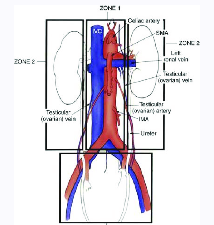
- Zone 1 - Upper midline central retroperitoneal haematoma
Retroperitoneal Haematoma
- Zones
- I - central (further subdivision based on apex of haematoma)
- Supracolic
- Juxtarenal or suprarenal aorta, SMA, coeliac
- Infracolic
- Aortic bifurcation/IMA
- Transperitoneal or left medial visceral rotation approach
- Left or right
- If apex is right of midline and primarily venous bleeding
- Right medial visceral rotation
- Access to Infrarenal IVC, aorta and portal vein
- If apex is left of midline and primarily arterial bleeding
- Left medial visceral rotation
- Access aorta, coeliac, SMA, splenic A/V, left renal A/V
- Mobilise kidney to access posterior wall of aorta
- If apex is right of midline and primarily venous bleeding
- Supracolic
- II - Lateral x 2
- Leave alone if not expanding
- Renal injuries - non operative and selective embolization approach
- If penetrating injury or expanding - explore
- Need to rule out occult post colonic and ureteric injury
- To prevent abscess/urinoma formation
- III - Pelvic
- If stable and CT proven blush - Angioembolise
- If found at surgery and non-expanding - Leave alone
- If found at surgery and expanding - Manage
- Extraperitoneal pelvic packing
- Pelvic binder is on
- Grasp peritoneal edge of inferior 25% of laparotomy wound
- Develop Retzius space (pre-peritoneal plane) all the way round to posterior rectum on both sides
- Evacuate clot
- Packs x 2-3 on each side
- If fails then culprit Internal Iliac Artery ligation
- If works then to IR for assessment/embolization
- Pelvic fixation/binder
- Extraperitoneal pelvic packing
- I - central (further subdivision based on apex of haematoma)
Major Abdominal Vessels
Aorta
- Supracoeliac Aortic cross clamping
- Retraction
- Left Liver lobe → Right shoulder
- Body of Stomach → Left hip
- Dissection
- Lesser omental window (Pars flaccida)
- Crus peritoneum incised to expose muscle fibres which are split
- Retract oesophagus to patients left
- Adventitia of Aorta exposed, develop bilateral planes and clamp
- Satinsky clamp placed supracoeliac
- Retraction
- Suprarenal aortic exposure
- First do a supracoeliac clamp
- In context of central supracolic haematoma
- Then left medial visceral rotation
- Exposes coeliac/SMA/L renal vessels/iliacs
- SMA and coeliac nerve plexuses require dissection to access them
- Distal Aorta
- Retraction
- SB right | Transverse Colon superior | Descending Colon left
- Retraction
- Injury Treatment of Aorta
- Aortic injuries need repair
Coeliac Axis
- Proximal control - supracoeliac aortic clamping or anterolateral thoracotomy
- Left medial visceral rotation and may need to mobilise left triangular lig and segments 2+3
- Difficulties
- Dissection coeliac plexus of nerves - dense and fibrous
- Short origins of left gastric, splenic and common hepatic
- All can be tied off if SMA and IMA are intact - collateralization
- How
- 3.0 prolene suture repair
SMA
- Left medial visceral rotation or root of SB mesentery
- Comes off aorta distal to coeliac and proximal to left renal A
- If damage control → Shunt it
- If can’t be repaired then should be replaced with graft to infrarenal aorta
- Graft can kink if proximal end is placed too high
- Keep it away from pancreas
- SMV can be shunted or ligated
IMA
- Can be tied off
- Colon viability needs assessment
Renal Artery
- Repair via infrarenal or medial rotation access
- Down time poorly tolerated - 45mins = Complete loss of function
- Therefore may need nephrectomy at that time
Iliacs
- Proximal and distal control
- Mobilise sigmoid colon and caecum
- May need groin access for distal control
- Beware of the ureters crossing the iliac bifurcation
- Veins closely adhered to back of arteries - Don’t dissect, just enough to clamp
IVC
- Suprahepatic ? chest access
- Not controlled by Pringle’s maneuver
- Controlled with direct downward pressure of liver onto IVC
- Exposure and clamping of infrarenal IVC
- Divide coronary ligaments to displace superomedially and see supra IVC
- Infrahepatic
- Right medial visceral rotation
- Kocher’s will show infrahepatic IVC well and right renal vascular pedicle
- Direct pressure with swabs above and below
- With penetrating injury it may be through and through
- But all posterior injuries do not require repair
- If it does, repair them transcaval as can’t get posterior on outside easily
- Caval injuries below Renal Veins can be ligated if extensive but prefer lateral repair
- Above Renal Veins should be repaired - ligation is unsurvivable
Portal Vein
- In free edge of lesser omentum - Portal Triad
- Pringle’s maneuver
- If more proximal: Kocher’s
- If damage control: Shunt
- Portocaval shunting is a high mortality option to consider
Shunting
- Indications
- Can’t be repaired
- Damage control situation
- Rationale
- Allows flow to occur
- Rules
- Diameter of tube - ⅔ circumference of vessel
- Length of tube – 3 x length of defect
- What to use
- NGT/ETT/chest drain/anything
- Other issues
- Ends should be beveled
- Tie silk around tube - ⅓ | ⅔ division
- Technique
- Proximal and distal control
- Proximal control applied
- Place ⅔ end in
- Use suture to pull back to distal end
- Secure with ties
- Last for 2-3 days
Bowel, Rectum and Diaphragm
General
- Assess from diaphragm/OGJ to rectum and systematically with 2 hands and 4 eyes
- Common areas for missed injury
- Diaphragm/GOJ
- Lesser and greater curve
- Posterior stomach and pancreas
- SB
- Retroperitoneal colon/rectum
- If there’s one defect, look for a second defect
Diaphragm repair
- With penetrating injury without herniation, diaphragm injury cannot be ruled out with CT
- Needs trauma laparoscopy or laparotomy
- With soiling, may need to extend the wound radially to wash the chest out, close the diaphragmatic defect by pulling ends up with allis and suturing with continuous non-absorbable suture then placing a chest drain, then dealing to abdo washout
- Left side more common due to absence of liver on this side
- And right-handed assailant
- Be suspicious with thoracoabdominal injury below T5
- Large defects may need PTFE patch best placed via thorax
- Later complications include hernia with incarceration
Stomach
- Babcocks to grasp and inspect
- NGT in situ useful - can be grasped and used as a retractor
- Assess posterior wall either via anterior injury or in lesser sac along with pancreas
- 3.0 PDS repair full thickness
- Simple - debride edges and close
- Complex - Non-anatomic resection, reconstruct at relook
Duodenum
- Kocherise it if there is a haematoma as it is mandatory to inspect the posterior aspect
Small bowel
- Stable pt - repair or resect and anastomosis
- Unstable - staple off and leave ends in
- With missiles like shotguns (multiple small pellets), run SB through water ? gas leak
Colon
- Broadly similar to SB management
- Colostomy as definitive procedure rather than anastomosis dependent on patient factors at time of relook/definitive surgery
- Stomas should be placed laterally and at the level of the umbilicus, away from wounds
Rectum
- Intraperitoneal upper ⅓ and distal ⅔ extraperitoneal
- Mx with repair and diversion if upper and diversion alone if lower
- Flexi sig and rectal contrast CT useful
Mesentery
- Tie off bleeders/oversew and watch out for bowel ischaemia
Liver and Biliary System
Liver
General
- Cantile’s Line - GB to IVC - Left and right functional lobes
- Glisson’s Capsule = Visceral peritoneum of liver and covers porta hepatis structures
- Portal triad runs WITHIN segments
- Hepatic veins run BETWEEN segments (true on the right - not completely true on the left)
- Also have no valves, no Glisson’s capsule protection and are majority intraparenchymal (8-12cm length with 1-2cm extrahepatic)
- Retrohepatic IVC is encase by liver – important re: mobilisation
- 3-11 short hepatic veins go from liver direct to IVC
- Also small branches from caudate lobe direct to IVC
- Bile ducts rely on Hepatic Artery for supply
- Liver parenchyma mainly relies on portal vein so is ok with embolisation/ligation
- Blunt injury = Direct compressive or shear
- Arteries resistant, veins and biliary ducts also but less so
- Parenchyma is weak
- Tears along segmental fissures mainly (where hepatic veins are)
- Regardless of liver injury grade - physiology determines management
- Hepatorhapphy or resectional debridement should be done if can’t control bleeding
Disposition
- Stable without peritonism – non-op
- Otherwise can have FAST or CT with IV contrast in arterial and portal venous phase
- Unstable - operate
- If bleeding → OT
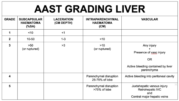
AAST Liver Injury Grading
- Imaging based but criteria also exist for operative findings and pathological criteria
- 5 grades
- First 3 based on haematoma and laceration
- Last 2 based on disruption of parenchyma and vascular injury
- Laceration = Depth
- Subcapsular = Surface Area %
- 5 grades
Management
- Non-operative - Over 80% of liver injuries managed this way
- Requirements for Non-Op management:
- Haemodynamic stability
- Absence of peritoneal irritation and confidence that isolated injury
- High quality CT-scan with experienced radiologist
- 24/7 availability of monitoring, repeat clinical assessment and intervention
- Can use IR for embolization if blush present
- Subcapsular haematomas as long as they are not expanding
- Requirements for Non-Op management:
- Operative
- Majority is venous bleeding manage with packing/pressure
Operative Approach to Liver Trauma
- Initially compress right and left lobes together then push backwards for compressive effect
- i.e. Push, Pack, Pringle, then Total Vascular Isolation
- Options
- Packing | Pringle’s | Liver clamp | Diathermy | Haemostatic agents/glue | Hepatic suture | Hepatorrhaphy and non-anatomic resection
- Packing
- Dry, folded packs with radioopaque marker
- Remove within 1-3 days - Can cause infection
- Complications
- Reduced CO - Too tight, compress IVC
- Respiratory deterioration - increased pressures from diaphragm reduced compliance
- Other options
- Hepatic tourniquet
- Tract tamponade balloon - with Sengstaken Blakemore tube is ideal
- Pringle’s
- Diagnostic - Hepatic Artery/Portal Vein vs Hepatic Vein/Caval bleeding
- Therapeutic
- Timing
- 1 hr max
- Less ischaemia with 10 mins on 5 mins off
- Timing
- Sometimes need to finger fracture to get access to deeper bleeders within parenchyma
- Can staple off or ligasure bleeding segments along anatomical planes
- Anatomical lobectomy acutely has high mortality
- Situations where anatomical lobectomy is needed
- Left lateral segment extensive injury - can’t pack
- Non-viable liver segment post-packing
- Almost free segment
- Devitalized liver at pack removal
- Situations where anatomical lobectomy is needed
- Seldom have to mobilise the liver fully, acutely and without specialist help
- Hepatic isolation
- Inflow - Pringle
- Outflow - Clamp IVC infrahepatic and suprahepatic
- Thoracic or abdominal approach
- Complications of severe liver injuries
- Perihepatic infection
- Biloma
- Biliary fistulae
Spleen
- Relevant Anatomy
- Mobilisation of ligaments
- Gastrosplenic
- Splenorenal
- Splenocolic
- Mobilisation of ligaments
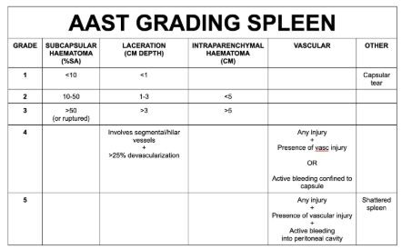
Grading
- Similar to liver with regards to 5 grades, haematoma, laceration, disruption and vascular injury
Management
- HD stable and grade 1-3 → NOM
- Re-bleed rate 1-8% - acceptably low
- Angioembolization is useful adjunct
- Indications
- Evidence of ongoing bleeding
- Significant Hb drop
- Tachycardia
- Contrast extravasation on CT-A (blush)
- Pseudoaneurysm
- NB: Prophylactic embolisation is useful and should be performed, i.e. arterial blush but patient is haemodynamically stable
- Indications
- Benefit of NOM
- Avoid morbidity of non-therapeutic laparotomy
- Fewer intra-abdominal complications
- Reduced transfusion risk
- No risk of post-splenectomy OPSI and pancreatic leak
- HD unstable → Open Splenectomy
- Indications
- HD unstable
- Other injury requiring laparotomy
- Evidence of continued splenic haemorrhage
- Replacement of > 50% of pts blood volume
- Indications
- Surgical Approach
- Midline laparotomy/bookwalter/right tilt/headlight
- Left hand pulls spleen anteriorly and medially
- Divide splenorenal and splenophrenic ligs
- Divide gastrosplenic lig with short gastrics closer to spleen sides
- Can pack laterally to bring spleen closer
- Can leave a closed suction drain if concern for pancreatic tail injury
- Vaccinations
- If urgent splenectomy give vaccines within 14 days (/on discharge)
- 2 weeks pre-op or post-op
- To cover Haemophilus influenzae B, pneumoccoccus and Neisseria meningitidis
- Risk of OPSI
- Opportunistic Post-Splenectomy Infection
- Encapsulated organisms can cause overwhelming infection
- Prevent by getting regular vaccinations including annual flu vaccine and COVID vaccines
- Take abx at the earliest sign of infection incl. suspected viral illness Pathogenesis of OPSI
- Insufficient opsonization filter function
- Delayed and impaired production of immunoglobulin
- Lack of splenic macrophages
- Minimal tufts production
- Reduced number of memory B-Cells – usually produce IgM to promote clearance of polysaccharide-encapsulated bacteria
- Other
- Medic alert bracelet
- Emergency abx script - amoxil 3g initially then 1g TDS
- Flu and covid vaccine
- Children get Amoxil until 18yrs old
- Document on splenic vaccination register
- Vaccination/Abx splenectomy spiel
- Prevent risk of OPSI
- 2 weeks before or 1 week after splenectomy
- Unvaccinated patients
- Place on National immunisation register
- Medic alert bracelet
- 5 vaccines
- Pneumoccocal (4)
- Dose 1 conjugate
- Dose 2-4 polysaccharide
- Timing = 2mths, 5yrs, 5yrs or age 65 (later)
- Meningococcal (lifelong)
- Doses = conjugate + B
- Timing - 2mths then 5yrly for life
- Haemophilus influenza B (Hib) (1)
- One off dose
- Annual flu vaccination
- Pneumoccocal (4)
- Covid vaccination
- Abx
- Amoxil 250mg daily for 3 years
- Lifelong if immsupp, prev sepsis or splenic malignancy
- Allergy - Roxi 150 daily
- Prevent risk of OPSI
Pancreas
- Anatomy
- Closely related to IVC/Aorta | SM vessels | Renal vessels
- Mechanisms of Injury
- Blunt - High impact - in a well-protected position
- Penetrating - Can cause duct disruption
- Diagnosis
- Clinical assessment, serum lipase and USS minimally helpful
- Grey-Turner sign - Flank ecchymosis
- Cullen sign - Periumbilical ecchymoses
- Lipase not a reliable marker
- CT useful but can miss injuries esp. early
- MRCP is the best way to assess but can’t be done on unwell pt - delayed
- ERCP is useful to visualise ductal injury and localise it but can risk pancreatitis
- Best reserved for stable patients with equivocal MRCP
- Could place PD stent but mixed evidence
- Clinical assessment, serum lipase and USS minimally helpful
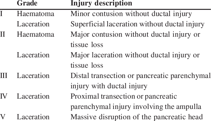
Pancreas Grading AAST
Management
- NOM
- For grade 1 and 2 found on imaging and at time of surgery
- Adjunctive delayed ERCP can be considered for stenting
- Operative
- For distal injuries without duct injury or any proximal injury - Drain
- For distal injuries with duct disruption - Distal Pancreatectomy
- For grade 3 and 4 found on imaging and at time of surgery
- Consider exclusion for high grade injuries with Pyloric Exclusion and Gastrojejunostomy
Operative managment
- Operative Findings
- Central haematoma
- Bile staining
- Peripancreatic oedema
- Saponified fat - from pancreatic enzymes
- Surgical Approaches
- Via Lesser Sac
- Kocher’s
- Right and left medial visceral rotation
- All allow the entire pancreas to be assessed from all surfaces
- PD assessment
- Give Secretin to stimulate PD secretions
- IOC via GB, duodenal defect, anterior duodenotomy
- Contusion and Parenchymal Injuries
- If there’s duct disruption and left of SM vessels
- Ligate duct
- Distal pancreatectomy
- Splenectomy as well if pt is unstable
- Extra time required to preserve spleen
- If duct is not disrupted
- Leave a suction drainage device
- Risks with distal pancreatectomy
- Fistula/leak
- Diabetes
- If there’s duct disruption and left of SM vessels
- HOP and duodenal injuries
- Damage control surgery
- Drains/packing
- Tube into duct directly
- Pyloric Exclusion with Gastrojejunostomy
- Why
- Minimise exocrine pancreatic stimulation by Gastric Acid and Duodenal filling
- Pylorus usually spontaneously opens in 2-3 weeks then the GJ anastomosis normally closes down on its own too
- Why
- T-tube drainage (duodenal injury)
- Whipple’s is an option but should be done in a staged approach
- Indications
- Massive disruption of the pancreaticoduodenal complex (Gd5)
- Duodenal devascularization
- Extensive duodenal injury involving ampulla or distal CBD
- Indications
- Damage control surgery
Pancreatic Trauma Complications
- Early
- Pancreatitis (duct disruption) - Self limiting
- Fistula
- Low output - Self limiting
- High output
- Mx
- Nutrition - Distal enteral feeds ideally
- Drainage - Percutaneous/suction drain
- ERCP and Pancreatic Duct stenting for duct disruption
- Mx
- Abscess
- True pancreatic abscesses are rare and require debridement
- Late
- Pseudocyst
- Mx
- Cystgastrostomy
- Endoscopic stenting
- Resection
- Mx
- Exocrine and endocrine dysfunction
- Unlikely with distal resection - only need 20% functioning gland
- Insulin and Creon
- Pseudocyst
Summary of Pancreas Trauma
- Duct not involved
- All - leave a drain
- Duct involved
- Distal
- Distal Pancreatectomy
- Proximal
- Damage control
- Consider whipple’s
- Consider Pyloric Exclusion
- Distal
Duodenum
- General
- Relatively well-protected like the pancreas
- If it’s injured then very likely for other things to be injured too
- Doesn’t present with peritonitis making dx hard
- Mechanism of Injury
- Penetrating (more common and seldom isolated)
- Blunt (associated pancreatic injury, steering wheel/handle bar/chance # of L1/L2)
- Presentation
- Dependent on location of injury and where it drains
- Retro | Lesser Sac | Greater Sac
- Signs of duodenal injury at surgery
- Retroperitoneal haematoma
- Peri-duodenal air bubbles
- Peri-duodenal haematoma
- Bile staining of tissues
- Investigation
- CT
- Laparotomy Duodenal Injury Scale
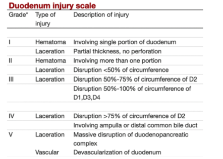
Operative management
- Techniques for repair of a duodenal injury
- Surgical Approach
- Key factors
- Proximity to ampulla
- Injury characteristics - Simple vs complex
- Circumference of involved duodenum
- Associated injuries - Biliary tree, pancreas, vascular
- Can’t really dissect D2 away from pancreas - closely apposed and inter-reliant on blood supply
- D3 exposure via left medial visceral rotation - Cattell Brasch and SB mesentery mobilisation
- D4 exposure by mobilising ligament of Treitz
- Intramural haematoma
- Leave alone if possible
- Can cause duodenal obstruction but should settle with time
- If doesn’t - warrants exploration
- Key factors
- Operative Possibilities
- Simple suture repair
- Patch
- Duodenal diversion
- Suction drainage peri-duodenal
- Damage control preceded Whipple’s
- Options
- Laceration and perofation
- Simple suture repair
- Closure of longitudinal duodenotomy transversely if length <50% is of circumference
- Omental or serosal patch
- Simple suture repair
- Complete transection
- Primary anastomosis often possible if mobility permits (D1,D3,D4)
- May need reconstruction
- Proximal to ampulla - antrectomy and Bilroth II
- Distal to ampulla - R&Y duodenojejunal anastamosis and closure of distal duodenum
- D2 - R&Y end-to-side duodenojejunal anastamosis
- Pyloric exclusion with gastrojejunostomy
- Consider this in high risk repairs to divert gastric content and improve chance of healing + make a leak easier to manage
- Other options
- Duodenal Diverticulation - this is dated
- This is Bilroth II gastrectomy + closure of duodenal injury + placemnet of a decompressive catheter into the duodenum (also described is performing a truncal vagotomy and biliary drainage at the same time to decrease the fluid in the repair)
- Triple tube decompression
- Damage control surgery followed by Whipple
- Duodenal Diverticulation - this is dated
- Laceration and perofation
- Consider cholangiogram if close to ampulla/bile duct Difficult duodenum
Renal Injuries
- General
- Blunt > Penetrating
- Haematuria (5 RBC/HPF) in 95% of renal injury but its absence does not exclude
- Diagnosis
- Macrohaematuria
- Microscopic haematuria - Urinalysis
- Unstable pt → OT
- Stable pt
- Triple phase CT
- Grade injury to guide ideally NOM (up to and incl. Grade 4 without vascular injury)
- Contrast-enhanced and duplex USS can ID active intra-renal bleeds/AV fistula
- Triple phase CT
- IVP = Give IV contrast (2 mg/kg) then do x-ray of renal tract 10 mins after contrast
Renal Injury Grading
grade I
- subcapsular haematoma and/or contusion, without laceration
grade II
- superficial laceration ≤1 cm depth not involving the collecting system (no evidence of urine extravasation)
- perirenal haematoma confined within the perirenal fascia
grade III
- laceration >1 cm not involving the collecting system (no evidence of urine extravasation)
- vascular injury or active bleeding confined within the perirenal fascia
grade IV
- laceration involving the collecting system with urinary extravasation
- laceration of the renal pelvis and/or complete ureteropelvic disruption
- vascular injury to segmental renal artery or vein
- segmental infarctions without associated active bleeding (i.e. due to vessel thrombosis)
- active bleeding extending beyond the perirenal fascia (i.e. into the retroperitoneum or peritoneum)
grade V
- shattered kidney
- avulsion of renal hilum or laceration of the main renal artery or vein: devascularisation of a kidney due to hilar injury
- devascularised kidney with active bleeding
Management
- Unstable
- Findings of renal injury at laparotomy - Retroperitoneal haematoma
- Options
- Single shot on table IVP
- Ensure kidney functionality
- Exploration of injured kidney
- Pack and get out (DCS)
- Stable
- Grade 1 or 2 injuries - NOM
- Grade 3 - Major lacs through cortex and into medulla/CD
- May need drainage
- Grade 4 - Multiple renal lacs and vascular injury
- Usually require nephrectomy
- If HD stable could consider partial
- Grade 5 - Pelviureteric junction injuries - avulsed or lac (incomplete tear)
- Nephrectomy
- Kidney is designed to facilitate non-op mx
- Segmental pattern of division of blood supply
- Haematoma displaces renal tissue but doesn’t disrupt blood vessels
- Closed retroperitoneal space to tamponade in
- Kidney rich in tissue factor - activates extrinsic coagulation cascade - haemostasis
- Options
- Drainage and embolization!
- Urine - Ureteric stenting
- Collection/Urinoma - Percutaneous drainage
- Embolization
- Surgical Approach
- Need to get vascular control
- Good exposure of the Aorta and IVC
- Left and right medial visceral rotation and Kocher’s
- Need to see both ends of renal arteries and veins
- Also need to mobilize the kidney to get behind it
- Try and keep Gerota’s fascia intact until you have vascular control
- Origin of Right Renal Artery is adjacent to IVC therefore need to dissect between the 2
- Exposure of the Right Kidney
- Kocherise Duodenum
- Mobilise Right Colon
- Try and repair the Right Renal Vein if possible
- Lack of collaterals
- 2 ways to improve access to Left Renal Artery
- Ligate the Left Renal Vein tributaries
- Adrenal | Gonadal | Lumbar
- Ligate the Left Renal Vein distally (close to IVC)
- Collateral drainage via the 3 more proximal tributaries
- Ligate the Left Renal Vein tributaries
- With proximal control → Ischaemic time should be limited to 30 mins
- Allows time to open Gerota’s, debride, suture and partial nephrectomize
- Repair
- Suture close the medulla then the cortex/capsule
- Can use omentum/mesh to fill gap of large defect or suture over repair
- Close Gerota’s
- Leave a suction drain to monitor for urine leak
- Can consider nephrostomy tube/ureteric stents later if needed
- Adjuncts
- Pledgets - Closing capsule can be hard
- Sealants
- Drains
- Need to get vascular control
- Complications
- Urinomas (infected or non-infected) - Perc/transureteric
- Perinephric abscess - Perc/transureteric
- Delayed bleeding - IR
- HTN - Late
Ureteric Injuries
- General
- Usually missed or late dx
- Diagnosis
- Scans can miss it
- Intra-op methylene blue testing can help
- Surgical Approach
- As described earlier for vessel access gives good access to ureters
- Do not denude them over long segments - Segmental supply
- Proximal injuries accessed via rotations
- Distal injuries accessed through local peritoneal incisions and mobilising bladder
- Management
- Unstable
- Leave alone
- Stent
- Ligate (nephrostomy tube later)
- Nephrectomy (esp. if concurrent colonic injury)
- Stable
- Injury between PUJ and Pelvic Brim
- End to end spatulate ureteric anastomosis
- Interrupted and over a ureteric stent
- PUJ injuries
- Same but add a nephrostomy tube
- Pelvic brim ureter injuries
- Anastomose ureter to neobladder with a reflux mechanism (specialised)
- Boari flap
- Or anastomose ureter to other ureter
- End ureterostomy (if loss of length)
- Nephrectomy
- Anastomose ureter to neobladder with a reflux mechanism (specialised)
- Ensure that retroperitoneal drains, urethral/SP catheters are left
- Injury between PUJ and Pelvic Brim
- Unstable
- Complications
- Stricture and hydronephrosis
- Anastomotic leak
- Urinoma
- Infection
Bladder Injuries
- General
- Associated with pelvic #’s/fixation
- Diagnosis
- Lower urinary tract symptoms
- Intraperitoneal injuries - high Cr, high urea, low Na - peritoneum absorbs urine
- USS/CT
- FF | bladder clots | change in filling shape of bladder
- CT cystogram ideal - ? intra or extraperitoneal
- Retrograde cystography in ED - Inject contrast diluted into bladder
- Get AP and lateral X-rays
- Do post voids too
- Does contrast go into abdo and can see SB loops
- Or into EP plane around scrotum/pelvic bones etc.
- Management
- Urgent surgery
- All intraperitoneal
- Some extraperitoneal injuries
- Delayed surgery for failure of NOM
- Most blunt extraperitoneal injuries will heal with NOM
- Non-Operative Management
- What is it
- Urethral IDC or SPC for 2 weeks then Cystogram
- If ongoing leak → Surgery
- Contraindications
- Bladder neck injury
- Presence of bony fragments in bladder wall
- Infected urine
- Associated female genital tract injury
- What is it
- Pitfalls of extraperitoneal repair during laparotomy for other reasons
- Opening into a tamponaded pelvic haematoma
- Takes time - DCS setting
- Urgent surgery
- Surgical Approach
- All repairs intraperitoneally from within the lumen of the bladder
- Longitudinal incision on the anterior surface of the bladder
- Repair method
- EP injury - single layer mass closure
- IP injury - separate layer closure
- Consider option of catheterise ureteric orifices to prevent iatrogenic injury when suturing in the vicinity
- All repairs intraperitoneally from within the lumen of the bladder
- Leave drain in Retzius space and IDC/SPC for 2 weeks
- If can’t get SPC in easily, inflate bladder with H20 with a needle under USS guidance first to facilitate insertion
Urethral Injuries
- General
- Associated with pelvic fractures
- Relevant Anatomy
- Posterior urethra
- Prostatic and membranous
- Anterior urethra
- The remainder
- Posterior urethra
- Diagnosis
- Suspicion → Mechanism | Blood at Meatus | Haematuria | High Riding Prostate
- Retrograde cystography - catheter in Fossa Navicularis
- SPC is for dx and subsequent tx
- Management
- Principles
- Divert urinary stream
- Refer to specialist for delayed reconstruction/repair
- SPC
- Don’t enter pelvic haematoma (may need to do intraperitoneal insertion)
- Surgical Options
- Urethral Repair
- Immediate repair indications
- Penetrating injuries of posterior urethra and most of anterior
- Posterior injuries with rectal and bladder neck injuries
- Wide separation of urethral ends
- Penile fracture
- Types
- End to end anastomosis
- End realignment and stenting over foley catheter
- Can be manipulated by flexi cystoscopy
- Immediate repair indications
- Urethral Repair
- Principles