- General
- Injuries are often missed, underestimated or have high rate of mortality
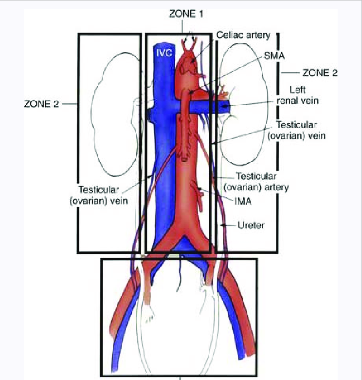
- Zones
- I - Central
- Between both psoas muscles and contains the abdominal aorta, inferior vena cava, pancreas, and midline duodenal structures
- II - Lateral x 2
- Lateral to the psoas muscles on bilateral sides and contains the kidneys, ureters, and portions of the colon
- III - Pelvic
- Management
- Zone 1 - Upper midline central retroperitoneal haematoma
- All should be explored
- Potential sites of injury
- Aorta/IVC
- Kidneys, Ureters, Renal vessels
- Duodenum/Pancreas
- Colon
- Zone 2 - Lateral retroperitoneal haematomas
- Only explore if expanding, thought to have injury to Colon or Ureter
- Usually related to renal bleed which should tamponade
- Zone 3 - Pelvic
- Non - expanding - don’t explore
- Ongoing bleeding
- External fixation of pelvis
- Angioembolization
- If angio is unavailable and enlarging pelvic haematoma/ongoing bleeding
- Extra-peritoneal pelvic packing
- General
- Unstable and peritonitis - Laparotomy
- Observe if stable, not too sore and imaging is reassuring
- Serial abdominal examinations
Retroperitoneal Haematoma
- Zones
- I - central (further subdivision based on apex of haematoma)
- Supracolic
- Juxtarenal or suprarenal aorta, SMA, coeliac
- Infracolic
- Aortic bifurcation/IMA
- Transperitoneal or left medial visceral rotation approach
- Left or right
- If apex is right of midline and primarily venous bleeding
- If apex is left of midline and primarily arterial bleeding
- II - Lateral x 2
- Leave alone if not expanding
- Renal injuries - non operative and selective embolization approach
- If penetrating injury or expanding - explore
- Need to rule out occult post colonic and ureteric injury
- To prevent abscess/urinoma formation
- III - Pelvic
- If stable and CT proven blush - Angioembolise
- If found at surgery and non-expanding - Leave alone
- If found at surgery and expanding - Manage
- Extraperitoneal pelvic packing
- Pelvic binder is on
- Grasp peritoneal edge of inferior 25% of laparotomy wound
- Develop Retzius space (pre-peritoneal plane) all the way round to posterior rectum on both sides
- Evacuate clot
- Packs x 2-3 on each side
- If fails then culprit Internal Iliac Artery ligation
- If works then to IR for assessment/embolization
- Pelvic fixation/binder