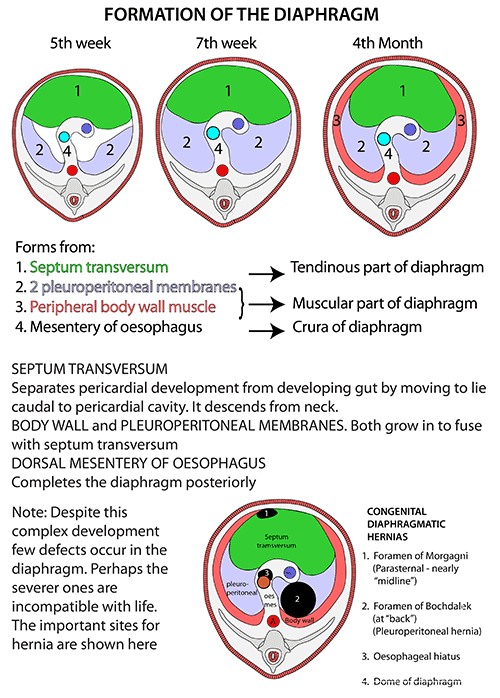
- Develops from 4 sources
- Septum Transversum – gives rise to Central Tendon
- Pleuroperitoneal Membranes (Mesodermal folds) – close the connection between the thoracic and abdominal parts of the Coelom
- Oesophageal Mesentery – also contributes to the development of the Diaphragm
- Cervical Myotomes
- Muscle cells derived from the 3rd, 4th and 5th Cervical Myotomes
- Muscle cells carry their own nerve supply with them – hence the motor supply from the Phrenic Nerves
- Failure of Pleuroperitoneal Membrane development – most common cause of Congenital Diaphragmatic hernia
- The defect (Bochdalek’s Foramen) is posteriorly placed
- Clinically manifests more often on left side probably due to the presence of the liver on the right
- Morgagni’s Foramen – possible small hernia site at the junction of the costal and xiphoid origins
Overview
- Derivative of the Inner (Transversus) layer of the muscles of the body wall
- Fibres arise in continuity with Transversus Abdominis from within costal margin
- Completed behind the costal margin by fibres arising from the Arcuate Ligaments and the Crura
- From circumference of oval origin, fibres arch upwards into pair of domes and then descend to a Central Tendon which lies at the level of the xiphisternal joint
- Right dome – ascends in full expiration as high as nipple line (4th space)
- Left dome – ascends as high as 5th rib
- Viewed from side: diaphragm resembles inverted J – long limb extending up from Crura (upper lumbar vertebrae) and the short limb attached to xiphisternum (T8 level)
- About 55% of muscle fibres (and 65% of intercostal muscle fibres) are slow twitch fatigue-resistant type
See: Diaphragmatic hernia