Overview
- Lies mainly in left hypochondrial, epigastric and umbilical regions
- Much of it under cover of lower ribs
- Completed invested by peritoneum
- Lesser Omentum – Double layer of peritoneum from the Lesser Curvature to the Liver
- Greater Omentum – Double layer of peritoneum hanging down from the Fundus and Greater Curvature
- Anterior relations include Diaphragm, Anterior Abdominal Wall and Left Lobe of the Liver
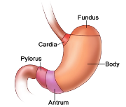
Parts of the Stomach
- Cardia
- Gastro-oesophageal junction
- Most fixed part
- Lies 2.5cm to the left of the midline at the level of T10
- Behind the 7th Left Costal Cartilage
- 40cm from the incisor teeth
- Fundus
- Projects above the level of the Cardia
- In contact with the Left Dome of the Diaphragm
- Body
- Largest part
- Extends from the Fundus to the Angular Incisure (Notch) on the lower part of the Lesser Curvature
- Variable amounts will be above and below the costal margin and in contact with the diaphragm and abdominal wall
- Lowest part of Greater Curvature may be above or below the Umbilicus
- Pylorus
- Extends from the Angular Incisure (Notch) to the Gastro-duodenal junction
- Proximal Pyloric Antrum which narrows distally as the Pyloric Canal
- Circular muscle of distal end of Pyloric Canal forms the Pyloric Sphincter
- Position indicated on anterior surface by the Prepyloric Vein of Mayo
- NB: This indicates the junction with the Duodenum
- Position indicated on anterior surface by the Prepyloric Vein of Mayo
- Pyloric Canal lies on the Head and Neck of the Pancreas
- Circular muscle of distal end of Pyloric Canal forms the Pyloric Sphincter
- In the recumbent position with the stomach empty, this is usually a little to the right of the midline at the level of L1 (but may be much lower)
Stomach Bed
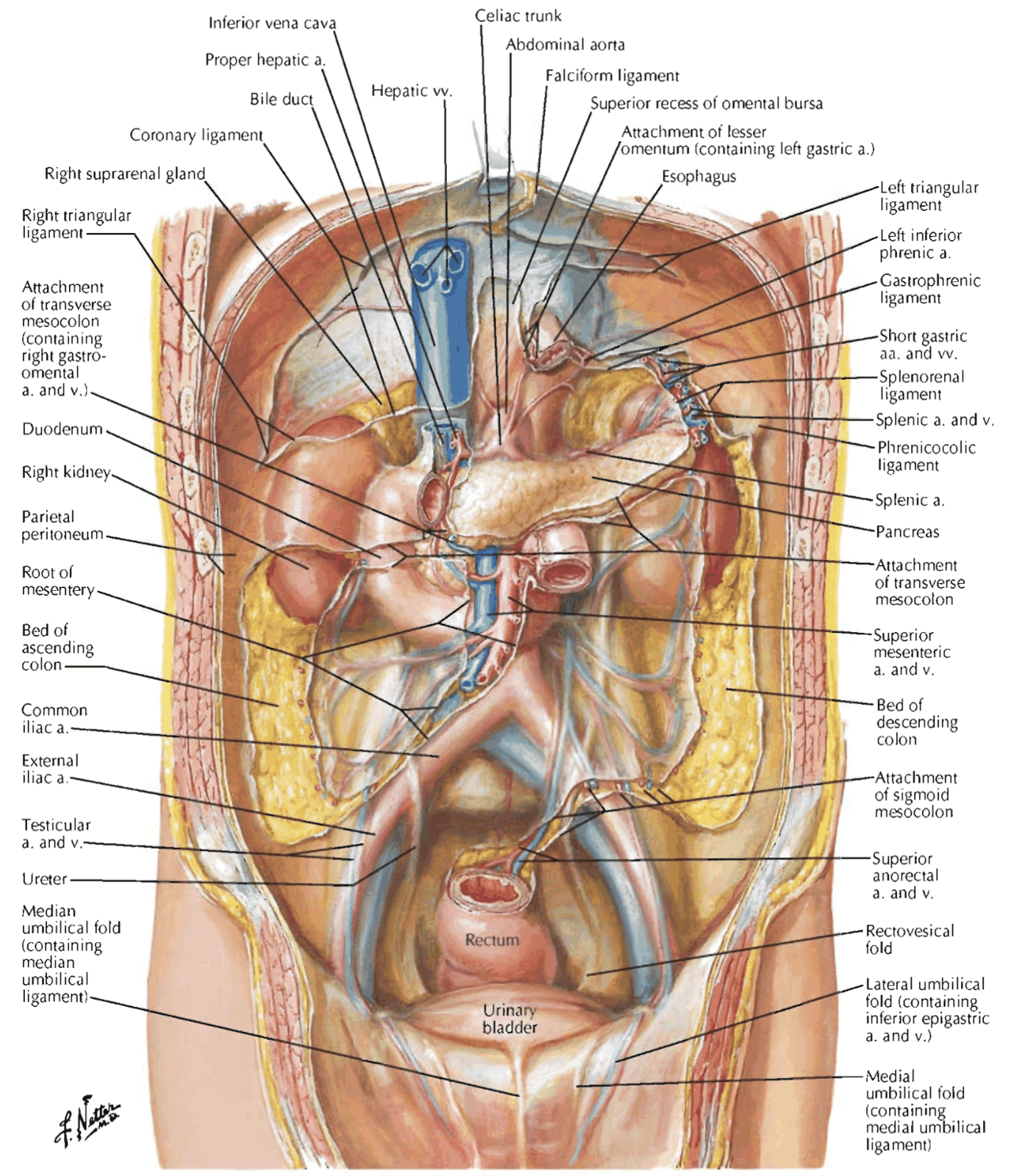
- Posterior wall of stomach covered by peritoneum of the anterior wall of the Lesser Sac
- Stomach bed covered by Lesser Sac’s posterior wall
- Contents of Stomach Bed:
- Left Crus and Dome of Diaphragm
- Splenic Artery
- Pancreas Body
- Transverse Mesocolon
- Left Kidney (Upper Part)
- Left Suprarenal Gland
- Spleen
- Splenic Flexure of Colon
- To the right of the Lesser Curvature in the midline lies:
- Aorta with the Coeliac Trunk
- Coeliac Plexus and Ganglia
- Coeliac Lymph Nodes
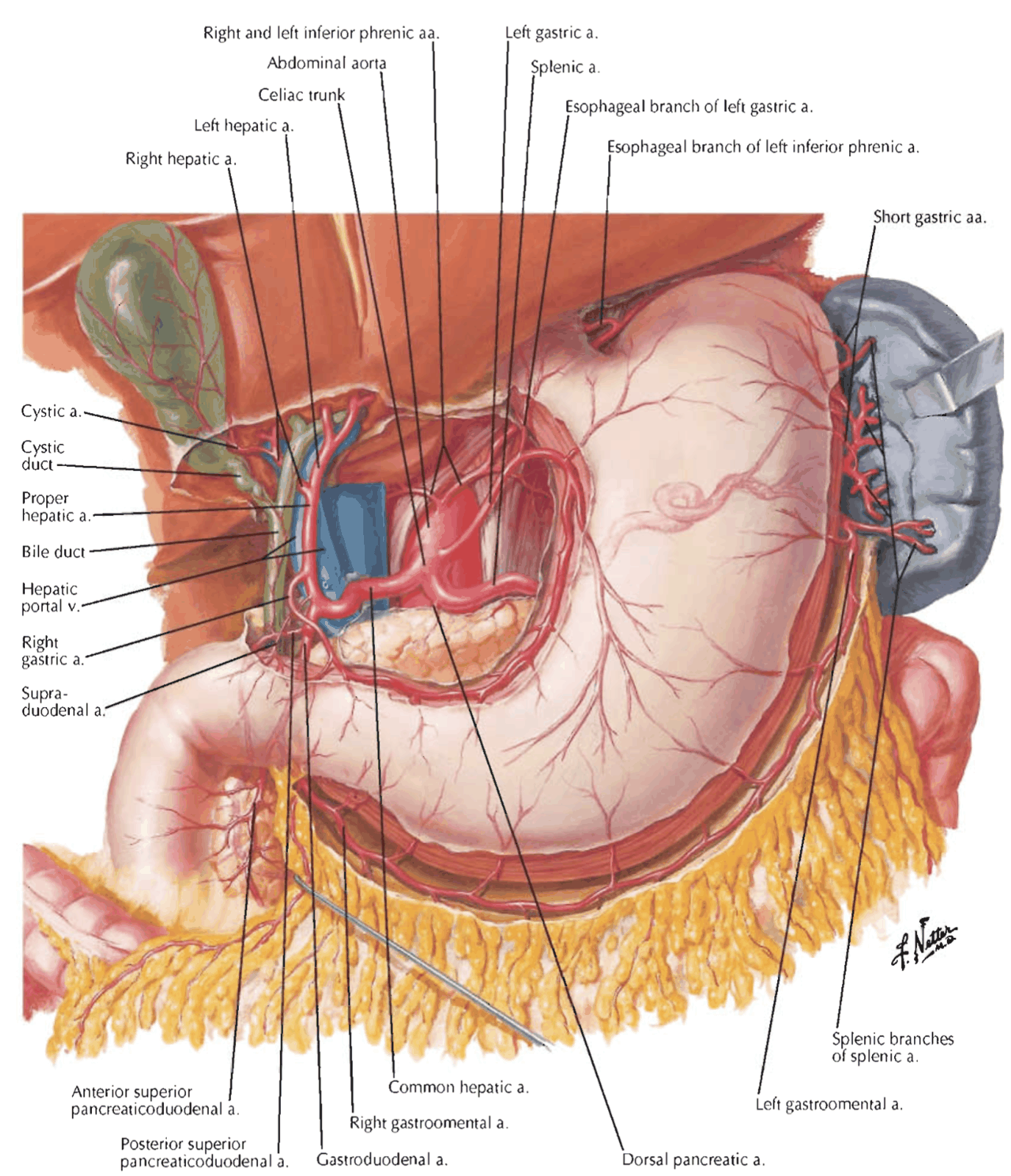
Blood Supply
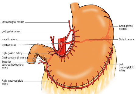
Branches from the Coeliac Trunk
- Left Gastric Artery
- Travels along the lesser curvature between the 2 layers of Lesser Omentum
- Anastomoses with the Right Gastric Artery
- Right Gastric Artery
- Short Gastric Arteries
- Originate from the Splenic Artery in the Gastrosplenic Ligament
- Usually ~6
- Left Gastroepiploic Artery
- Right Gastroepiploic Artery
- Gastroepiploic arteries supply the Greater Curvature
- Run between the 2 layers of the Greater Omentum and anastomose with each other
- Right Gastroepiploic Artery closer to the Greater Curvature than the Left
- During partial gastrectomy, the Greater Omentum is divided below the Right and above the Left Gastroepiploic Arteries
- Larger omental branches of the Left Gastroepiploic Artery preserved thus keeping the blood supply to the omentum intact
- Posterior Gastric Artery
- Branch of the Splenic Artery as it runs along the upper border of the Pancreas
- Veins of same names accompany arteries and drain into the Portal Vein itself or its Splenic Vein and Superior Mesenteric Vein tributaries
- Prepyloric Vein of Mayo (unaccompanied by an artery) drains into the Right Gastric Vein
Alternative Description of Blood Supply by Stomach Portion
- Stomach comprised of Greater Curvature, Lesser Curvature, Cardia, Fundus, Body, Pyloric Antrum and Pyloric Canal [WB]
- Arterial supply from 3 major branches of the Coeliac Trunk – Common Hepatic, Splenic and Left Gastric Arteries
- Fundus and Upper Left Part of Greater Curvature:
- Short Gastric Arteries (Branches of the Splenic Artery) – run in the Gastrosplenic Ligament
- Lesser Curvature:
- Left Gastric Artery
- Right Gastric Artery (Branch of the Hepatic or Gastroduodenal Artery)
- Run between the two layers of Lesser Omentum
- Greater Curvature:
- Left Gastroepiploic Artery (Branch of the Splenic Artery)
- Right Gastroepiploic Artery (Branch of the Gastroduodenal Artery)
- Run between the two layers of the Greater Omentum 1cm from the gastric wall
- Both sets of arteries anastomose with each other on their respective gastric curves and then again with the supply of the other curve via rich anastomosing network at 3 levels in the gastric wall – mucosal, submucosal and intra-muscular
- Network well developed everywhere except along the Lesser Curvature when anastomosing channels are of a finger calibre and more likely to be end arteries
- Network well developed everywhere except along the Lesser Curvature when anastomosing channels are of a finger calibre and more likely to be end arteries
Application of Arterial Anatomy in Oesophagectomy
- Short Gastric, Left Gastroepiploic and Left Gastric Arteries divided to mobilise upper portion of Stomach so it can be pulled up to form an oesophageal substitute and allow clearance of the Lesser Curve lymph nodes [WB]
- Short Gastric Arteries ligated away from the Greater Curvature to avoid damage to the intramural network
- Left Gastric Artery ligated at its origin for oncological reasons – no evidence that its branches need to be preserved to maintain viability of the stomach
- Right Gastroepiploic and Right Gastric Arteries preserved to maintain viability of stomach
Lymphatic Drainage
- All lymph eventually reach Coeliac Nodes after passing through various groups
- Lymph vessels anastomose freely in the stomach wall
- Valves in vessels direct lymph in such a way that a line drawn parallel to the greater curvature and 2/3rds of the way down the anterior surface indicates a ‘watershed’
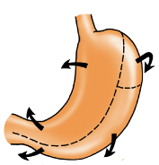
- Largest zone above and to the right of this line ⇒ Lymph passes to the left and right Gastric Nodes along the Left and Right Gastric Arteries
- Upper left quadrant ⇒ Lymph flows via left Gastroepiploic nodes and directly to Pancreaticosplenic nodes at the Splenic Hilum and at the upper border and posterior surface of the Pancreas, accompanying the Splenic Artery
- Rest of the greater curvature ⇒ Lymph reaches nodes along the Right Gastroepiploic vessels ⇒ Drain to Subpyloric nodes near the Gastroduodenal Artery
- Pyloric part of stomach ⇒ Drain to Hepatic nodes in the Porta Hepatis and to Subpyloric and Right Gastric nodes
- Troisier’s Sign/Virchow’s Node: Involvement of Left Supraclavicular nodes in cases of gastric carcinoma, presumably due to spread along the Thoracic Duct
Nerve Supply
Sympathetic Fibres
- Vasomotor and cause Pyloric Sphincter contraction
- Accompanied by Afferent (pain) fibres
- Run with various arterial branches to the stomach Parasympathetic Fibres
- From Vagus
- Controls motility and secretion
- 90% of Vagal fibres below the Diaphragm are afferent for reflex activities, not pain
- Anterior Vagal Trunk:
- Comprised mainly left vagal fibres from the Oesophageal Plexus in the posterior mediastinum
- Lies in contact with anterior oesophageal wall (usually nearer to its right margin)
- Sometimes trunk is double or triple
- Each trunk gives off one or two hepatic branches
- ⇒ Run in upper part of the Lesser Omentum to join the plexus on the Hepatic Artery and Portal Vein
- ⇒ Turn down in anterior wall of the Epiploic Foramen to reach the Pylorus
- Also gives off several gastric branches which supply Fundus and Body
- Gives off the Greater Anterior Gastric Nerve or Anterior Nerve of Latarget
- ⇒ Runs down in Lesser Omentum near the Lesser Curvature with the Left Gastric Artery
- ⇒ Subdivides in manner of Crow’s foot to supply Antrum and Pyloric Sphincter
- NB: Anterior Surface of the stomach innervated by the Left Vagus Nerve
- Posterior Vagal Trunk:
- Comprised mainly of right vagal fibres
- Lies in loose tissue a little behind and to the right, not in contact with the posterior surface of the oesophagus
- Gives Coeliac branches that run backwards along the Left Gastric Artery to the Coeliac Plexus
- Coeliac Plexus supplies the large bowel up to the Transverse Colon
- Gives Gastric branches to the Fundus and Body
- Gives off the Greater Posterior Gastric Nerve or the Posterior Nerve of Latarget
- ⇒ Runs in the Lesser Omentum behind the Anterior Trunk to reach the Antrum (but NOT the pyloric sphincter)
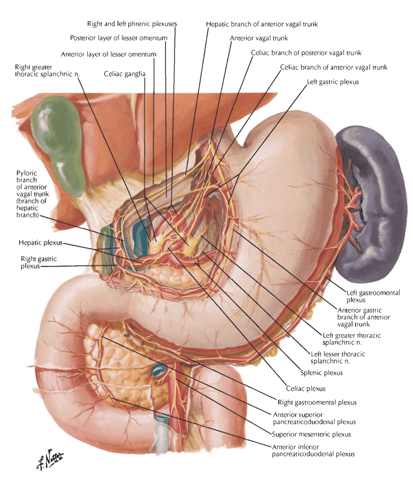
- ⇒ Runs in the Lesser Omentum behind the Anterior Trunk to reach the Antrum (but NOT the pyloric sphincter)
Possible Nerve-Related Operations
- Truncal Vagotomy – Cutting the trunks at the level of the abdominal oesophagus
- Consequences:
- Reduced gastric acid secretion due to removal of the acetylcholine-mediated secretion of acid from the Parietal Cells
- Accelerated emptying of liquids due to removal of relaxation of the gastric Fundus
- Decreased emptying of solids due to removal of relaxation of the Pylorus
- Consequences:
- Selective Vagotomy – Anterior trunk cut distal to Hepatic branch and Posterior trunk cut distal to Coeliac branches
- Both are effective in reducing gastric acid secretion
- However: Often accompanied by gastric stasis. Thus, antral drainage procedure often required.
- Highly Selective Vagotomy – Cuts branches only to Fundus and Body
- Leaves Antral and Pyloric branches intact
- Attempts to avoid gastric stasis
- Arterial branches run in lesser curvature transversely while nerve fibres run obliquely.
- Ligating vessels will inevitably sever some nerve branches but not all because not all nerves accompany vessels closely
Structure
- Angular notch – usually taken as the dividing line between the body and pyloric parts of the stomach
- This does not necessarily indicate the division between the body-type of mucosa (i.e. Parietal cells which secrete acid) and the pyloric-type of mucosa (i.e. G-cells producing gastrin)
- Muscle layers – inner circular, outer longitudinal, innermost oblique (incomplete)
- Innermost layer – fibres loop around the fundus – being thickest at the notch between the oesophagus and stomach, thus, helping to maintain the angle here
Greater Omentum
- Double sheet of peritoneum folded on itself to form 4 layers
- Anterior 2 layers descend from the Greater Curvature of the Stomach (where they are continuous with the peritoneum on the anterior and posterior surfaces of the Stomach)
- Overlie coils of intestines
- Turn around and ascend up to the Transverse Colon
- Loosely blend with the Peritoneum on the anterior surfaces of the Transverse Colon and Transverse Mesocolon above it
- Four layers of the Greater Omentum below the Transverse Colon fuse to form an integral structure
- Contains adipose tissue, and numerous macrophages
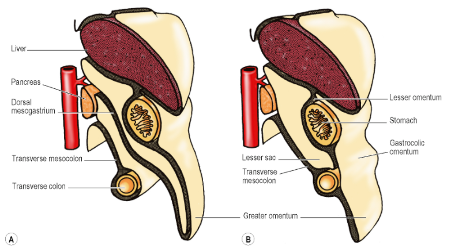
- Gastrocolic Omentum:
- The part of the Greater Omentum between the Stomach and Transverse Colon
- Right and Left Gastroepiploic Vessels run between the layers, close to the Greater Curvature of the Stomach
- Lesser sac can be accessed through the Gastrocolic Omentum
- Other routes of access:
- Through the Lesser Omentum
- Through the Transverse Mesocolon
- Other routes of access:
- Below Stomach, left border of the Greater Omentum envelops Spleen (except for a small bare area at the Hilum)
- Two double-layered folds of Peritoneum, the Gastrosplenic and Splenorenal Ligaments connect the hilum of the Spleen to the Greater Curvature of the Stomach and anterior surface of the Left Kidney, respectively
- Splenorenal Ligament:
- Contains the Pancreatic Tail and Splenic Vessels
- Gastrosplenic Ligament:
- Contains the Short Gastric and Left Gastroepiploic Vessels
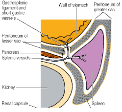
- The right border extends as far as the origin of the Duodenum
- NB: Greater Omentum is indeed attached to the Left Kidney