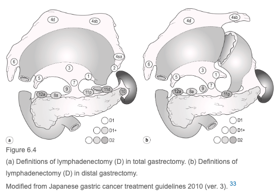
D1 - 1-7 D1+ - 1-9 D2 - 1-12 (+/- 10 for all)
Spread of cancer:
-
Spread of gastric cancer – predominantly lymphatic way at earliest stage, leading to other ways (peritoneal, haematogenous and direct)
-
Peritoneal metastasis most common type of failure after radical surgery. More common in diffuse-type than intestinal
-
Surgery can cause cancer spread (intraoperative spillage)
-
Classified into stations numbered 1 to 20, plus stations 110, 111 and 112
-
Stations 1 to 12 and 14v are considered Regional Stations
-
The other lymph node stations are considered Distant Stations and classified as M1.
-
Lymph node stations 16, 19, 100, 110 and 111 are considered Regional Stations in the case of direct invasion of the Oesophagus by the tumour.
-
Peri-Gastric Lymph Nodes
- Station 1 – Right Para-Cardial Nodes
- At the right side of the cardia, along the first ramification of the ascending branch of the Left Gastric Artery
- Station 2 – Left Para-Cardial Nodes
- At the left side of the cardia, along the Cardio-Oesophageal branch of the Left Inferior Phrenic vessels
- Station 3 – Lesser Curvature Nodes
- Along the descending branch of the Left Gastric Artery and along the Right Gastric Artery distal to the first gastric branch
- Station 4 – Greater Curvature Nodes
- Divided into two groups separated by the Von Ghoete point – where Right and Left Gastroepiploic Arteries meet each other – a left group (4s) and right group (4d)
- Left group subdivided into proximal (4sa) and distal (4sb)
- LNs of 4sa are located around the short gastric vessels
- LNs of 4sb are located along the Left Gastroepiploic Vessels
- Right group LNs are located along the Right Gastroepiploic vessels, distal to the first gastric branch (Boundary with Infrapyloric LNs)
- Station 5 – Supra-Pyloric Nodes
- At the Lesser Curvature immediately above the pylorus, along the Right Gastric Artery and its origin
- Station 6 – Infra-Pyloric Nodes
- At the Greater Curvature, immediately below the pylorus at the confluence of the Right Gastroepiploic Vein and the Anterior Inferior Pancreaticoduodenal Vein
- Station 7 – Left Gastric Artery Nodes (although this is now included in a D1 resection)
- Between its roots from the Coeliac Trunk till the origin of its ascending branch on the Lesser Curvature
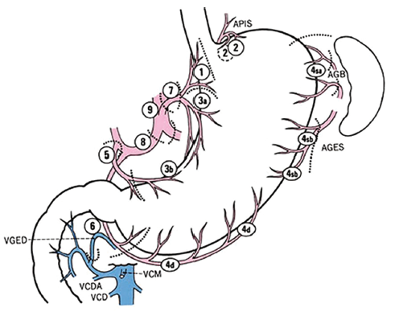
- Between its roots from the Coeliac Trunk till the origin of its ascending branch on the Lesser Curvature
- Station 1 – Right Para-Cardial Nodes
-
Second Tier Lymph Nodes
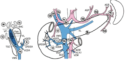
- Station 8 – Common Hepatic Artery Nodes
- Between its root from the Coeliac Trunk to the branching off of the Gastroduodenal Artery
- Divided into anterior part (8a) and posterior part (8p)
- Station 9 – Coeliac Trunk Nodes
- Immediately near the origins of the Left Gastric Artery, the Common Hepatic Artery and the Splenic Artery
- Station 11 – Splenic Artery Nodes
- Distinguished into a proximal group (11p) and a distal group (11d)
- Group 11p – from the origin of the Splenic Artery from the Coeliac Axis to the rise of the Posterior Gastric Artery – about halfway between its origin and the pancreatic tail end
- 11d – from the origin of the Posterior Gastric Artery to the end of the pancreatic tail
- Station 8 – Common Hepatic Artery Nodes
-
Second or Third Tier or M nodes (according to site of the primary tumour)
- Station 10 – Splenic Hilum Nodes
- The vascular boundary between 10 and 4sb is the first gastric ramification of the Left Gastroepiploic Artery
- Station 12 – Hepatoduodenal Ligament Nodes
- Divided into 3 separate groups based on this ligament: 12a, 12b and 12p
- 12a – Left Hepatoduodenal Ligament LNs – located along the Hepatic Artery Proper in the caudal half between the confluence of the Right and Left Hepatic Ducts and the upper pancreatic margin
- 12b – Posterior Hepatoduodenal Ligament LNs – Nodes located along the Bile Duct in the caudal half between the confluence of the Right and Left Hepatic Ducts and the upper pancreatic margin
- 12p – Posterior Hepatoduodenal Ligament LNs – Nodes located posteriorly to the Portal Vein in the caudal half between the confluence of the Right and Left Hepatic Ducts and the upper pancreatic margin
- Station 13 – Retropancreatic Nodes
- Posterior surface of the Pancreatic Head cranial to the Ampulla of Vater along the Superior and Inferior branches of the Posterior Pancreaticoduodenal Artery
- Left lateral border of this location is marked by the Portal Vein
- Upper border is the origin of the Posterior Hepatoduodenal Ligament LNs
- Station 14 – Superior Mesenteric Vein and Artery Nodes
- Subdivided into 14v and 14a
- 14v – along the confluence of the SMV into the Portal Vein
- 14a – along the origin of the SMA at the root of the mesentery
- Lateral border – confluence of the Gastrocolic Vein into the SMV
- Lower border – Confluence of the Middle Colic Vein into the SMV
- Upper border – Origin of the SMA from the Aorta at the lower hedge of the Pancreas
- Station 15 – Middle Colic Nodes
- In the Transverse Mesocolon around the Middle Colic Artery and Vein, from their origin/confluence from/into the Superior Mesenteric Vessels, until the Mesocolic hedge of the Transverse Colon
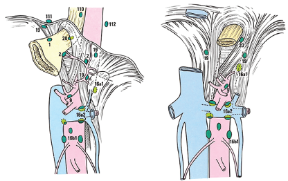
- Station 16 – Aortic Hiatus, Middle and Caudal Para-Aortic Nodes
- Four separate groups around the Abdominal Aorta and Inferior Vena Cava
- 16a1 – Around the Diaphragmatic Aortic Hiatus, over the anterior side of the Aorta – from the inferior hedge of the hiatus to the upper border of the Coeliac Artery
- 16a2 – Over the anterior surface of the Aorta, from the Coeliac Artery to the lower border of the Left Renal Vein
- 16b1 – Around the anterior surface of the Aorta and IVC, from the lower border of the Left Renal Vein to the upper border of the origin of the Inferior Mesenteric Artery – Right and left borders are defined by the Right border of the IVC and by the Left Gonadal vessels
- 16b2 – Around the anterior surface of the Aorta and IVC, between the upper border of the IMA origin and the aortic bifurcation
- Station 17 and 18 – Peripancreatic Nodes
- 17 - Along the Superior Anterior Pancreaticoduodenal vessels, on the anterior surface of the Pancreatic Head beneath the pancreatic sheath
- 18 – Along the inferior border of the Pancreatic Body
- Station 19 – Infradiaphragmatic Nodes
- Along the Subphrenic vessels
- Station 20 – Paraoesophageal Nodes
- Paraesophageal region I the Diaphragmatic Oesophageal Hiatus
- Stations 100,111 and 112 – Lower Thorax Nodes
- 100 – Lower thorax
- 111 – Supradiaphragmatic space separate from the Oesophagus
- 112 – Posterior mediastinal space separate from the Oesophagus and Oesophageal Hiatus
- Station 10 – Splenic Hilum Nodes
Lymph node stations:
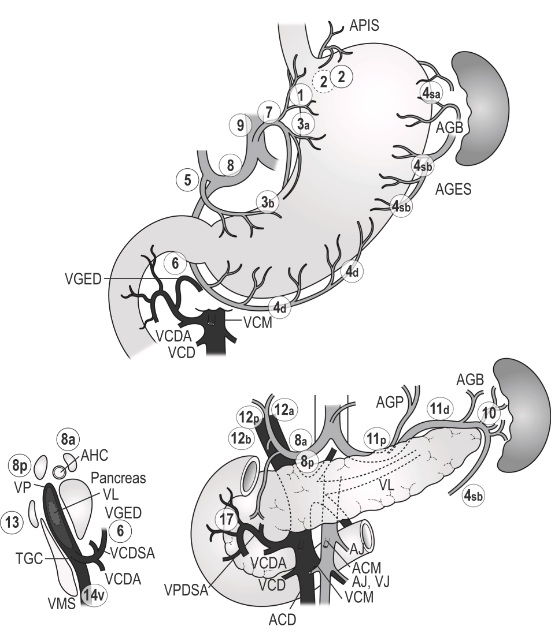
| 1 | Right paracardial region of stomach | First branch of ascending left gastric artery |
| 2 | Left paracardial region of stomach | Esophagogastric branch of left subphrenic artery |
| 3a | Lesser curvature | Left gastric artery |
| 3b | Lesser curvature | 2nd branch and distal part of the right gastric artery |
| 4sa | Left greater curvature | Short gastrics |
| 4sb | Right greater curvature | Left gastroepiploic artery |
| 4d | Right greater curvature | 2nd branch and distal part of the right gastroepiploic artery |
| 5 | Suprapyloric | 1st branch and proximal part of the right gastric artery |
| 6 | Infrapyloric | 1st branch and proximal part of the right gastroepiploic artery |
| 7 | Left gastric artery | |
| 8a | Anteriosuperior | Common hepatic artery |
| 8b | Posterior | Common hepatic artery |
| 9 | Celiac artery | |
| 10 | Splenic hilum | Splenic artery distal to pancreatic tail and at roots of short gastrics |
| 11p | Along margin of pancreas | Proximal splenic artery from origin to halfway from origin to pancreatic tail |
| 11d | Along margin of pancreas | Distal splenic artery from halfway from origin to pancreatic tail to end of pancreatic tail |
| 12a | Hepatoduodenal ligament | Proper hepatic artery from confluence of the right and left hepatic ducts and upper border of the pancreas |
| 12b | Hepatoduodenal ligament | Along bile duct caudal half the confluence of the right and left hepatic ducts and upper border of the pancreas |
| 12p | Hepatoduodenal ligament | Along portal vein caudal half the confluence of the right and left hepatic ducts and upper border of the pancreas |
| 13 | Posterior surface of the head of the pancreas | |
| 14V | Root of the mesentery | Superior mesenteric vein |
| 15 | Colonic | Middle colic vessels |
| 16a1 | Paraaortic in diaphragmatic hiatus | |
| 16a2 | Paraaortic | Upper margin of the origin of the celiac artery and lower border of the left renal vein |
| 16b1 | Paraaortic | Lower border of the left renal vein and upper border of the origin of the inferior mesenteric vein |
| 16b2 | Paraaortic | Between the upper border of the origin of the inferior mesenteric artery and the aortic bifurcation |
| 17 | Anterior surface of the pancreatic head beneath the pancreatic sheath | |
| 18 | Along the inferior border of the pancreatic body | |
| 19 | Infradiaphragmatic | Predominantly along the subphrenic artery |
| 20 | Paraesophageal in the diaphragmatic esophageal hiatus | |
| 110 | Paraesophageal in the lower thorax | |
| 111 | Supradiaphragmatic, separate from the esophagus | |
| 112 | Posterior mediastinal, separate from the esophagus and the esophageal hiatus |