Overview
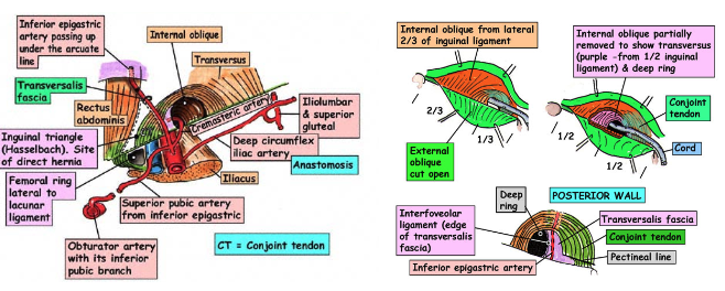
- Definition: Oblique intermuscular slit located above the medial half of the inguinal ligament
- Origin: Deep inguinal ring
- Termination: Superficial inguinal ring
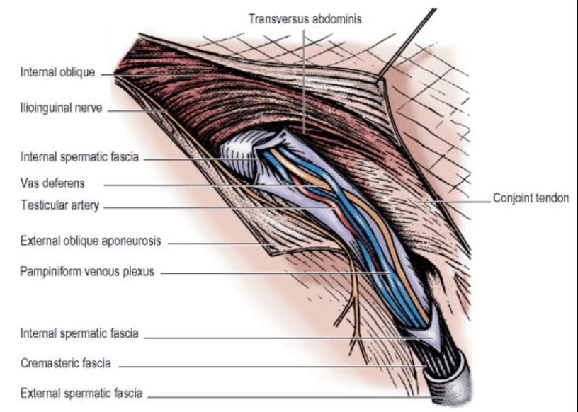
- Contents: Ilioinguinal nerve and spermatic cord (if male) or round ligament (if female)
Boundaries
- Anterior Wall
- External oblique aponeurosis and laterally assisted by internal oblique
- Floor
- Inguinal ligament (in-rolled lower edge) reinforced medially by Lacunar Ligament
- Roof
- Internal oblique and transversus abdominis – arch over from in front of the cord laterally to behind the cord medially where the conjoined aponeurosis (conjoint tendon) inserts into pubic crest and pectineal line of pubic bone.
- Posterior Wall
- Conjoint tendon medially and weak transversalis fascia throughout.
- Structures deep to posterior wall:
- Inferior epigastric artery
- Crosses posterior wall at medial edge of deep inguinal ring
- Vas deferens or round ligament hook around the Interfoveolar ligament lateral to the artery
- Crosses posterior wall at medial edge of deep inguinal ring
- Inferior epigastric artery
Ligaments
- Integrity depends on strength of:
- Anterior wall in lateral part
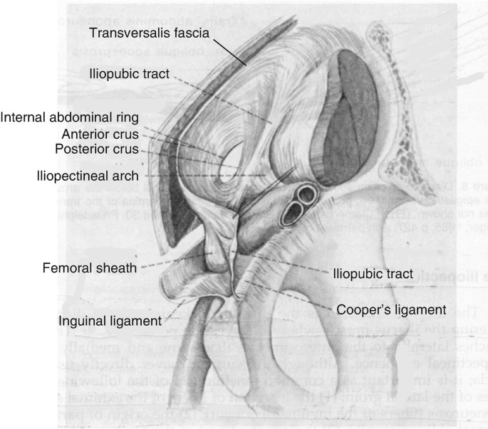
- Transversalis fascia in posterior wall reinforced by interfoveolar ligament (i.e. tendinous/muscular fibres derived from the transversus abdominis muscle)
- Arches down from lower border of transversus around the vas to the inguinal ligament
- Constitutes the functional medial edge of the deep ring
- Posterior wall in medial part
- Conjoint tendon reinforces posterior aspect of superficial inguinal ring
- Posterior wall in medial part
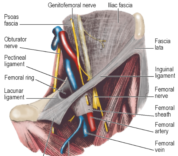
- Divides space deep to inguinal ligament
- Medial fascia of iliopsoas
- Fascia blends with inguinal ligament in front and pectineal ligament and pectineus fascia behind
- Thus the only way under the ligament here is beneath iliopsoas fascia
- Medial to iliopectineal arch is a space through which iliac vessels pass
- Thickening of the Transversalis Fascia extending from the Iliopectineal Arch to the Pubic Ramus
- Curves around the medial surface of the Femoral Sheath to attach to the Pectineal Ligament
- Not the same as the Inguinal Ligament
- Inguinal Ligament is the rolled inferior aspect of the External Oblique Aponeurosis
- Inguinal Ligament more superficial and not seen in a laparoscopic extra-peritoneal approach
Lacunar Ligament (of Gimbernat)
- From the medial end of the Inguinal Ligament, the triangular Lacunar Ligament extends horizontally backwards to the pectineal line on the pubis
- NB: Is NOT attached to the Pubic Bone in continuity with the Adductor Longus
- The free lateral edge of the Lacunar Ligament forms the medial margin of the Femoral Ring
- NB: Lacunar Ligament in the erect position has the spermatic cord on its superoanterior surface
Pectineal Ligament(of Astley Cooper) extends laterally further along the pectineal line of the pubis
Superficial Inguinal Ring
- Oblique, triangular gap in the External Oblique aponeurosis
- Above and lateral to the pubic tubercle
- Base of gap: Pubic Crest
- Margins: Crura of the ring
- The reflected part of the Inguinal Ligament are the fibres that can be traced upwards and medially behind the Spermatic Cord that interdigitate in the linea alba with those of the opposite side
- Intercrural fibres run at right angles to those of the aponeurosis near the apex of the Superficial Inguinal Ring to prevent the crura from separating
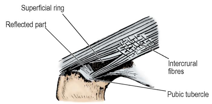
Deep Inguinal Ring
- Definition: Opening in the transversalis fascia located above midpoint of inguinal ligament
- Transversalis fascia projects along canal as internal spermatic fascia
Spermatic Cord
- Contents (in males):
- 3 Arteries
- Artery to Vas deferens (derived from the Superior or Inferior Vesical Artery)
- Testicular artery
- Cremasteric artery (derived from the Inferior Epigastric Artery)
- NB: There is NO cremaster muscle within the Spermatic Cord in the Inguinal Canal
- 3 Nerves
- Genital branch of genitofemoral nerve
- Cremasteric nerve
- Autonomic nerves
- 4 Other things
- Vas deferens (usually lies in the lower and posterior part of the cord)
- Testicular veins/pampiniform plexus
- Obliterated processus vaginalis
- Lymphatics (those from testis draining to Para-Aortic Nodes, and those from the coverings to the External Iliac Nodes)
- 3 Arteries
- Contents (in females):
- Round ligament
- Obliterated processus vaginalis
- Lymphatics from the uterus
- Ilioinguinal nerve does not enter inguinal canal through deep ring – it pierces internal oblique muscle, i.e. enters canal from the side not the back.
Relevant Nerves
-
- Enters inguinal canal by piercing internal oblique muscle, i.e. enters from the side, not the back
- Thus, not within spermatic cord
- Lies in front of the cord
- Exits inguinal canal via superficial ring
- Supplies: Skin of inguinal region, upper part of thigh, anterior third of scrotum (or labium majus) and root of penis
- Enters inguinal canal by piercing internal oblique muscle, i.e. enters from the side, not the back
- Thus, not within spermatic cord
-
- Leaves with the spermatic cord
-
Surgical Practical Points:
- Nerves affected by inguinal hernia surgery include:
- Open repair:
- Ilioinguinal Nerve
- Iliohypogastric Nerve
- Genital branch of the Genitofemoral Nerve
- Femoral Nerve (rare)
- Laparoscopic repair:
- Femoral branch of the Genitofemoral Nerve
- Lateral Femoral Cutaneous Nerve
- Femoral Nerve (rare)
- Open repair:
- Nerves affected by inguinal hernia surgery include:
Triangles
Triangles of Doom and Pain
-
Triangle of Doom:
- Boundaries:
- Medial: Vas Deferens
- Lateral: Testicular Artery
- Apex: Deep Ring
- Posterior: Peritoneal fold
- Importance: Location of external iliac artery and vein
- NB: Also contains one nerve – Genital Branch of Genitofemoral
- Boundaries:
-
Triangle of Pain:
- Boundaries:
- Medial: Testicular Vessels
- Superolateral: Iliopubic Tract
- Lateral: Reflected peritoneal edge
- Importance: Contains 3 nerves:
- Lateral femoral cutaneous nerve
- Femoral branch of the genitofemoral nerve
- Femoral nerve
- NB: Also contains one artery – Deep External Pudendal Artery
- Boundaries:
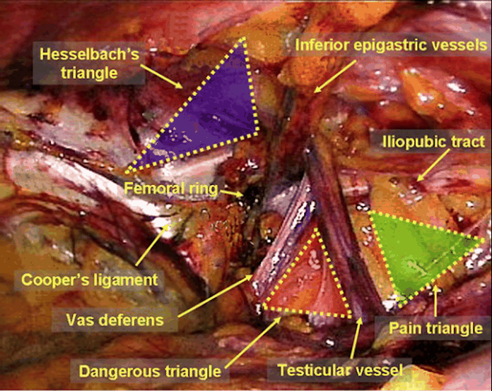 Figure: Laparoscopic view of Right hand side
Hesselbach’s triangle
- Media – lateral border of the rectus abdominis muscle.
- Lateral – inferior epigastric vessels.
- Inferior – inguinal ligament.
Inguinal Lymph Nodes
- Superficial Inguinal nodes
- Drainage from large lymphatic vessels accompanying Great Saphenous Vein from the foot, leg and thigh
- Consists of about 10 nodes
- NB: Said to consist of medial, lateral and vertical groups
- Proximal group = Just distal to inguinal ligament
- Lateral Nodes of Proximal group receive lymph from:
- Buttock
- Flank
- Back below the waist
- Medial Nodes of Proximal group receive lymph from:
- Umbilicus and anterior abdominal wall below it
- External genitalia of both sexes (excl. testis but including lower vagina)
- Lower Anal Canal
- Perineum
- Small proportion of drainage from Uterus via lymphatics accompanying Round Ligament
- Distal group = Lies vertically along terminal GSV
- Receives lymph from all superficial lymphatics of lower limb
- EXCEPT posterolateral part of calf
- Superficial inguinal nodes ⇒ Drain to External Iliac lymph nodes by efferent lymphatics, some of which pierce the Cribriform Fascia
- Lateral Nodes of Proximal group receive lymph from:
- Deep Inguinal Nodes
- Lie deep to Fascia Lata
- Medial to Femoral Vein
- One or two in the Femoral Canal
- Receive lymph from:
- Lymphatics that accompany the femoral vessels from the Popliteal Fossa
- Glans penis (or clitoris)
- Few efferent lymphatics from the Superficial Inguinal nodes
- Deep Inguinal nodes ⇒ Drain to External Iliac Nodes through Femoral Canal
Inferior Epigastric Artery
- Origin: External Iliac Artery at the Inguinal Ligament
- Course:
- Pass upwards behind the Conjoint Tendon
- Slips over the Arcuate Line to enter the Rectus Sheath
- Veins drain to External Iliac veins
TEP
Totally Extraperitoneal Laparoscopic Inguinal Hernia Repair Critical Landmarks for Orientation
- Iliopubic Tract – demarcates the landmark for safe conduct of dissection
- Structures below Iliopubic Tract (External Iliac Vessels and Gonadal Vessels and Nerves) must not be damaged by placing tacks into them
- Pubic Symphysis
- Inferior Epigastric Vessels
- External Iliac Vessels
- Gonadal Vessels
- Vas Deferens
Critical Landmarks for Hernia Classification
- Deep Internal Ring:
- Above Iliopubic Tract
- Lateral to the Inferior Epigastric Vessels
- Indirect sac pass into deep ring lateral to epigastrics
- Direct Space:
- Above the Iliopubic Tract
- Medial to Inferior Epigastric Vessels
- Femoral Ring:
- Below Iliopubic Tract
- Medial to External Iliac Vessels