Liver
- Large gland in RUQ of abdomen. Curves superiorly convex by diaphragm and ribs.
- Concave inferior surface which abuts abdominal organs.
- Invested in peritoneum, apart from posterior surface (bare area) and gallbladder bed and porta hepatis.
- 2 surfaces: Diaphragmatic and visceral
Liver Embryology
- Starts as liver bud aka hepatic diverticulum, 3 weeks of development as outgrowth of endoderm.
- Grows into septum transversum (mesoderm) – which contributes certain cells e.g. Kuppfer cells, stroma/connective tissue of liver.
- After 30 days, connection from liver to duodenum narrows to form bile duct.
- Hepatic cells from diverticulum form cords which mix with viteline and umbilical veins to form hepatic sinusoids.
- Viteline veins fuse to from portal vein, SMV and splenic veins.
- Paired umbilical veins carry oxygenated blood to fetus through umbilicus.
- These initially drain through sinus venosus which is connection between portal vein and hepatic veins, allowing fetal blood to bypass liver.
- Left umbilical vein regresses to form ligamentum teres.
- Continuation of this, Ductus venosus becomes ligatamentum venosum.
Ligaments
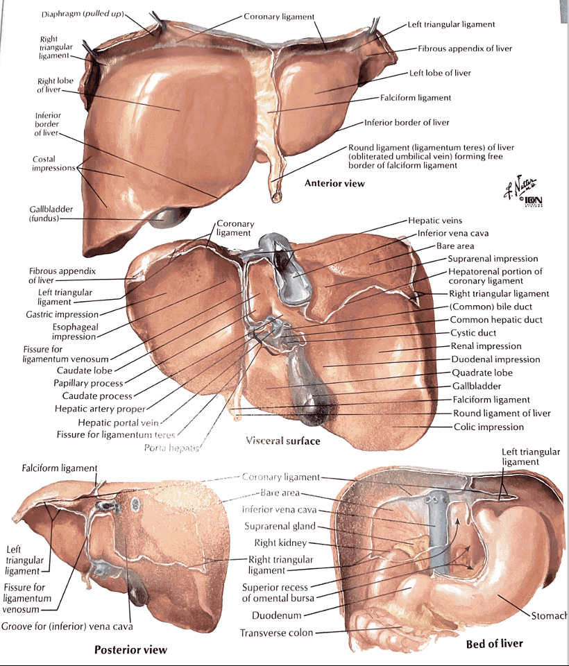
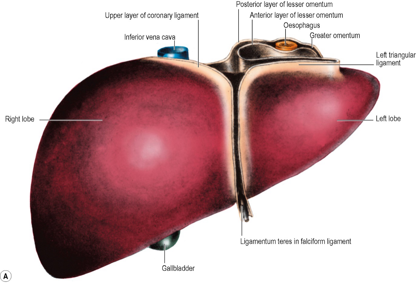
- Falciform ligament
- Anchors the liver to the anterior abdominal wall and the diaphragm
- Superioloy just to the right of the median plan
- Attaches between the anatomical left and right lobes
- Sweeps to the right in front of the IVC to become the right coronary ligament
- Ligamentum teres
- Inferior border of falciform ligament (round ligament of the liver)
- Remnant of the left umbilical vein which drains blood into the left portal vein.
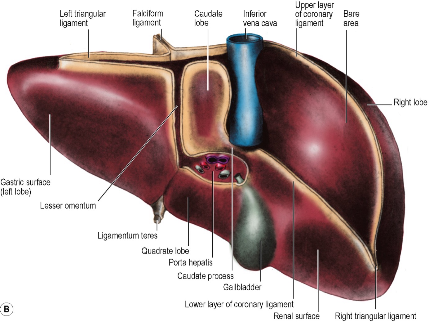
- Coronary ligaments
- Anterior leaf attach the liver to the inferior surface of the diaphragm
- Posterior leaf attaches the liver to the posterior abdominal wall, right kidney and right adrenal gland
- The two leaves border the bare area
- Triangular ligaments
- Formed by the coronary ligaments
- Left and right, coronary ligaments form triangular ligaments
- Ligamentum venosum
- In embryo the ductus venosum was responsible for shunting blood from the left portal vein to the left hepatic vein, thereby bypassing the hepatic circulation.
- It passes from the portal vein in a fossa to the IVC
Anatomical division
- Divided in to a small left lobe and a large right lobe by the falciform ligament
- Segment II and III are left lobe and the remaining are right
- In the functional division segment I and IV are included in the left lobe
Functional division
- Functional division of halves - line from tip of gallbladder to just right of IVC
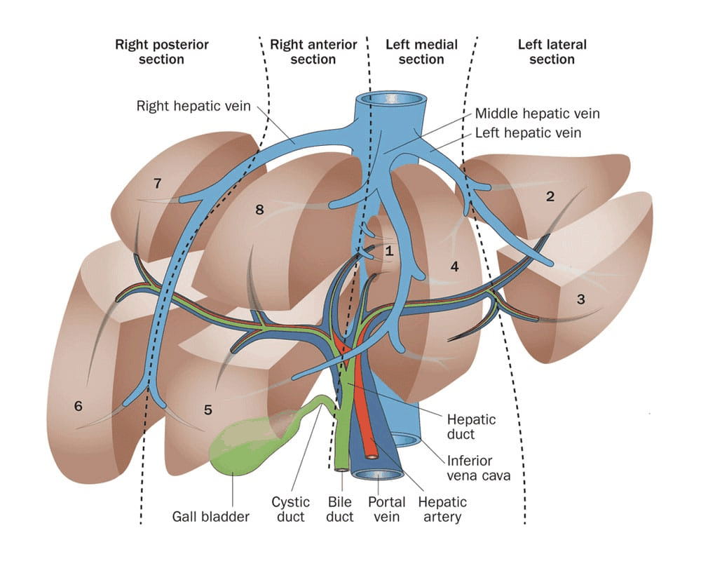
- The liver is divided into four sectors based on internal vascular anatomy
- Right anterior
- Right posterior
- Left medial
- Left lateral
6 fissures
- Main (midplane of the liver )
- Middle hepatic vein
- Separates right and left sides. Separating segment V/VIII from IV.
- Surface landmarks are the tip of the gallbladder back to the midpoint of the inferior vena
- Left
- Left hepatic vein - runs in between Seg 2/3 for most of its course then joins the left intersectional plane and receives the umbilical vein before uniting with the middle hepatic vein and forming a common channel to enter the IVC.
- Runs between segment II and III
- Right (right intersectional plane)
- Right hepatic vein
- Divides the right hemi-liver into posterior (lateral) and anterior (medial) sectors
- Separates right anterior from posterior sector. Right hepatic vein runs in this.
- Umbilical (left intersectional plane)
- Has the umbilical branch of the left hepatic vein
- Marks the division between the left lateral and medial sectors of the liver
- Marked by the attachment of the falciform ligament
- It contains the umbilical portion of the left portal vein and the final divisions of the left hepatic duct and the left hepatic artery branches.
- Venous (dorsal)
- The venous fissure is a continuation of the umbilical fissure on the under surface of the liver and contains the ligamentum venosum.
- It lies between the caudate lobe and segment IV.
- The deeper continuation of this plane is the dorsal fissure.
- Fissure of Gans
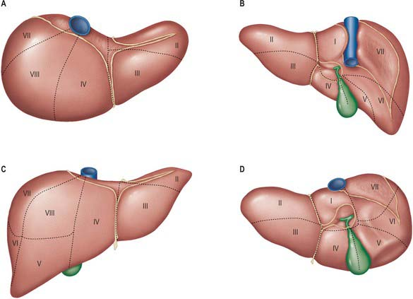
Sectors and segments
- Functional anatomy can be dived in two ways
- Based on hepatic vein and portal (this is done in Last’s Anatomy)
- Left lateral - II
- Left medial III and IV +/- I
- Right anterior (medial) - V and VIII
- Right posterior (lateral) VI and VII
- As done by Couinaud (based on the Hepatic artery and Biliary tree anatomy)
- The difference being II and II are included as the left lateral segment
- Based on hepatic vein and portal (this is done in Last’s Anatomy)
The Brisbane 2000 Terminology of Liver Anatomy and Resections
- Met and created accepted terminology - essentially accepted Couinaud’s nomenclature
- “The watersheds of the hepatic artery, bile duct and portal vein are identical except for the second-order division of these structures on the left side of the liver. The arterial and biliary watersheds divide the left side of the liver through the umbilical fissure. All anatomists, including Couinaud and Healey, agree on this point. The portal vein watershed divides the left side of the liver through the plane between segments 2 and 3 (Couinaud). Since both modes of division of the left side of the liver are currently accepted as anatomically correct, two terminologies are required for the second-order division of the liver. It should be noted that, on the right side of the liver, the second-order watersheds of the hepatic artery, bile duct and portal vein are identical. Therefore, on the left side of the liver the two terminologies describe anatomically distinct structures, whereas on the right side they describe the same anatomical structures.”
- Portal vein and hepatic vein divide the liver through the left hepatic fissure (segment 2/3)
- Hepatic artery and bile duct divide the liver through the umbiliacal fissure (segment 4/2,3)
- Summary:
- The liver can be divided functionally into left and right and further subdivided into sectors/sections and segments based on the Watersheds of the hepatic artery, bile duct and portal vein. The liver is divided into right and left hemi-livers through the main hepatic fissure which contains the middle hepatic vein. The right and left hemi-livers are independent in terms of portal and arterial vascularization and biliary drainage. The second order division on the right is straightforward as the watershed areas for the hepatic artery, bile duct and portal vein are the same however on the left the watersheds differ for the portal vein compared to the hepatic artery and bile duct.
- In 2000 The Scientific Committee of the IHPBA published a consensus paper caller the Brisbane 2000 Terminology of Liver Anatomy and Resection. This paper outlined the following:
- The liver is divided based on watersheds of the hepatic artery and bile duct
- Right:
- Divided by the right intersection plane in anterior section and posterior section
- The watersheds of the hepatic artery, bile duct and portal vein are identical and therefore divide the liver
- The right hepatic vein runs in the right intersection plane
- Left
- The arterial and biliary watersheds divide the left side of the liver through the umbilical fissure into the left medial section and left lateral section.
- The portal vein watershed divides the left side of the liver through the plane between segments 2 and 3.
- The left hepatic vein runs in between segment 2/3 for most of its course then joins the left intersectional plane and receives the umbilical vein before uniting with the middle hepatic vein and forming a common channel to enter the IVC.
- Alternatively the left liver can be divided based on the portal vein watershed (and left hepatic vein) into left lateral (segment 2) and left medial (segment 3, 4a and 4b).
- The Brisbane 2000 Terminology name these as “sectors” to delineate them from “sections” and include this terminology as an addendum.
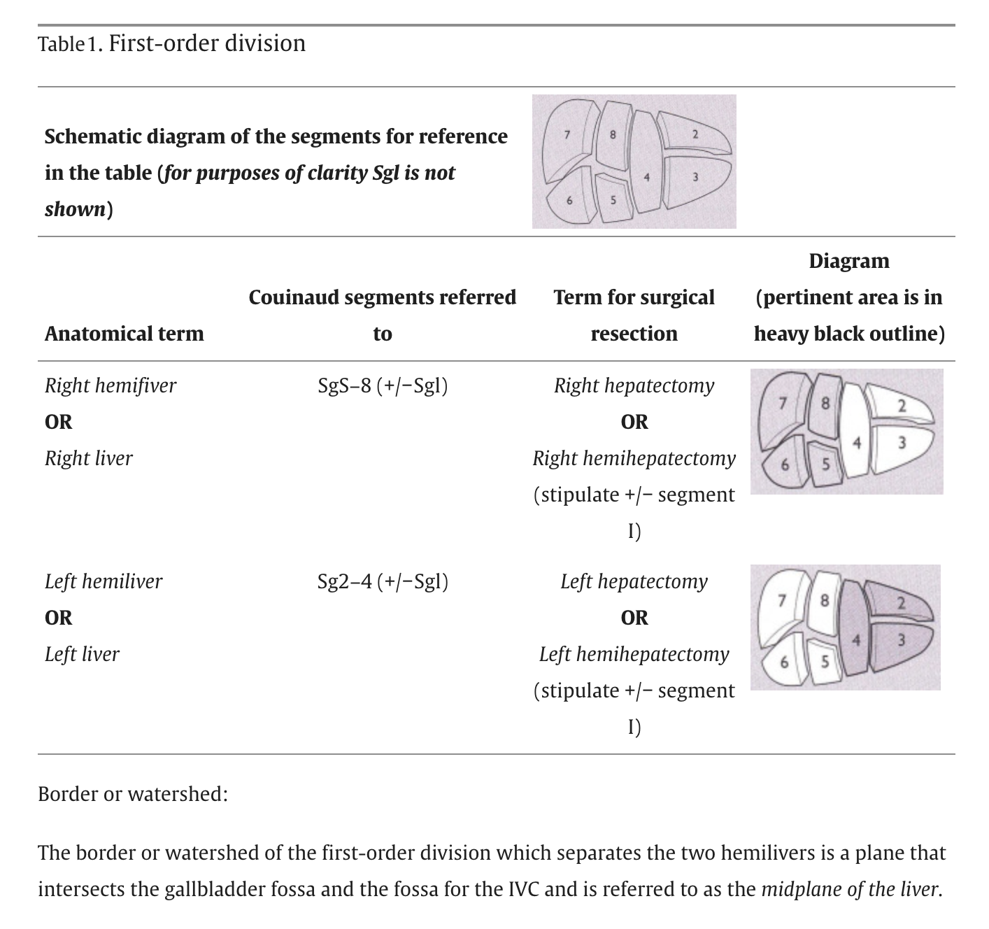
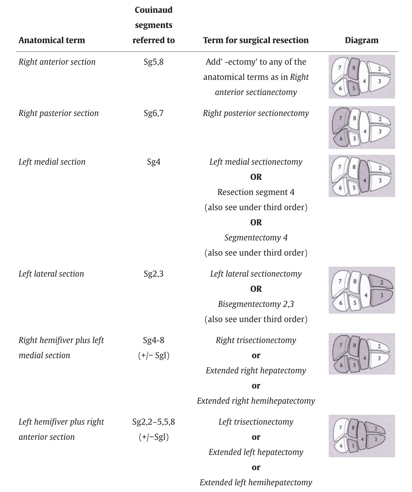
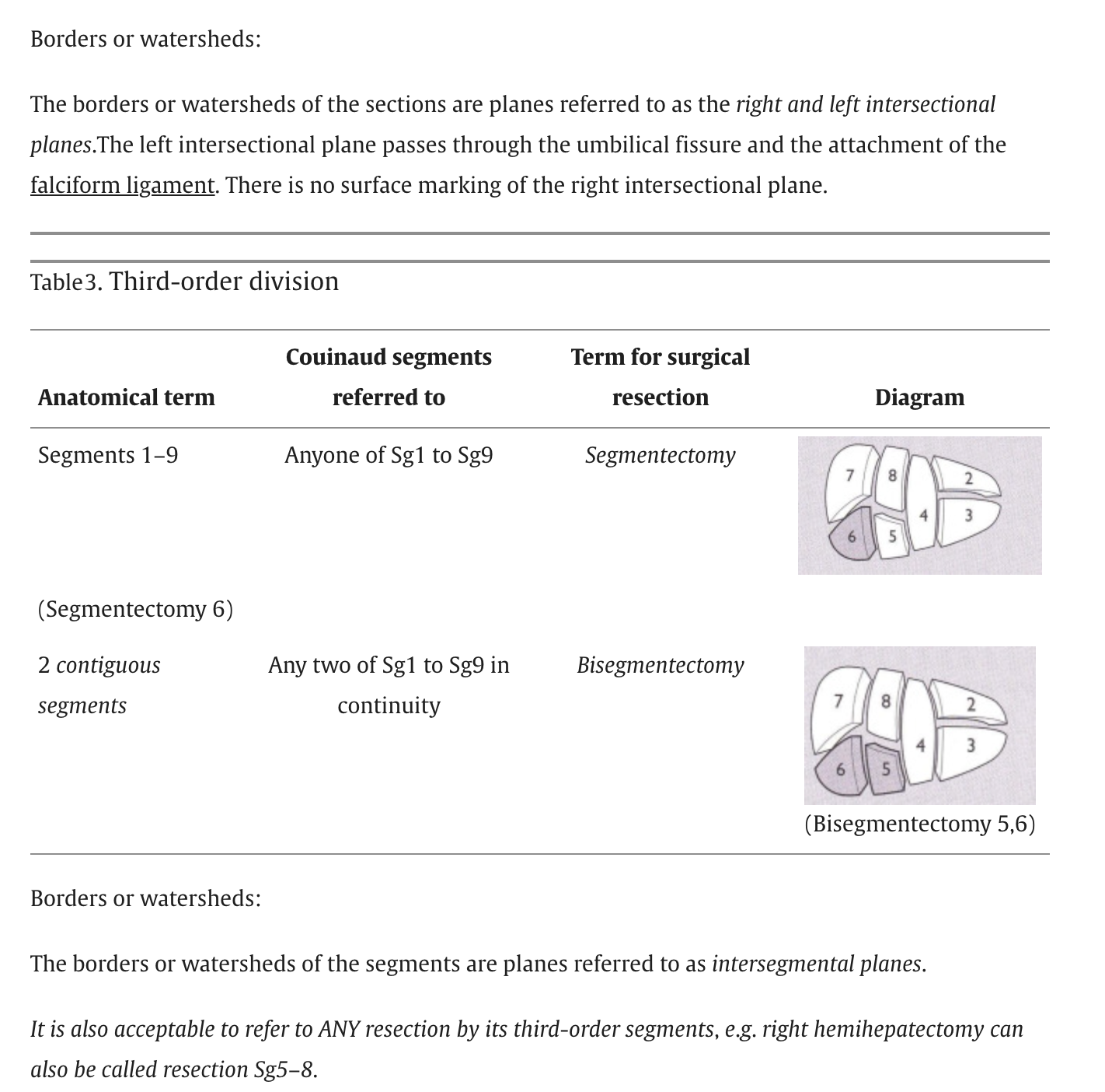
- The Brisbane 2000 Terminology name these as “sectors” to delineate them from “sections” and include this terminology as an addendum.
- In 2000 The Scientific Committee of the IHPBA published a consensus paper caller the Brisbane 2000 Terminology of Liver Anatomy and Resection. This paper outlined the following:
- The liver can be divided functionally into left and right and further subdivided into sectors/sections and segments based on the Watersheds of the hepatic artery, bile duct and portal vein. The liver is divided into right and left hemi-livers through the main hepatic fissure which contains the middle hepatic vein. The right and left hemi-livers are independent in terms of portal and arterial vascularization and biliary drainage. The second order division on the right is straightforward as the watershed areas for the hepatic artery, bile duct and portal vein are the same however on the left the watersheds differ for the portal vein compared to the hepatic artery and bile duct.
And the addendum with the alternative division is below
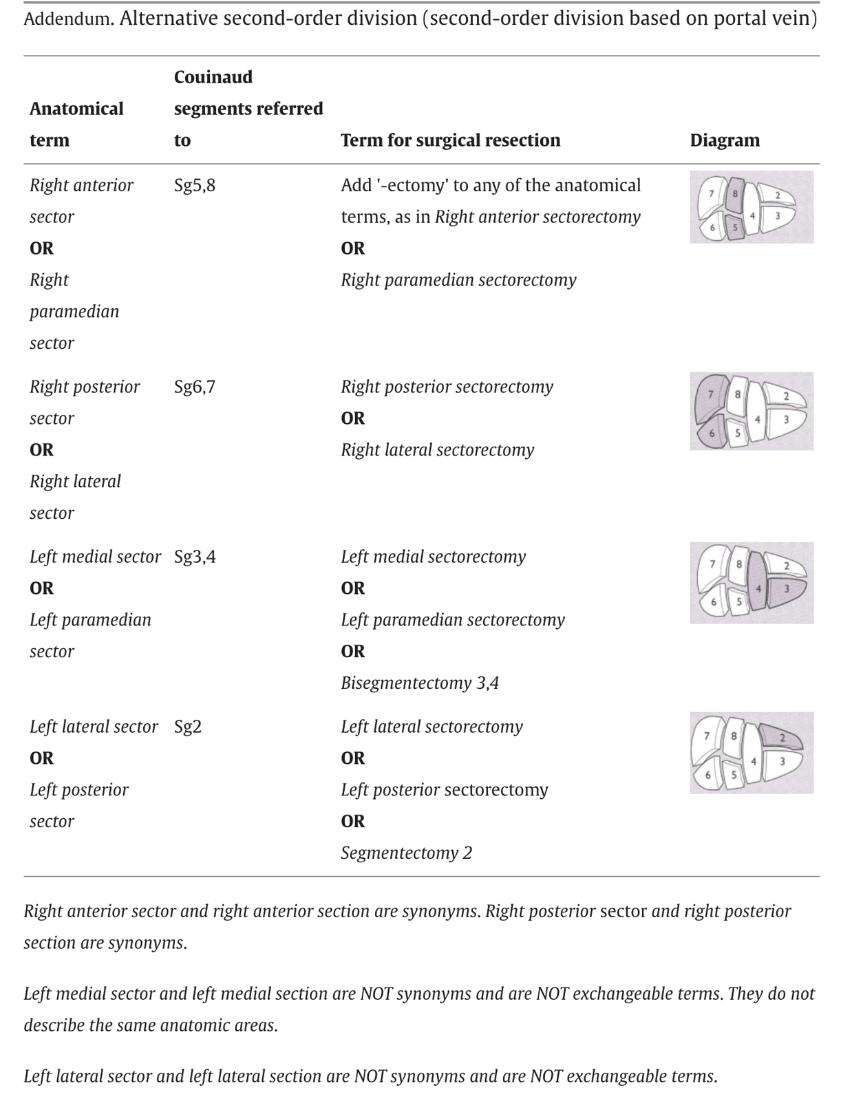
First order division (hemiliver)
- Right hemiliver
- Left hemiliver
Second order division (section)
- Left lateral section - II and III
- Left medial section - III and IV
- Right anterior section - V and VIII
- Right posterior section - VI and VII
Third order division (segments)
Segment I (caudate lobe)
- Posterior (dorsal) to segment IV with its left half directly posterior to segments II and II and its medial half surrounded by major vascular branches.
- Receives vessels independently from the left and right portal veins and hepatic arteries
- Drains independently into the inferior vena cava by multiple small branches
Segment II
- Lies postero-lateral to the left fissure.
- Drains into the left hepatic vein.
Segment III
- Between the umbilical fissure and the left fissure
- Drains into the left hepatic vein
Segment IV
- Between the umbilical fissure and the main fissure, anterior to the dorsal fissure and segment I.
- Main venous drainage segment is into the middle hepatic vein; the segment can also drain into the left hepatic vein through the vein of the falciform ligament.
Segment V
- inferior segment of the right anterior sector and lies between the middle and the right hepatic veins.
- Venous drainage is into the right and middle hepatic veins.
Segment VI
- Segment VI forms the inferior part of the right posterior sector
- Posterior to the right portal fissure.
- Venous drainage is normally into the right hepatic vein
Segment VII
- Segment VII forms the superior part of the posterior sector and lies behind the right hepatic vein.
- Venous drainage is into the right hepatic vein
Segment VIII
- Segment VIII is the superior part of the right anterior sector. T
- The venous drainage is to the right and middle hepatic veins.
Microscopic structure
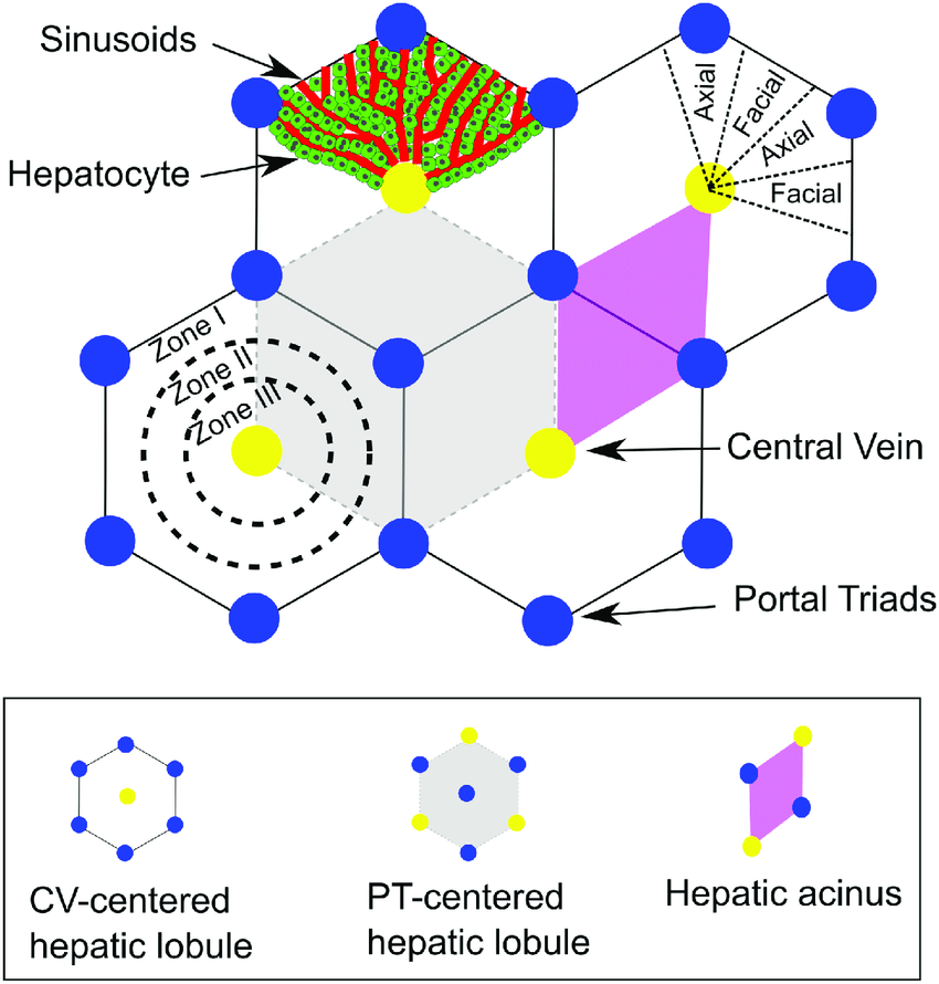
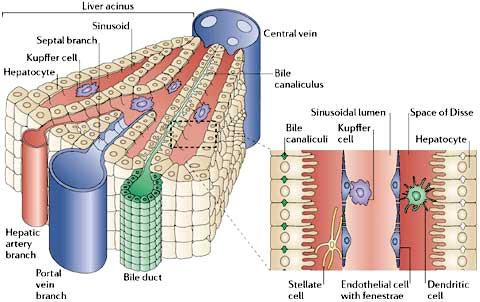
Hepatic Lobules
- Portal triad in the centre and tributaries of hepatic veins at boundary.
- Sinusoids between cords of hepatocytes are lined by endothelial cells.
- Kupffer cells are phagocytes part of endothelial lining.
- Bile canaliculi form meshwork draining into ductulus then ducts.
- Lined by thin capsule
- Lined by thin capsule
Relationships
Liver
- Visceral surface related to oesophagus, stomach, duodenum, hepatic flexure of colon, right kidney
Bare area
- Only area of the liver without peritoneal covering
- Attaches directly to the diaphragm
- Lies between the two leaves of the coronary ligament and the triangular ligaments
Caudate lobe
- At the back
- Lies between the IVC and the fissure for the ligamentum venosum
- Connected to the right lobe (to the right of the IVC) by an isthmus of liver substance – ‘caudate process’
- In the lesser sac
- Recives blood supply from both right and left hepatic atery
- Drains into right and left hepatic ducts
- Drain directly into IVC
Arterial supply
- Divisions
- First order division hepatic proper supply right & left
- Second order supply 4 sections
- Third order supply segments
- Caudate lobe is from branches of right & left
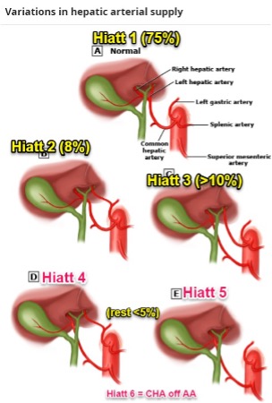
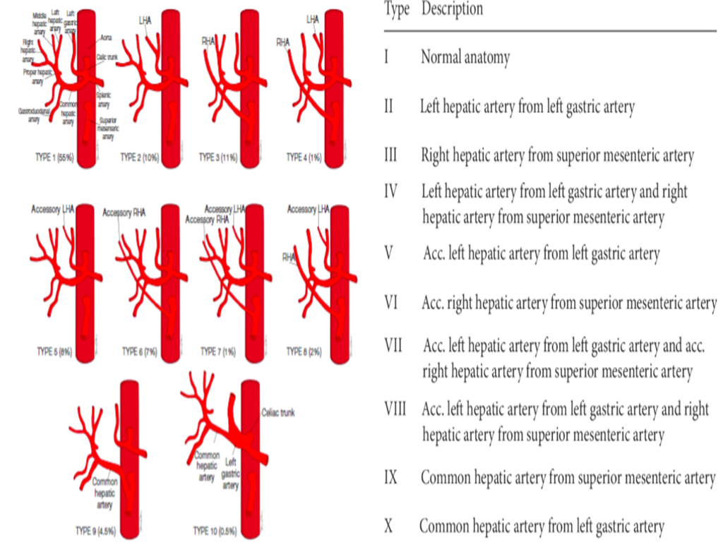
- Anomalies
- 75% normal
- Replaced or accessory right hepatic off SMA
- Runs behind lower end of CBD and courses on right posterior border.
- Replaced or accessory left hepatic artery arises from left gastric
- Courses in lesser omentum in conjunction with vagal branches to liver
- Can also be from coeliac trunk, right hepatic, splenic or SMA
Venous drainage
- 3 main veins to IVC.
- May have small accessory veins joining IVC below main veins, including separate vein from caudate.
- Anastomosis between portal venous channels in liver and azygos system above diaphragm across bare area of liver.
Lymphatic drainage
- 3-4 nodes in porta hepatis.
- Downwards along hepatic artery to pyloric nodes and directly to coeliac nodes.
- Lymphatics from bare area communicate with extraperitoneal lymphatics which perforate diaphragm and drain to nodes in posterior mediastinum.
- Similar communications exist along left triangular and falciform ligaments.
Nerve supply
- Sympathetic: coeliac ganglia, with nerves running with vessels in free edge of lesser omentum.
- Vagus: from hepatic branch of anterior vagal trunk via lesser omentum.
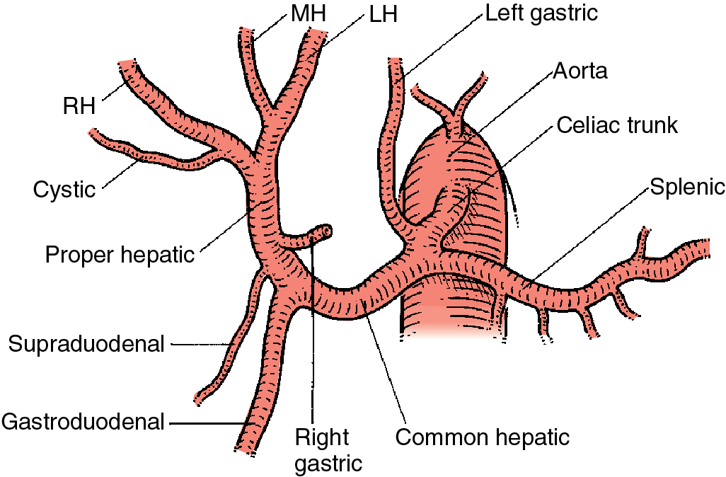
Coeliac artery
Coeliac artery
- Left gastric
- Splenic
- Common hepatic
- Passes over upper baorder of pancreas along the posterior aspect of the less sac
- Turns forward and at the free edge of the less omentum
- Runs in front of the portal vein and to left of the bile duct
- Branchs
- Right gastric artery
- Given off just before the pylorus
- Turns back to run along the less curve
- Right gastric artery
- Gastroduodenal
- Just in front of the portal vein at D1
- Courses behind D1, in front of pancreas in a groove
- Can come off higher
- Commonly vessel involved in a bleeding duodenal ulcer posteriorly - can be ligated
- Terminates as the right gastroepiploic and the superior pancreaoduodenal
- Proper hepatic
- Enters the hepatoduodenal ligament an
- Left side of the common bile duct (CBD)
- Terminates by dividing into the right and left hepatic arteries,
- Right immediately passing behind the common hepatic duct
- But can pass in anterior in 20%
Portal vein
- 70% blood supply
- Most posterior structure in portal triad
- Formed by Splenic vein and SMV behind neck of pancreas
- Divides into left and right portal
- On the left, the PV consists of transverse portion and umbilical portion, with both parts lying on the surface within fissures, terminating in ligamentum teres. The ligamentum venosum (remnant of ductus venosum) divides the two parts.
Variations (uncommon):
- Absence of right portal vein.
- Right posterior and anterior sectoral veins originate independently from main portal vein.
Extra details
Overview Liver
-
Weighs ~ 1500g
-
Receives ~ 1.5L of blood per minute
-
Two surfaces: Diaphragmatic and Visceral
- Diaphragmatic Surface: divided into anterior, superior, posterior and right surfaces
- Anterior Surface: Related to diaphragm, lung, pleura and ribs
- Diaphragmatic Surface: divided into anterior, superior, posterior and right surfaces
-
Superior Surface: Related to heart and pericardium centrally and lung and pleura laterally
-
Right Surface: Extends from ribs 7-11
- Top 1/3: Related to ribs, diaphragm, pleura and lung
- Middle 1/3: Ribs, diaphragm and pleura
- Lower 1/3: Ribs, diaphragm
-
Posterior Surface: Straddles IVC, continuation of Superior and Right surfaces, transitions to visceral surface
-
Visceral Surface:
-
Related, with peritoneum intervening, to the Stomach, Oesophagus, Duodenum, Right Kidney, Right Adrenal Gland, Hepatic Flexure
-
Most of the main vessels and ducts enter/leave at the Porta Hepatis on the visceral surface - Hepatic Veins emerge from the posterior surface
-
Organs may leave impressions
-
Surface slopes upwards and backwards to merge with the posterior surface - Omental Tuberosity - slight bulge to the right of the Gastric Impression - In contact with the Lesser Omentum – separates it from a similar eminence on the Body of the Pancreas
-
Sharp inferior border separates right and anterior surfaces from the visceral surface
-
Liver suspended by the Hepatic Veins and the IVC
- Hepatic veins entirely intra-hepatic
Liver Ligaments
- Peritoneal duplications extending from the Liver
Falciform Ligament
- Connects anterior Liver to Diaphragm, abdominal wall, and Umbilicus
- Contains Ligamentum Teres in inferior edge
- Obliterated Left Umbilical Vein
- Passes superiorly from anterior surface to superior surface
- Splits into Left and Right leaves near the superior surface
- Left Leaf forms Left Triangular Ligament
- Right Leaf (Anterior to IVC) forms Upper layer of Coronary Ligament
- Does not prevent ptosis of the Liver
Coronary Ligaments
- Connect Liver to Diaphragm
- Upper layer and lower layers meet laterally/medially to make Left and Right Triangular Ligaments
- Left Triangular Ligament:
- Needs to be divided before the Left Lobe of the Liver can be retracted to the right to expose the abdominal Oesophagus and upper Stomach
Lesser Omentum
-
NB: Strength of liver attachment mostly comes from Hepatic Veins, not ligaments
-
Posterior and visceral surface contain an H-shaped pattern of structures
-
Centrally/Cross Limb: Porta Hepatis
-
Right limb of H – Inferior Vena Cava (on posterior surface) and Gallbladder (on inferior surface)
-
Left limb of H – Continuity of the fissures for the Ligamentum Venosum and Ligamentum Teres
- NB: Fissure for the Ligamentum Teres extends to the Porta Hepatis
- But NOT to the Right End of the Porta Hepatis
- NB: Fissure for the Ligamentum Teres extends to the Porta Hepatis
-
Caudate Lobe Superiorly
-
Quadrate Lobe Inferiorly
-
Right of the IVC: Triangular Bare Area. Boundaries are:
- Base: IVC
-
Sides: Superior and inferior layers of the Coronary Ligament
-
Right Triangular Ligament: where these two layers meet
- Bare area in contact with the Diaphragm and the Right Adrenal Gland
- NB: The Liver is still separated from the Right Adrenal Gland by the Renal Fascia
- Bare area in contact with the Diaphragm and the Right Adrenal Gland
-
From the inferior layer, the line of peritoneal attachment passes in front of the IVC and then up along its left side to the summit of the liver
-
At the summit, it meets with the right leaf of the Falciform Ligament
-
The two peritoneal layers are attached to the bottom of a deep groove that runs to the left from the IVC and lodges the Ligamentum Venosum
-
This fissure and ligament turn at a right angle and run downwards on the posterior surface to the left of the Porta Hepatis
-
Caudate Lobe – the rectangular area outlined by the above
- Two layers of Lesser Omentum attached to the bottom of the fissure along the left margin of the Caudate Lobe
- Due to the depth of the fissure passing obliquely into the liver substance, the Caudate Lobe is partly separated from the rest of the liver
- Caudate Lobe’s anterior surface forms the posterior wall of the fissure
- Caudate lobe thus lies behind the right part of the Lesser Omentum and is a content of the Lesser Sac
- A narrow Caudate Process extends to the right between the Porta Hepatis and the IVC
-
At the Porta Hepatis, the 2 layers of the Lesser Omentum deviate to the right to enclose the Right and Left Hepatic Ducts, the Right and Left branches of the Hepatic Artery and the Portal Vein
- Ducts, Arteries, Vein (Anterior to Posterior – DAV)
- Ducts in front and thus more accessible to surgery
- Porta also contains several lymph nodes and nerves
-
Gallbladder lies on visceral surface
- The Cystic Duct is close to the right end of the Porta Hepatis
- The Quadrate Lobe lies between the Gallbladder and the fissure for the Ligamentum Teres
Surface Marking
- Upper margin of Liver: Level with the Xiphisternal Joint
- On the left: Reaches the 5th Intercostal Space 7-8cm from midline
- On the right: Reaches 5th Rib ⇒ Curves down to right border ⇒ Extends from ribs 7 to 11 in the Midaxillary Line
- Inferior border: Along a line joining the right lower and upper left extremities
- Right 11th Rib to Left 5th Intercostal Space
- Some of it lies level with the right Costal Margin
- Centrally, some of it crosses behind the upper abdominal wall between the Costal Margins and ~ a hand’s breadth below the Xiphisternal Joint
Liver Lobes
-
Anatomical division: Divided by the Falciform Ligament anteriorly and the fissures for the Ligamentum Teres and Ligamentum Venosum on the visceral surface
-
Functional division: Divided by the oblique plane that runs through the centre of the Gallbladder bed and the groove for the IVC
- a.k.a. Cantiles Line
- Middle Hepatic Vein lies in this plane
Liver Segments
-
Four main hepatic sectors/sections
-
Right, Middle and Left Hepatic Veins divide the liver in the vertical plane and the Portal Vein bifurcation divides the liver in the horizontal plane
-
NB: 6 segments lie on right side of Falciform Ligament and 2 on left side [SEAM Module]
- Left lateral
- Lies left of the Falciform Ligament and the groove for the Ligamentum Teres and Ligamentum Venosum
- Contains Segment II posteriorly and Segment III anteriorly
- Left Hepatic Vein lies between them
- Left medial
- Between the above line and the planes of the Gallbladder and IVC
- Right medial
- No external marking dividing right medial and lateral sectors
- Oblique division
- Right Hepatic Vein lies in this plane
- Contains Segment V and Segment VIII
- Right lateral
- Contains Segment VI and Segment VII
- Left lateral
-
Four sectors further subdivided into 8 segments
Segment I: Caudate Lobe
- Despite lying on the left of the plane between the two functional lobes, it is an autonomous segment
- Lies between IVC and fissure for Ligamentum Venosum
- Receives blood from the Right and Left Branches of the Hepatic Artery and Portal Vein
- Drains bile into the Right and Left Hepatic Ducts
- Has independent venous drainage into the IVC
- In Budd-Chiari Syndrome (i.e. Thrombosis of the Hepatic Vein): Compensatory hypertrophy of the Caudate Lobe occurs ⇒ Lobe enlargement
- Subdivided into Right and Left and the Caudate Process
- Only part of the posterior liver surface in the Lesser Sac
Segment IV
- Divided into Segment IVa (superior) and Segment IVb (inferior)
- Segment IVb corresponds to the Quadrate Lobe
-
Lies between Gallbladder Fossa and Fissure for Ligamentum Teres
-
NB: Right Hepatic Artery does NOT supply the Quadrate Lobe
-
Bounded by the fissure for the Ligamentum Teres NB: It is NOT bounded by the fissure for the Ligamentum Venosum
-
NB: Is NOT in close contact with the Right Kidney
-
Blood Supply
- Two sources – Hepatic Artery and Portal Vein
Hepatic Artery
-
Divides into Right and Left branches in the Porta Hepatis
- Divides in a Y-shaped manner
-
Right Branch normally passes behind the Common Hepatic Duct
- Divides in the Liver into Medial and Lateral Sectoral Branches
-
Left Branch also divides in the liver into Medial and Lateral Sectoral Branches
-
Sometimes, Common Hepatic Artery arises from the SMA or Aorta (instead of the Coeliac Trunk)
- In such cases, it usually runs behind the Portal Vein
-
Right Hepatic Artery may arise from the SMA (15%)
-
Left Hepatic Artery may arise from the Left Gastric Artery (20%) as aberrant or accessory arteries
Portal Vein
-
Approximately 8cm long
-
Upward continuation of the Superior Mesenteric Vein
-
Name changed to Portal Vein after it receives the Splenic Vein behind the Neck of the Pancreas at the level of L1
-
Lies anterior to the Inferior Vena Cava as it lies behind the Pancreas and the 1st part of the Duodenum
-
Loses contact with the Inferior Vena Cava by entering between the two layers of the Lesser Omentum
-
Runs almost vertically in the free edge (where the Lesser Omentum forms the anterior boundary of the Epiploic Foramen)
-
Lies behind the Bile Duct and the Hepatic Artery
-
Divides in the Porta Hepatis into Right and Left branches which then give sectoral branches like the arteries
- Division happens at a higher level than the division of the Hepatic Artery
- Divides via a T-junction
-
Also receives tributaries from:
- Left Gastric Vein
- Right Gastric Vein
- Cystic Veins – (when present) join the Right Branch of Portal Vein
- Oesophageal Veins
- Paraumbilical Veins – (Running with the Ligamentum Teres) join the Left Branch of Portal Vein
- Superior Pancreaticoduodenal Veins
- Remains of the embryonic umbilical veins
-
Portal Vein and its Tributaries are Valveless
-
NB: Does NOT have a valve at its commencement
-
Five sites of Portal-Systemic Anastomosis are:
- Lower end of the Oesophagus
- Upper end of the Anal Canal
- Bare Area of the Liver
- Peri-umbilical Region
- Retroperitoneal Region
-
In Portal Hypertension – 80% of Portal blood may be shunted into collateral channels so that only 20% reaches the Liver
- However, the opening up of the collaterals does not decrease the level of hypertension
-
No communication exists between Right and Left halves of the Liver
- Even within each half, the arteries are end arteries
-
In presence of disease, there are often enough anastomoses with Phrenic vessels (e.g. across the Bare Area) to provide collateral circulation sufficient to allow ligation of the Hepatic Artery
Venous Drainage
- Venous return involves mixing of Right and Left halves of the Liver
- Three main Hepatic Veins drain into the IVC
- Middle Hepatic Vein – receives blood from both halves
- Middle Hepatic Vein frequently joins the Left Hepatic Vein near the IVC
- All veins have no extra-hepatic course
- Hepatic Veins drain into IVC just below the Central Tendon of the Diaphragm
- There is some anastomosis between Portal Venous Channels in the Liver and the Azygos System of veins above the Diaphragm across the Bare Area of the Liver
- Liver is supported by the Hepatic Veins
Lymph Drainage
- Drain into 3 or 4 nodes that lie in the Porta Hepatis (Hepatic Nodes)
- Hepatic Nodes also receive lymphatics from the Gallbladder
- Hepatic Nodes drain downwards alongside the Hepatic Artery to Pyloric Nodes as well as directly to Coeliac Nodes
- Lymphatics from the bare area of the liver communicate with extra-peritoneal lymphatics which perforate the diaphragm and drain to nodes in the Posterior Mediastinum
- Similar communications exist along the left triangular and falciform ligaments from the adjacent liver surfaces
Nerve Supply
- Derived from Sympathetic and Vagus fibres
- Sympathetic fibres via the Coeliac Ganglia
- Nerves run with the vessels in the free edge of the Lesser Omentum and enter the Porta Hepatis
- Vagal fibres via the Hepatic Branch of the Anterior Vagal Trunk
- Reach the Porta Hepatis via the Lesser Omentum also
Structure Hepatic Lobule
-
Hexagonal shape
-
Central vein
-
Plates/cords of Hepatocytes separated by vascular spaces or Sinusoids radiating from the vein to the periphery of the lobule
-
Portal triads located at the corners – i.e. small branches of the Hepatic Artery, Portal Vein and Bile Ductules
-
In humans, such a classical lobule not usually present
-
Arrangement is actually a polygonal territory with a portal triad at the centre and tributaries of the Hepatic Veins at the boundary
-
Is termed a Portal Lobule – equivalent to sections of at least 3 ‘Classic’ Lobules
-
-
Sinusoids lined by Endothelial Cells with frequent intercellular spaces and fenestrations
- Allows blood to enter the Peri-Sinusoidal spaces between the Endothelium and Hepatocytes
- Allows exchange of materials between plasma and Hepatocytes
-
Kupffer Cells – lines the Endothelium and capable of phagocytic activity
Development
- Stems from endodermal cells
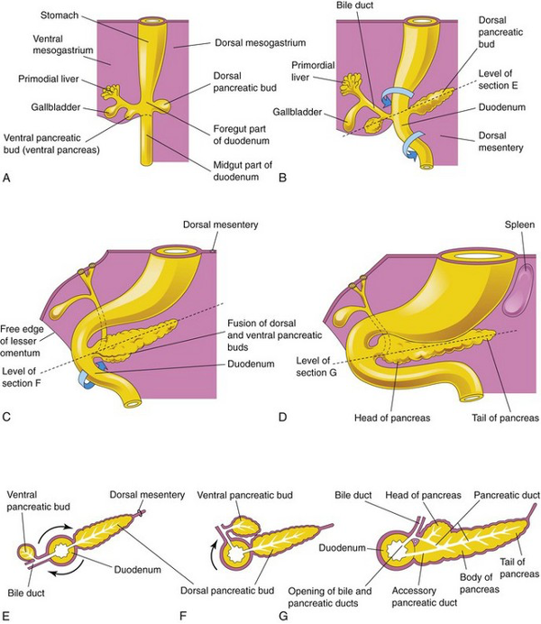
- Starts as hepatic diverticulum (Liver Bud) around the 4th week of development
- Develops from proliferation of cells from the blind ends of a Y-shaped diverticulum which grows from the Foregut into the Septum Transversum (structure that divides the heart from the abdominal cavity – later contributes to diaphragm formation)
- Caudal part of Septum Transversum becomes the Ventral Mesogastrium which the liver grows into
- Connection between hepatic diverticulum and future duodenum narrows to form bile duct
- Outpouching of the bile duct forms into gallbladder and cystic duct
- Diverticulum differentiates into primordium of liver or gallbladder
- Liver develops into hepatic cords that anastomose around spaces lined by endothelium (intermingle with Vitelline and Umbilical Veins) – forming primordium of hepatic sinusoids
- Portal vein is the central vessel in which the hepatic cords form around
- Original diverticulum from the Endoderm of the Foregut becomes the Bile Duct
- The Y-shaped bifurcation produces the Right and Left Hepatic Ducts
- Blind diverticulum from the Bile Duct becomes the Cystic Duct and Gallbladder
- Hepatocytes create bile at around 12th week
Mesoderm of Septum Transversum
- Gives off haematopoietic cells, Kuppfer cells and connective tissue
- Connects liver to ventral abdominal wall and foregut
- Stretches into thin membranes to become Falciform Ligament and Lesser Omentum
- Mesoderm on surface of liver becomes visceral peritoneum
- Except superiorly, where contact is kept between liver and mesoderm (future diaphragm)
- Forms bare area
Role in Foetal Circulation
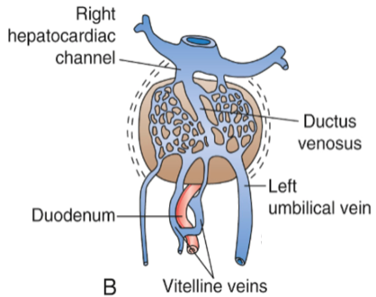
- Names of Structures
- Vitelline Veins
- Initially carry blood from Yolk Sac to Sinus Venosus
- Eventually carry blood from gut to sinusoids
- Turn into Portal Vein, SMV and Splenic Vein
- Sinus Venosus
- Empties into foetal heart
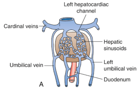
- Becomes hepatocardiac channel
- Eventually becomes Hepatic Veins and retro-hepatic IVC
- Umbilical Veins
- Carry oxygenated blood from mother to foetus
- Initially paired
- Initially drain straight into Sinus Venosus
- At 5 weeks begin to drain into hepatic sinusoids
- Eventually right is obliterated, and left drains straight into the hepatocardiac channel
- Bypasses sinusoids, through the Ductus Venosus
- Left obliterated in adulthood, becomes Ligamentum Teres
- Ductus Venosus
- Connection of left umbilical vein into hepatocardiac channel
- Creates a bypass for oxygenated blood from mother to bypass liver sinusoids
- Ductus Venosus obliterated in adulthood, becomes Ligamentum Venosum
- Vitelline Veins
Timeline: 5 weeks
- Hepatic sinusoids have developed
- Vitelline (from gut) and umbilical veins (from mother) are beginning to drain into them
- Although there are channels that bypass these sinusoids
Second month
- Vitelline Veins (from gut) drain directly into the hepatic sinusoids
- Ductus Venosus has formed and accepts oxygenated blood from the Left Umbilical Vein
- Bypasses the hepatic sinusoids
- Directly enters the hepatocardiac channel
- Right umbilical vein begins to shrink
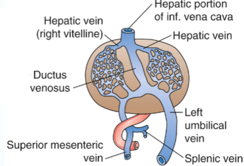
Third month
- Vitelline Veins have formed into the Portal system
- Splenic Vein
- SMV
- Portal Vein
- Right Umbilical Vein disappeared
- Left Umbilical Vein (future Ligamentum Teres) drains into the Ductus Venosus
- Then into Sinus Venosus/hepatocardiac channel
- Bypassing the hepatic sinusoids
- Note development of IVC + hepatic veins
Biopsy, Resection and Transplantation Liver Biopsy
- Usually done through the right 8th or 9th intercostal space in the midaxillary line
- The needle path is below level of lung but traverses the Costodiaphragmatic Recess of the Pleura before going through the diaphragm and crossing the peritoneal cavity
- Needle must not penetrate > 6cm from the skin to avoid entering IVC
- Misplaced needle could damage: Kidney, Colon, or Pancreas
Liver Resection
- Right (Hemi)Hepatectomy
- Dividing liver tissue along a line from the left of the Gallbladder to the right edge of the IVC
- Segments V – VIII and gallbladder removed
- Middle and Left Hepatic Veins preserved
- Left (Hemi) Hepatectomy
- Segments II – IV and most of the Caudate Lobe (I) are removed
- Gallbladder may be removed or left intact
- Line of resection at the back is level with the left edge of the IVC
- Right and (usually) Left Hepatic Veins preserved
- If more extensive, Left Hepatectomy is combined with removal of Segments V and VIII or Right Hepatectomy is combined with removal of segment IV
Liver Transplantation
- Patient’s liver is removed usually with the attached segment of the IVC
- Utilizes a venovenous bypass between the Portal Vein and the Left Femoral Vein to the Left Axillary Vein
- Suprahepatic IVC of the donor liver is sutured to the patient’s followed by the infrahepatic caval anastomosis and then the portal vein anastomosis
- Venous clamps released in the same order and venovenous bypass interrupted
- Hepatic Artery anastomosis is made
- Biliary continuity established by end-to-end CBD anastomosis or by anastomosis donor Bile Duct to recipient’s Jejunum