Section: Hepatobiliary Sub-section: Pancreas
Pancreas embryology
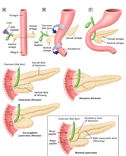
- Two separate buds, outgrowths of endoderm
- Ventral bud from ventral mesogastrium (with outgrowth of bile duct) – uncinate process, part of head and main duct.
- Dorsal bud from dorsal mesogastrium – tail, body, neck and part of head and accessory duct.
- Both buds rotate around, ventral rotates further.
- Anomaly: Pancreatic divisum – failure of two ducts to fuse
- Anomaly: Incomplete rotation results in annular pancreas.
Overview
- Composite gland with exocrine acini and endocrine cells (Islets of Langerhans)
- Length: 15cm
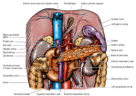
- Head and Tail incline towards the Paravertebral Gutters while Neck and Body curve boldly forward over the Inferior Vena Cava and Aorta in front the L1 vertebra
- Transpyloric Plane (L1)
- Guide to surface marking
- Neck lies on the plane
- Passes across the Head and Body
- Passes below the Tail
Head of Pancreas
- Anterior to:
- Inferior Vena Cava
- Right and Left Renal Veins
- Mainly at the Level of L2
- Bile Duct (terminal part)
- Uncinate Process
- Hook shaped extension of the Pancreatic Head
- Extends upwards and to the left
- Posterior to the Superior Mesenteric Vessels
- Anterior to the Aorta
- Above the 3rd part of the Duodenum
- A tumour in the Uncinate Process can compress the above structures
- Transverse Mesocolon attaches across the anterior surface
- Thus, Head of Pancreas lies in both supracolic and infracolic compartments
Neck of Pancreas
- Anterior to:
- Commencement of the Portal Vein by the union of the Splenic Vein and Superior Mesenteric Vein
- At lower margin – Superior Mesenteric Vein is embraced between the Neck and the Uncinate Process
- Transverse Mesocolon attaches towards the lower border
Body of Pancreas
- Anterior to:
- Left Renal Vein
- Inferior Mesenteric Vein as it joins the Splenic Vein in front of the Left Renal Vein where it lies over the Left Psoas Muscle
- Aorta
- Left Crus of the Diaphragm
- Left Psoas Muscle
- Lower part of the Left Suprarenal Gland
- Reaches hilum of Left Kidney
- Triangular in cross section with posterior, anterosuperior and anteroinferior surfaces separated by superior, anterior and inferior borders
- Superior border
- Crosses Aorta at the origin of the Coeliac Trunk
- Splenic Artery passes to the left along the upper border of the Body and Tail
- Inferior border
- Crosses the origin of the Superior Mesentery Artery
- Superior border
- Omental Tuberosity – slight convexity that projects upwards from the right end of the Superior border; above the lesser curvature of the stomach and touches the left lobe of the Liver with Lesser Omentum intervening.
- Transverse Mesocolon attached to anterior border
- Thus, body lies behind the Lesser Sac where it forms part of the Stomach bed
- NB: Majority of the pancreas lies in the supracolic compartment.
Tail of Pancreas
- Anterior to:
- Left Kidney (NB: Left Renal Hilum)
- Accompanied by the Splenic Artery, Splenic Vein and lymphatics
- Lies within the two layers of the Splenorenal Ligament
- Reaches the Hilum of the Spleen
Vessels Behind the Pancreas
- Splenic Artery
- Splenic Vein
- Inferior Vena Cava
- Renal Veins
- Portal Vein
- Inferior Mesenteric Vein
- Superior Mesenteric Vein
- Aorta
Pancreatic Ducts
Pancreatic Duct (of Wirsung)
- Runs from Tail to Head
- Gradually increases in diameter as it receives tributaries
- Joined at an angle of about 60° by the Bile Duct at the Hepatopancreatic Ampulla
- Drains most of the Pancreas except for the Uncinate Process and Lower part of the Head
Accessory Pancreatic Duct (of Santorini)
- Drains the Uncinate Process and Lower part of the Head
- Opens into the Duodenum at the Minor Duodenal Papilla located 2cm proximal to the Major Papilla
- Two ducts communicate with one another
Blood Supply
- Arterial:
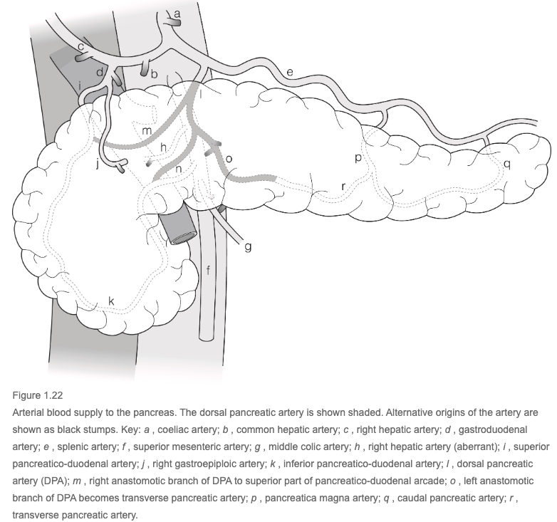
- Two system (neck is watershed)
- Head/uncinate process
- Superior and inferior pancreaticoduodenal arteries
- Body and tail
- Splenic artery
- Dorsal pancreatic artery
- Anastomoses with the pancreatico-duodenal arcade
- Pancreatica magna artery
- Caudal pancreatic artery
- Dorsal pancreatic artery
- Splenic artery
- Head/uncinate process
- Two system (neck is watershed)
Venous Return
Drainage
- Splenic vein
- Superior pancreaticoduodenal veins
- Anterior superior - into Gastrocolic trunk of Henle
- Posterior superior - Into portal vein
- Inferior pancreaticoduodenal veins
- Anterior and posterior are smaller often only draining the uncinate process. They pass horizontally to the left and drain into the first jejunal vein or directly into the superior mesenteric vein at the caudal margin of the pancreas
Lymphatic Drainage
- Follow course of arteries
- To the left of the Neck ⇒ Pancreaticosplenic nodes that accompany Splenic Artery
- From the Head ⇒ Upper part into Coeliac Group and Lower part and Uncinate Process into Superior Mesenteric Group of Pre-aortic Lymph Nodes
Nerve Supply
- Parasympathetic Vagal Fibres
- Stimulates exocrine secretion
- Reaches gland mainly from Posterior Vagal Trunk and Coeliac Plexus
- Sympathetic Vasoconstrictor Impulses
- Derived from spinal cord segments T6-T10 via Splanchnic Nerves and the Coeliac Plexus
- Post-ganglionic fibres running to the gland with its blood vessels
- Pain fibres
- Accompany Sympathetic supply
- Thus, pain radiates to T6-T10 dermatomes
Structure
- Lobulated gland
- Serous Acini producing exocrine secretion
- Cells show cytoplasmic basophilia typical of protein-secreting cells
- Stimulated by Secretin and CCK produced by Neuroendocrine cells of the Small Intestine
- Secrete Trypsin and Lipase
- Centroacinar cells produce bicarbonate
- Endocrine cells
- Islets of Langerhans, aiding in carbohydrate metabolism.
- α-(alpha-)cells - secreting glucagon
- β-(beta-)cells - Insulin
- δ-(delta-)cells - somatostatin
- γ-(gamma-)cells - pancreatic polypeptide
Surgical Approach
- Kocher’s Manoeuvre
- Mobilizing Head of Pancreas and Duodenum by incising peritoneum along the right edge of the 2nd part of the Duodenum and turning the Duodenum medially
- Gives access to posterior surface of Duodenum, Head of Pancreas and to lower part of Bile Duct
- Caution: Inferior Vena Cava, Ureter, and Gonadal Vessels must not be damaged