Overview
- Flat, triangular formation on the front of Piriformis
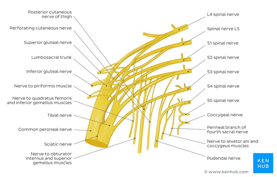
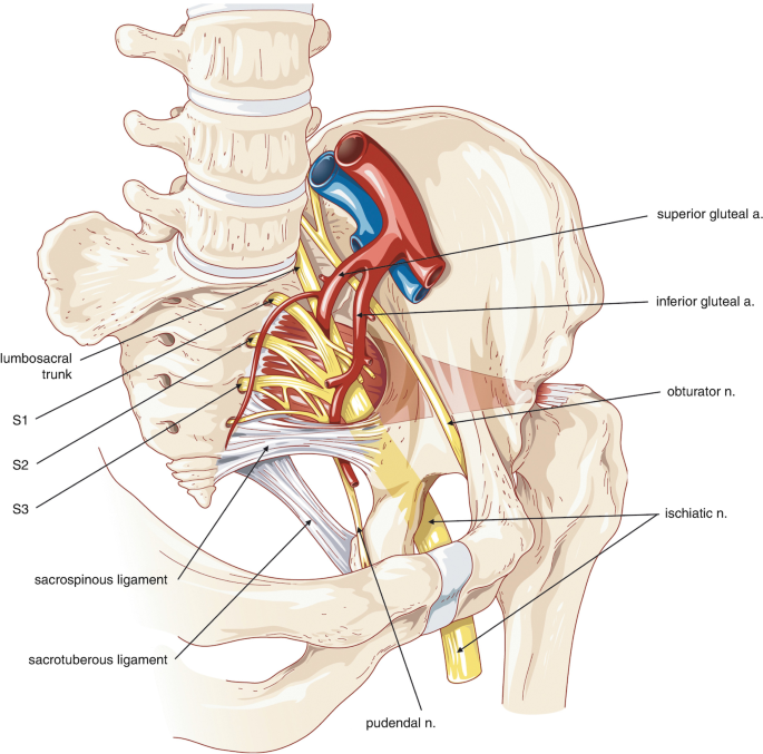
- Formed from Lumbosacral trunk (L4, L5) and upper 4 anterior Sacral Rami (S1, S2, S3, S4)
- Rami divide and unite as they converge to the Greater Sciatic Foramen forming the branches of the Sacral Plexus
Branches
- Within the pelvis
- Nerves to Piriformis (S1, S2)
- Perforating Cutaneous Nerve (S2, S3)
- Posterior Femoral Cutaneous Nerve (S1, S2, S3)
- Pelvic Splanchnic Nerves (S2, S3, S4)
- Pudendal Nerve (S2, S3, S4)
- Perineal Branch of S4
- Muscular Branches of S3 and S4
- After gone through the greater sciatic foramen
- 2 Anterior
- Nerve to Quadratus Femoris (L4, L5, S1)
- Nerve to Obturator Internus (L5, S1, S2)
- 2 Posterior
- Superior Gluteal Nerve (L4, L5, S1)
- Inferior Gluteal Nerve (L5, S1, S2)
- 2 Anterior
- Both anterior and posterior
- Sciatic Nerve
- Common Peroneal/Fibular Nerve (L4, L5, S1, S2)
- Deep Peroneal Nerve
- Superficial Peroneal Nerve
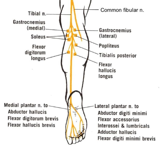
- Tibial Nerve (L4, L5, S1, S2, and S3)
- Common Peroneal/Fibular Nerve (L4, L5, S1, S2)
- Sciatic Nerve
Branches
Branches within the pelvis prior to passing through the greater sciatic foramen (6 all start with P)
- Nerves to Piriformis (S1, S2)
- Perforating Cutaneous Nerve (S2, S3)
- Pierces Sacrotuberous Ligament and fibres of Gluteus Maximus
- Supply small area of skin on lower medial side of buttock
- Posterior Femoral Cutaneous Nerve (S1, S2, S3)
- Runs down below Piriformis on the Sciatic Nerve
- From the lower border of Gluteus Maximus, it runs down in the posterior midline beneath the fascia as far as the lower ends of the Gastrocnemius bellies
- Supplies a strip of deep fascia and skin between anterior and posterior axial lines, from the buttock to the mid-calf, by a series of branches that pierce the deep fascia
- Gluteal Branches wind around Gluteus Maximus to supply skin over the convexity of the buttock
- Perineal Branch winds around the hamstrings and Gracilis origins and pierces the fascia lata at the medial convexity of the upper thigh
- Supplies the posterior two thirds of the Scrotum (labium majus)
- Pelvic Splanchnic Nerves (S2, S3, S4)
- Sacral parasympathetic outflow
- Fibres join Inferior hypogastric plexus
- Motor to the Bladder and to Large Intestine from Splenic Flexure Downwards
- Cause Penile/Clitoral Erection
- Afferent fibres include those of distension and pain from the bladder, lower cervix, lower colon and rectum
- Referred pain may be felt in the region of the Posterior Femoral Cutaneous Nerve
- Pudendal Nerve (S2, S3, S4)
- Runs down and curls around Gluteal Surface of Sacrospinous Ligament to enter Pudendal Canal
- The Nerve of the Perineum
- At the posterior end of the canal, it gives the Inferior Rectal Nerve which arches through the fat of the Ischioanal Fossa
- Supplies External Anal Sphincter, Anal Canal, and Peri-anal skin
- Terminal Branches: Perineal Nerve and Dorsal Nerve of the Penis/Clitoris
- Perineal Nerve:
- Emerges from the anterior end of the canal
- Supplies skin of the posterior 2/3 of the Scrotum and Vulva, mucous membrane of the Urethra and Vagina
- Motor to the Perineal muscles – Ischiocavernosus, Bulbospongiosus, Superficial and Deep Transverse Perineal and Sphincter Urethrae
- Dorsal Nerve of the Penis (Clitoris)
- Runs forwards deep to Perineal Membrane
- Pierces Perineal Membrane just below Pubic Symphysis to supply skin of the Penis (Clitoris)
- Perineal Branch of S4
- Pass between Levator Ani and Coccygeus to supply Puborectalis, Pubourethralis/Puboprostaticus, and Pubovaginalis parts of Levator Ani from below and the skin over the Ischioanal Fossa
- Muscular Branches of S3 and S4
- Supply Levator Ani and Coccygeus on their upper/pelvic surfaces
After gone through the greater sciatic foramen 2 Anterior
- Nerve to Quadratus Femoris (L4, L5, S1)
- Lies on Ischium deep to Sciatic Nerve and runs down deep to Obturator Internus to sink into the deep surface of Quadratus Femoris
- Also supplies Inferior Gemellus and Hip Joint
- Nerve to Obturator Internus (L5, S1, S2)
- Passes below Piriformis
- Curls around base of Ischial Spine
- Also supplies Superior Gemellus
2 Posterior
- Superior Gluteal Nerve (L4, L5, S1)
- Passes back through Greater Sciatic Notch above Piriformis
- Runs in the plane between Gluteus Medius and Gluteus Minimis
- Supplies both Gluteus Medius and Minimis and ends in the Tensor Fascia Lata
- Inferior Gluteal Nerve (L5, S1, S2)
- Passes back below Piriformis
- Sinks into deep surface of Gluteus Maximus
Both anterior and posterior
- Sciatic Nerve
- Largest branch of Sacral Plexus
- Formed at lower margin of Piriformis by union of its Tibial and Common Peroneal parts
- Passes into buttock lying on posterior surface of Ischium
- Superficial to nerve to Quadratus Femoris and deep to Posterior Femoral Cutaneous Nerve
- Passes vertically downwards into the hamstring compartment midway between the Greater Trochanter and the Ischial Tuberosity
- Lies on Obturator Internus and Gemelli, Quadratus Femoris and then on Adductor Magnus
- Overlaid by the Long Head of Biceps
- Main trunk supplies all Hamstrings and Ischial fibres of Adductor Magnus
- Then divides (at the upper angle of the Popliteal Fossa) into the Tibial and Common Peroneal Nerves
- Branches to the Long Head of Biceps, the two Semi- muscles and Adductor Magnus are from the Tibial part and the branch to the Short Head of Biceps comes from the Common Peroneal Part
- Common peroneal nerve (L4, L5, S1, S2)
- Supplies extensor and peroneal compartments of the leg and dorsum of foot
- Enters apex of Popliteal Fossa
- Runs medial to the Biceps tendon
- Crosses Plantaris, Lateral Head of Gastrocnemius and curves around neck of the Fibula through Peroneus Longus
- Divides into two terminal branches – Deep and Superficial Peroneal (Fibular) Nerves
- Most commonly injured nerve of the lower limb
- Common Peroneal Nerve itself supplies no muscles
- Three articular branches (Upper and Lower Lateral and Recurrent Genicular Nerves) supply knee joint
- Two cutaneous Nerves – Sural Communicating Nerve and the Lateral Cutaneous Nerve of the Calf
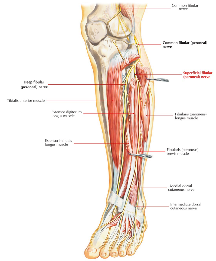
- Deep Peroneal nerve
- Formed in the substance of Peroneus Longus
- Spirals down over the Fibula deep to Extensor Digitorum Longus
- Reaches the Interosseous membrane
- Runs down lateral to the vessels
- Crosses the lower end of the Tibia and Dorsum of Foot
- Ends by supplying skin of the First Interdigital Cleft
- Supplies: Extensor Digitorum Longus, Tibialis Anterior, Extensor Hallucis Longus, and Peroneus Tertius and Extensor Digitorum Brevis
- Superficial Peroneal nerve
- Formed in the substance of Peroneus Longus
- Emerges from the anterior border 1/3 of the way down the leg
- Supplies Peroneus Longus and Brevis
- Perforates the fascia to supply the skin over the Peronei and Extensor muscles in lower 1/3 of the leg
- Above the ankle, it divides into a medial and lateral branch which supply the skin and deep fascia on the Dorsum of the foot
- Medial branch supplies the medial side of the big toe and the 2nd interdigital cleft
- Lateral branch supplies the 3rd and 4th clefts
- Tibial nerve (L4, L5, S1, S2, and S3)
- Supplies the calf and sole of foot around to the toenails
- Enters Apex of Popliteal Fossa
- Passes vertically down deep to the Heads of Gastrocnemius behind the knee joint
- Across the Popliteus muscle
- Runs beneath the Fibrous Arch in Soleus
- Branches in Popliteal Fossa:
- 3 x Genicular Nerves
- Branches to muscles: Plantaris, both heads of Gastrocnemius, Soleus and Popliteus
- Sural Nerve
- Lies in groove between the two heads of Gastrocnemius and pierces deep fascia halfway down the legs
- Runs alongside the Small Saphenous Vein behind the Lateral Malleolus
- Ends on the lateral side of the little toe
- Runs down with the Posterior Tibial Vessels deep to Soleus
- Neurovascular bundle lie in the groove between Flexor Hallucis Longus and Flexor Digitorum Longus
- Divides into its terminal Medial and Lateral Plantar branches behind the Medial Malleolus, beneath the Flexor Retinaculum
- Branches in the Calf:
- 4 x muscular branches – Soleus, Tibialis Posterior, Flexor Hallucis Longus, Flexor Digitorum Longus
- Medial Calcanean branches pierce the Flexor Retinaculum and supply the weight-bearing skin of the heel
- Medial and Lateral Plantar Nerves
- Medial Plantar: Medial part of sole, plantar surface of medial 3 ½ digits, Flexor Digitorum Brevis, Abductor Hallucis, Flexor Hallucis Brevis, and 1st lumbrical
- Lateral Plantar: Supplies the rest of the sole and other small foot muscles