Section: Skin and soft tissue Sub-section: Benign skin and soft tissue Curriculum: Curriculum, page 52
Definition
- Groups of melanocytes leading to a pigmented skin lesion
- Abnormal arrangement of normal cells
- Natural History
- Proliferation of melanocytes at dermal-epidermal junction
- Melanocytes drop into dermis = Naevus
- Then junctional activity cesses, leaving only dermal elements
- These then can disappear due to fibrosis in dermis
Incidence
- Occur from childhood
- Very common
- More common in fair-skinned people
Classification
Congenital Melanocytic Naevi
- Classified according to size
- Café au lait spots
- Speckled lentiginous naevus
- Naevus of Ota
- Mongolian spot
- Small congenitalnaevus
- Medium congenital naevus
- Giant naevus
- Hairy Congenital Naevi
- Definition
- Pigmented naevi present at birth
- Nevus cells often involve lower dermis (amongst collagen) & even fat or muscle
- Hamartoma
- Superficial part similar to compound nevus
- Deep part nests in sheaths of hair follicles, association with sebaceous glands & ducts
- New York Uni Registry Classification
- Based on projected size into adulthood
- Small ≤ 1.5cm in greatest diameter
- Medium: 1.5-19.9cm
- Small & medium – small risk of melanoma – mostly in adult life
- Large / Giant: ≥ 20cm
- Assoc with increased melanoma risk & also associated with neurocutaneous melanosis (e.g. CNS melanoma)
- Management
-
20cm – excise as infant/child
- < 10cm can observe
-
Dermoscopic classification
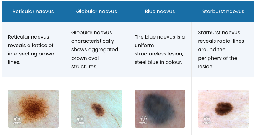
- Dermatoscopy has given rise to a new classification based on the pigment patterns of melanocytic naevi. Examples include:
- Reticular naevus
- Globular naevus
- Blue Naevi
- Starburst naevus
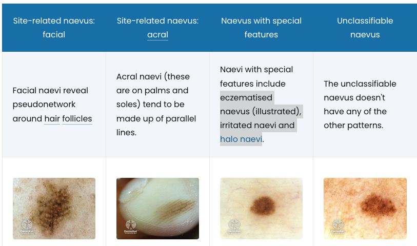
- Facial naevus
- Acral naevus
- Naevus with special features
- Eczematised naevus (illustrated)
- Irritated naevi
- Halo Naevi
- Unclassifiable naevus
Acquired melanocytic naevus
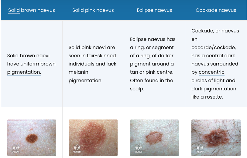
- Solid brown naevus
- Solid pink naevus
- Eclipse naevus
- Cockade naevus
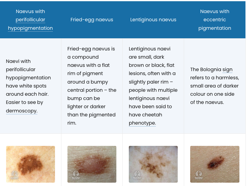
- Naevus with perifollicular hypopigmentation
- Fried-egg naevus
- Lentiginous naevus
- Naevus with eccentric pigmentation
Uncommon types of melanocytic naevi
- Spitz Naevi
- Agminated naevi
- Kissing naevus
Atypical melanocytic naevus
- Definition
- Varies
- A benign lesion that has some clinical or histopathological characteristics of melanoma
- A melanocytic naevus with specific characteristics: large (> 5 mm); ill-defined or irregular borders; varying shades of colour; with flat and bumpy components
- Or, any funny-looking naevus; large, or different from the patient’s other naevi. - Mole with unusual features
- Varies
- Cause
- Sporadic
- Familial
- Can be part of Familial Atypical Mole and Melanoma Syndrome
- Dysplastic naevus
- Best used for a naevus with a specific microscopic appearance
- Many histologically dysplastic naevi are clinically banale (eg, small in size, and uniform in colour and in structure)
- Histological dysplasia may be mild, moderate or severe
- Importance of atypical naevi
- if >5 then increased risk of melanoma
- Treatment
- Do not need removal
- Remove if unclear if melanoma
Aetiology of Melanocytic Naevus
- Congenital vs Acquired benign neoplasm
- ? Related to sun exposure
- Occasionally hereditary
Pathology of Melanocytic Naevus
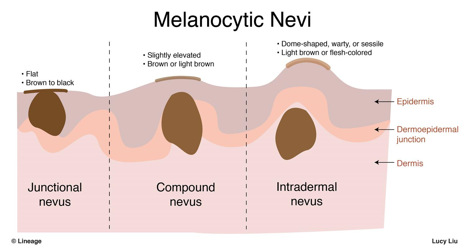
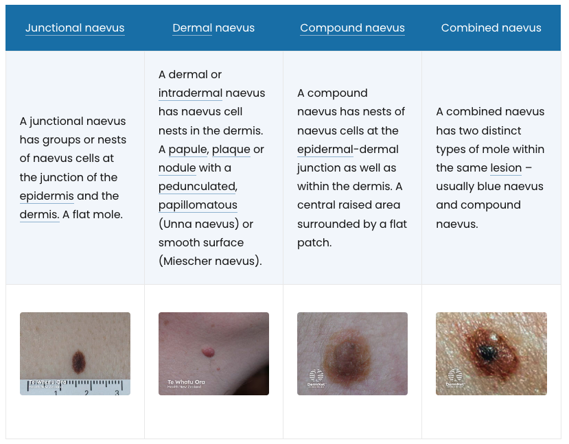
- The pathological classification of melanocytic naevi relates to where naevus cells are found in the skin.
- Lesions evolve with age
- Initial lesion being macular with nests of proliferating melanocytes confined to the dermoepidermal junction
- With time, nests extend into the dermis and lesions become elevated
- With further maturation junctional activity, is ceased and the naevus becomes intradermal.
Junctional Naevi
- Well-defined pigmented lesions
- Appear in infancy / pre-pubertal
- Flat / slightly elevated, brown, hairless
- Most typically seen on palms, soles & genitalia
- Proliferation of melanocytes at epidermal-dermal junction
- Low malignant potential
Compound Naevi
- Elevated, dome-shaped & brown ± hair
- Adolescents
- Melanocytes lie both in the epidermal-dermal junction & within the dermis
- Low malignant potential
Intradermal Naevi
- Dome-shaped (sometimes pedunculated)
- Fleshy to brownish pigmented moles
- Characteristically seen in adults
- Frequently contain hairs
- Micro:
- Melanocytes are entirely within the dermis
- Nests superficial, diffuse deeper
- Very low malignant potential
Management
- Should be excised if changes occur:
- Itching
- Inflammation
- Change in colour
- Halo formation
- ↑ Size
- Bleeding / ulceration
- If congenital naevi require excision, it has to be down to fascia
- Even then there is no guarantee that all the melanocytes have been removed
Prognosis
- Small risk of progression to melanoma
- True risk unknown ? 5%
- Congenital nevus > 20cm (10% risk of melanoma)
- If a melanoma is going to occur – 60% within first decade
Follow-up
- Lifetime surveillance for congenital naevi > 20cm
- Or if involves 2-5% of body area