Section: Skin and soft tissue Sub-section: Skin cancer Curriculum: Curriculum, page 52
Definition
- Malignant neoplasm of skin
- Arising from melanocytes (neural crest cells) in basement membrane
- At dermal/epidermal interface
Incidence
- 25% of all NZ cancers in 15-35 yo
- 1-3% of all malignancies
- Lifetime risk ≈ 5%
- Incidence highest in NZ and rising
- 50-90/100,000 in NZ (cf 34 in Austr)
- Incidence of thick melanomas ~ constant
- Mortality levelling/declining
- 95% occur on skin
- 4th most common cancer in NZ
- Median age at diagnosis = 50 years
- 60-80yo account for 40%
Classification
- Superficial Spreading 60%
- Nodular Malignant 28%
- Lentigo Maligna Melanoma 7%
- Acral Lentiginous 5%
- Desmoplastic/Neurotropic Malignant
- Spitzoid Melanoma
- Non-Cutaneous Melanoma 5%-10%
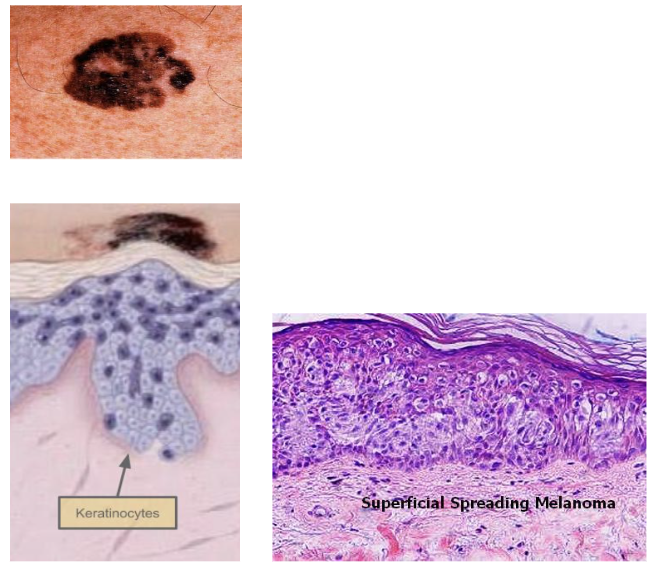
Superficial Spreading Melanoma
- Most common subtype - accounts for 70% of all melanomas
- F > M
- In sun exposed areas
- Often lower leg (women) or back (men)
- Common on trunk of naevus prone individuals
- Develop over 1-7 yrs
- Often in pts with Multiple Dysplastic Naevi
- Follows ABCD pattern usually
- Slight elevated, irregular but well-defined edge
- Stands out as “ugly duckling”
- Radial growth initially, then vertical
- Most are superficial, 5% deep invasion
- Microscopy
- Cells in nests epithelioid (“Pagetoid”)
- Large brown nuclei, prominent eosinophilic nucleoli
- Lots of cytoplasm filled with melanin granules
- Cells in nests 3-5 cells or singly along the dermal-epidermal junction
- May extend up to stratum corneum (uppermost layer)
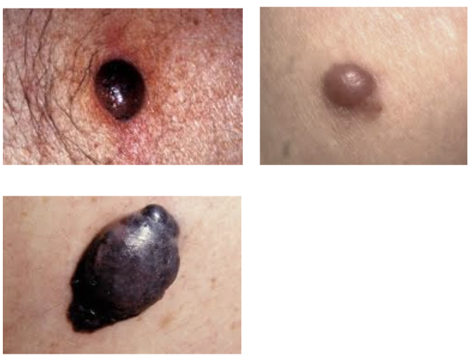
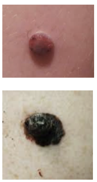
Nodular Melanoma
- Occurs in older males
- Most commonly on head, neck & trunk
- Severely sun damages sites
- De novo, rather than in a pre-existent naevus
- Less commonly associated with large numbers of naevi
- Doesn’t conform to ABCD rule
- Palpable: thicker, convex/raised shape ± ulcerated
- Dome-shaped; often resemble a blood-blister
- Symmetrical
- Can be amelanotic
- EFG – Elevated, Firm, Growing
- Aggressive
- Radial phase short
- No in-situ changes in local dermis
- Early and rapid vertical growth
- Lymph node metastasis commoner than in other types
- Prognosis poor, months to < 5yrs, due to depth of invasion
- Disproportionately contributes to melanoma deaths
Lentigo Maligna Melanoma
- Arises from lentigo maligna (in-situ disease)
- Hutchinson’s melanotic freckle
- Large irregular/asymmetric macular lesion
- Can be present for decades before invasion
- Blue-black flat lesion, or amelanotic
- Invasive disease (5%) often occurs
- On the face
- In elderly with other signs of sun exposure
- Least malignant potential of melanomas
- Histology: Diffuse continuous replacement of basal layer
- Spindle-shaped cells
- Lack of Pagetoid cells
- Cosmetically challenging, may require 2 stage procedure
- High recurrence rates after excision
- Good prognosis: 5-20 yrs or longer
Lentigo Maligna
- Treatment options:
- Surgical Excision – ideal margin 5-10mm
- Radiotherapy
- Cryotherapy
- Imiquimod – not yet fully evaluated
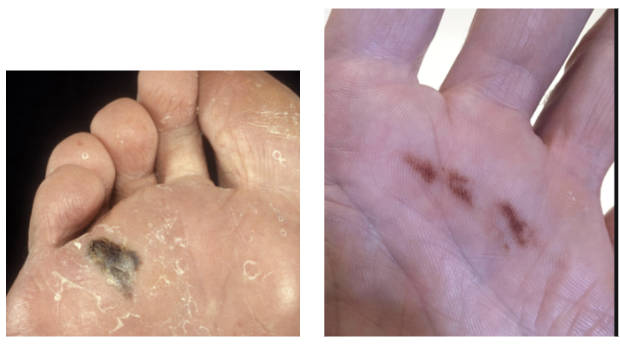
Acral Lentiginous Melanoma
- In thick glabrous (free from hair) epidermis
- Soles > Palms
- Subungual
- Perineal region
- More common in dark-skinned races
- Often large (> 3cm) at time of Dx
- Light, asymmetric, patchy pigmentation
- Can look like a bruise
- 30% hypomelanotic
- Histology:
- Prolonged radial growth phase
- No Pagetoid cells
- Marked junctional proliferation
- Large, atypical melanocytes with long dendritic processes
- Usually present as advanced lesions
- Poor prognosis:1-10yrs; 5yrs < 20%
Subungual Melanoma
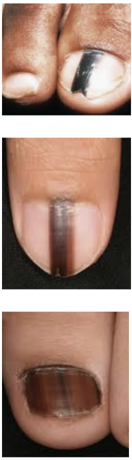
- Subset of Acral Lentiginous
- Presents as Longitudinal Melanonychia
- Full length pigment arising from nail matrix
- Band broadens over time
- Can get pigmentation of proximal or distal nail fold
- Common DDx is Subungual Haematoma
- Distinguish by presence of multiple reddish globules at periphery of pigmented area
- These grow out over months
Desmoplastic Melanoma
- Head & Neck most common site
- Sun damages skin
- Also lip, nose, ears etc.
- Can arise de-novo
- Neurtropism
- Or in association with pre-existing lentigo maligna
- More often amelanotic, firm, scar-like
- Deeply infiltrating
- Intense fibrous (desmoplastic) reaction or neural invasion
- Associated with high local recurrence rate (40-60%) with surgical excision alone
- Recurrence common at site of previously described benign biopsy
- Pathology hard to interpret
- Lower incidence of distant metastasis
Spitzoid Melanoma
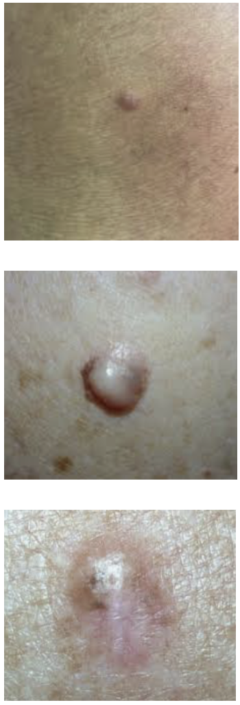
- Spectrum
- Spitz Naevi
- Atypical Spitz tumour
- Spitzoid Melanoma
- Benign naevus
- Young (< 20)
- Pink dome shaped papule with well-defined border
- Spitzoid melamonas tend to be
- Larger
- Asymmetrical with irregular border
- Pink to variegated in colour
- Challenging pathology, need entire lesion
Non-Cutaneous Melanoma
- Occur anywhere that neural crest cells migrate
- Eyes, mucosal surfaces etc.
- Any of these can be amelanotic
- Ocular (2-5%)
- Almost exclusively metastasizes to liver
- Always CT liver if ocular melanoma found
- Very rare to metastasize to LNs
- Mucous membranes (1-2%)
- Genitalia
- Anorectum
- Oropharynx, Sinuses
- Often diagnosed late
- Aggressive
- 5YS 25%
Aetiology/Risk Factors
- Host factos
- Melanocytic naevi
- Pre-existing disease
- Genetics
- Environmental factors
Host Factors
- Age
- Risk of developing melanoma for a 70yo in the next 10yrs ≈ 2%
- Male
- Skin colour, ability to tan vs burn, freckling
- Blue or green eyes, blood or red hair
- Melanocytic naevi
- Pre-existing disease
- Previous cutaneous Melanoma
- Second one occurs in up to 5%
- Risk highest in first 1-2yrs post 1st lesion
- FHx of Melanoma
- 10% have a 1° relative
- Genetics (≤ 2%) = Dysplastic Nevus Syndrome / Xeroderma Pigmentosum
- Immunosuppression
- HIV/AIDs etc.
- Evidence of actinic skin damage
Melanocytic Naevi
- Melanoma risk increases with naevus count
- Esp. > 100
- Atypical Naevi / Dysplastic Naevi
- More strongly associated with increased risk
- Especially >3-5 atypical naevi
- Arise in pre-existing naevi in 10-50% of cases
- More strongly associated with increased risk
- A congenital naevus > 20cm
- 2-30% risk of melanoma
Pre-existing Disease
- Multiple Dysplastic Naevi
- Can be AD, familial or sporadic
-
100 moles (tend to be 6-15mm, macular, colour variations & indistinct borders)
- Spitz Naevi
- Juvenile, spindle cell, epithelioid
- Rapidly growing, pink or brown
- Child to adolescent
- Stains with S100A6
Genetics
- Xeroderma Pigmentosum
- AR
- ‘Moon child’
- Rare Autosomal Recessive genetic disorder of DNA repair in which the ability to repair damage caused by ultraviolet (UV) light is deficient
- Familial Atypical Mole and Melanoma Syndrome
- p16 tumor suppressor protein (CDKN2A gene on chromosome 9p21) (inhibits CDK4)
- In 50%
- CDKN2A gene:
- Cyclin dependent kinase inhibitor 2A gene
- Codes for 3 different tumor suppressors, including p16
- p16 tumor suppressor protein (CDKN2A gene on chromosome 9p21) (inhibits CDK4)
Environmental Factors
- UV exposure
- UV-A longer wavelength and deeper penetration
- Found in tanning beds
- UV-B shorter wavelength and increased damage to superficial skin
- Both are attributed to melanoma
- UV-A longer wavelength and deeper penetration
- Sun exposure
- History of blistering sunburn
- Especially when young / < 18
- Cumulative – especially Lentigo maligna
- History of blistering sunburn
- Occupational exposure
- Sun beds
- Ionizing radiation
Clinical Presentation
General History/Exam
- Hx of naevus, risk factors for melanoma, hx of metastatic disease, family hx, prev melanoma
- Exam
- Exclude additional primary lesions
- Check of regional disease (in-transit = lymphatic metastasis along lymphatic channel deposit as subcut nodule + nodal disease)
- Distant disease
- Examine all LN basins, palpate liver
- PPV of clinical exam 6-20%
- Diagnostic accuracy increases 15x with dermoscopy
- Best predictor is
- Risk factors
- Change over time
- Symptoms of itching/bleeding
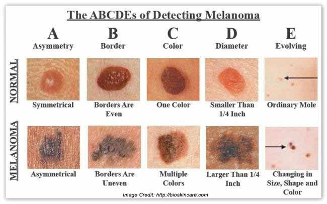
ABCDE Rule
- A – Asymmetry
- B – Border Irregularity
- C – Colour Variation
- D – Diameter > 6mm
- E – Evolution/Change over time
- May itch or ulcerate, leading to bleeding
- May have Regressed
- NB: Nodular melanoma does not fit
- May be symmetrical, and single colour or amelanocytic
- Often just look like a red nodule
- E - elevated
- F - firm
- G - growing
Other Cases
- Non-pigmented (Amelanotic) Melanomas ≈ 5%
- Nodular melanoma more likely to lack pigmentation
- Subungual melanomas
- Big toe & thumb more commonly affected
- Foot > Hand
- Usually associated with a longitudinal streak of melanin
- Due to a pigment-producing focus of melanocytes in the matrix
- Extension of the pigment onto the proximal/lateral nail fold
- Hutchinson’s sign
- Tend to occur in Blacks & pts 40-60yo
- Hutchinson’s sign
- Eye Melanoma: may be uveal or conjunctival
- Metachronous 5%
- Multiple melanomas in 1-4%
- Always look for a second primary
Pathology
Pathogenesis
- Malignant transformation of melanocytes
- May arise within Dysplastic Naevus
- Increased risk with increased dysplasia
- May arise within Dysplastic Naevus
- UV (esp. UV-B) light
- Direct mutagenic effects on DNA
- Decreases host defences
- Promotes ROS of melanin
- Cause DNA damage
- Suppress apoptosis
- BRAF mutation
- Most common oncogene mutations in melanoma (30% in NZ)
Phases of growth
Melanoma In-Situ
- Increased number of melanocytes
- Atypical melanocytes
- Melanocytes seen amongst more superficial layers of epidermis
- “Buck-shotting”
Radial Growth Phase
- Initial phase
- Predominantly intra-epithelial proliferation
- Lentiginous
- Diffuse replacement of basal layer
- Aggregated
- Nests of atypical melanocytes
- Pigmented flare seen clinically
- Accompanied by inflammatory infiltrate
- Lymphocytes & macrophages
Vertical Growth Phase
- Invasion of dermis
- Dense lymphocytic infiltration
- Loss of E-cadherin
- Loss of cell structure and tumour progression
- Melanoma cells express N-cadherin
- Allows adherence to fibroblasts & endothelial cells
- Allows migration through dermis & metastasis
Pathologic Slide Evaluation
- Histologic samples (and lymph nodes) should be serially sliced and stained with H&E
- Immunohistochemistry
- HMB-45
- S100
- SOX10
- Melan A
- Tyrosinase
Prognostic Information from Path Report
- Depth:
- Breslow Thickness
- Clark Level
- Margin
- Mitoses – rate/mm3
-
5 = High mitotic rate
-
- Ulceration (lack of epidermis)
- Important in staging
- LVI
- Regression
- Tumour-Infiltrating Lymphocytes (TILs)
- Predicts response to PD-1 inhibitor
Breslow Thickness
- Depth from the top of the granular layer of epidermis to the deepest tumour cell
- Correlates with 5-year survival
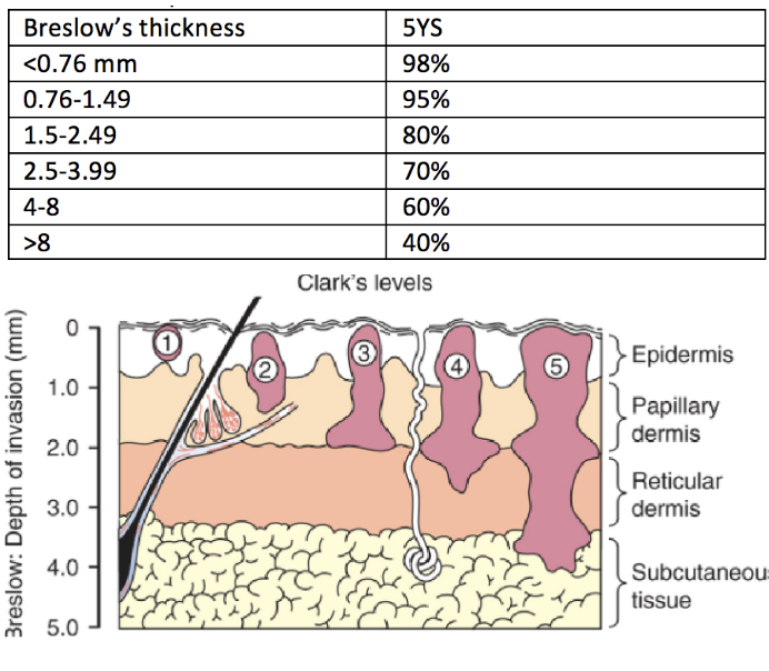
Clark’s Levels
- I - Confinement to epidermis premalignant/ melanoma in-situ
- All tumour cells above BM
- II - Invasion into Papillary Dermis
- III - Filling Papillary Dermis; cells at junction of papillary & reticular dermis
- IV - Invasion into Reticular Dermis
- V - Invasion into Subcutaneous Fat
- NB: Clark’s level is outdated, except in tumors ≤1mm - still retain their prognostic significance
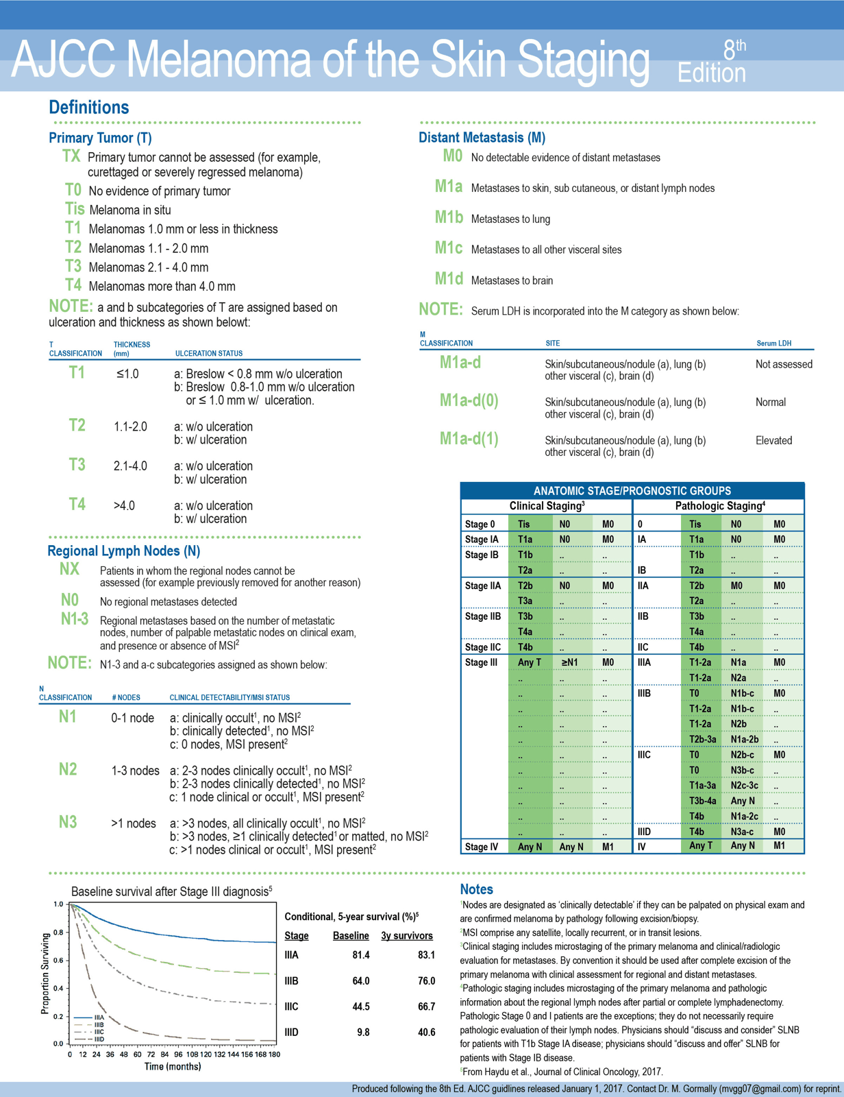
Staging
- Based on
- Breslow Thickness
- Ulceration
- NB: Mitotic rate has now been removed from AJCC 8th
- But should still be recorded
- Presence of LN and distant mets
- LDH level
T Stage
| Stage | Description |
|---|---|
| Tx | Tumor thickness cannot be assessed |
| T0 | No evidence of primary tumor (unknown primary or regressed tumor) |
| Tis | Melanoma in-situ |
| T1 | T1 Tumor <1 mm thick with unknown or unspecified ulceration T1a Tumor <0.8 mm thick without ulceration T1b Tumor 0.8 – 1 mm with/without ulceration OR Tumor < 0.8 mm with ulceration |
| T2 | T2 Tumor > 1-2 mm thick with unknown or unspecified ulceration T2a Tumor > 1-2 mm without ulceration T2b Tumor > 1-2 mm with ulceration |
| T3 | T3 Tumor > 2-4 mm thick with unknown or unspecified ulceration T3a Tumor > 2-4 mm without ulceration T3b Tumor > 2-4 mm with ulceration |
| T4 | T4 Tumor > 4 mm thick with unknown or unspecified ulceration T4a Tumor > 4 mm without ulceration T4b Tumor > 4 mm with ulceration |
N Stage
- All pts with nodal disease classified at least stage III
- Incl. microsatellites, satellites, and in transit disease
- N staging includes info such as
- A – Clinically occult
- B – Clinically detected
- C – Presence of in-transit, satellite, or microsatellite mets
N1 – Single node (or no node and in-transits) N2 – 2-3 nodes (or single node and in-transits) N3 – 4+ nodes (or 2+ nodes and in-transits)
| Stage | Description |
|---|---|
| Nx | Regional nodes not assessed, and no in-transit, satellite, or microsatellite metastases |
| N0 | No metastases to regional nodes and no in-transit, satellite, or microsatellite metastases |
| N1 | N1 One regional lymph node or any number of in-transit, satellite, or microsatellite metastases with no tumor involved regional node N1a One clinically occult lymph node and no in-transit, satellite, or microsatellite metastases N1b One clinically detected lymph node and no in-transit, satellite, or microsatellite metastases N1c No regional lymph node and any number of in-transit, satellite, or microsatellite metastases |
| N2 | N2 Two or 3 tumors involved nodes or any number in-transit, satellite, or microsatellite metastases with one tumor involved node N2a Two or 3 occult lymph nodes and no in-transit, satellite, or microsatellite metastases N2b Two or 3, at least one clinically detected node and no in-transit, satellite, or microsatellite metastases N2c One clinically occult or detected node and any number of in-transit, satellite, or microsatellite metastases |
| N3 | N3 Four or more tumor involved nodes or any number of in-transit, satellite, or microsatellite metastases with two tumors involved node or any number of matted nodes with or without in-transit, satellite, or microsatellite metastases N3a Four or more occult lymph nodes and no in-transit, satellite, or microsatellite metastases N3b Four or more, at least one clinically detected node and no in-transit, satellite, or microsatellite metastases N3c Two or more clinically detected and any number of in-transit, satellite, or microsatellite metastases or presence of matted nodes and any number of in-transit, satellite, or microsatellite metastases |
M Stage
- NB: Each M stage includes LDH level
- Not recorded
- Not elevated (0)
- Elevated (1)
- M1a
- To skin, soft tissue (including muscle), and/or non-regional LNs
- M1b
- Distant mets to lung
- M1c
- Distant mets to Non-CNS visceral sites
- M1d
- Distant mets to CNS
| Stage | Description |
|---|---|
| M0 | No distant metastases |
| M1a | M1a Distant metastases to the skin, soft tissue, including muscle and non-regional lymph node M1a LDH not available M1a (0) LDH not elevated M1a (1) LDH elevated |
| M1b | M1b Distant metastases to the lung with or without M1 site involvement M1b LDH not available M1b (0) LDH not elevated M1b (1) LDH elevated |
| M1c | M1c Distant metastases to non-CNS sites with or without M1 or M2 sites M1c LDH not available M1c (0) LDH not elevated M1c (1) LDH elevated |
| M1d | M1d Distant metastases to CNS sites with or without M1a, M1b, or M1c sites of involvement M1d LDH not available M1d (0) LDH not elevated M1d (1) LDH elevated |
Staging By TNM
- Stage I – Low risk 1° melanoma without evidence of regional or distant metastatic disease
- T1a, T1b, T2a
- Stage II – 1° melanoma with high risk for recurrence, but no evidence of locoregional or distant metastasis
- T2b, T3a, T3b, T4a, T4b
- Stage III – Pathologically documented involvement or regional LNs, or presence of in transit or satellite metastasis
- Further subdivided into A, B, C, D depending on depth and number of nodes involved
- NB: Melanoma metastasis (single) of unknown primary are classified as stage III
- Stage IV – Presence of distant metastasis
- CNS involvement esp. poor prognosis
Investigations
Initial Investigation
- Diagnostic excision biopsy for suspicious lesions
- 2mm margins
- Large facial or acral lesions, could consider full thickness punch
- Subungual melanoma
- Longitudinally orientated biopsy with nail bed sample
- If have clinically suspicious LN mets/distant mets
- FNA preferred to excision biopsy
- CT-PET
- Uptodate and Melanoma AUS/NZ recommends considering staging CT-PET for stage IIB and IIC tumours (thus T3b+ tumours)
- Interestingly, it doesn’t recommend staging for Stage IIIa (tumours <2cmm thick with clinically occult lymph node disease) unless you are planning a lymphadenectomy (in which case you should get a CT PET)
- Stage IIIB + tumours should all have a CT-PET with cranial imaging (MRI)
- For facial lesions you should include imaging of the neck
- CT-head or MRI brain
- For any patient with neurological symptoms, cranial imaging should occur
- Is not required for Stage II patients with no symptoms.
- Should be done in all stage III patients
- MRI brain is superior to CT head at detecting metastatic disease
- LDH
- This should be tested for in patients with metastatic disease - as it is a marker of more aggressive disease
Indications for Melanoma MDM
- Stage III and IV cutaneous Melanoma
- Desmoplastic Melanoma
- Difficult histology, and high local recurrence rates
- Melanoma < 18yrs old
Management
Prevention
- Avoid sun exposure
- Use clothing and sunscreen
- Need to block UV-A AND UV-B
- Abstain from sunbeds
- Statins and Fibrates may be protective
Wide Local Excision
- Rationale for completion WLE
- Decreases local recurrence rates
- Excision of occult cutaneous micromets
- Thickness of initial melanoma is the key factor in determining the clinical stage and recommended margin
- Most excisions are elliptical
- Required margin measured in the short axis of the ellipse
- Straight line closure along lines of natural tension
- Specimen should be orientated for pathologist
- WLE typically down to, but not necessarily including, muscle fascia
- No long-term benefits or decrease in local recurrence rates
- But some evidence may cause increased in transit of regional LN disease (? Removes barrier to spread)
- No long-term benefits or decrease in local recurrence rates
Resection Margins
- Data supporting resection margins based off surgeon “in-situ” measurements and not pathological measurements
- Data supported by multiple large clinical trials examining impact of surgical margins on local recurrence rates
- Tis - 0.5cm
- T1 - 1cm
- T2 1-2cm
- T3 - 2cm
- T4 - 2cm
Special Cases
- Desmoplastic / neurotropic lesions
- Consider adding ± 1 cm wider excision
- Subungual
- Amputation of digit at DIP
- Eye Melanoma:
- Refer (include for consideration of ‘eye conserving’ excisions)
- Oral or Anal Melanoma
- Excise to clear margins
- No evidence that WLE improves survival
Management of Regional LNs
Clinically Negative Nodes
- SLNB Rationale
- Sites of cutaneous melanoma have specific patterns of lymphatic spread
- One or more nodes are the first to be involved with metastatic disease
- If these are not involved, the entire nodal basin is likely to be free of tumour
- Status of the SLN reflects the status of the entire draining nodal basin
- MSLT-I showed improved 10yr disease-free survival from SNB vs observational approach (for intermediate and thick melanomas)
- SLNB may provide diagnostic information important for further treatment decisions
- To determine prognosis
- To select patients who may benefit from adjuvant therapy
- To select candidates for clinical trials
- SLNB should be offered for all tumours T1b or above
- Frozen section is not recommended due to difficulty in diagnosing micromets
Multicenter Selective Lymphadenectomy Trial I
- Phase 3 RCT
- Largest trial to address role of SLNB in determining prognosis and impact on survival
- Rationale
- Most important prognostic factor is regional lymph node status
- Lymph Node Dissection
- Comorbid & a lot will be negative
- Is SNB good at predicting regional node involvement?
- And how safe is it?
- 1661 randomised patients (1347 intermediate, 314 thick)
- WLE and SNB (with immediate completion lymphadenectomy if +ve)
- WLE and observation, with therapeutic lymphadenectomy if nodes picked up on observation
- Inclusions:
- Clark level 3 and > 1mm, or Clark 4-5 of any depth
- Primary study group was intermediate thickness (1.2mm-3.5mm)
- Findings
- 16% of SLNB were positive
- Same amount as the observation group would eventually go on to become clinically positive
- For SNB
- No significant difference in 10yr melanoma specific survival
- Mean 10yr disease-free survival significantly better
- For Intermediate melanomas (71.3 vs 64.7%)
- And for thick melanomas (50.7% vs 40.5%)
- Improved distant disease free survival
- Status of sentinel node was strongest predictor of recurrence or death from melanoma
- 16% of SLNB were positive
Indications for Sentinel LN Biopsy
- Not indicated
- Thin melanomas T1a (<0.8mm) without high risk features
- Overall risk of LN mets if < 5%
- Subsets of high risk thin melanomas can have higher rates of nodal disease
- Indicated
- T1b
- Thin melanomas T1b (0.8mm-1mm)
- or < 0.8mm with high risk features
- Ulceration, MR > 1/mm2, Clark 4/5, LVI
- T2 and T3
- Intermediate thickness melanomas (1-4mm)
- This group most likely to benefit from SNB
- T4
- Thick melanomas
- SNB offers prognostic benefit
- Reduced local recurrence
- No survival benefit in this group
- T1b
- Use Melanoma Institute Australia ‘Sentinel Node Metastasis Risk ‘Calculator’. Offer SLNBx if Risk > 10% and consider in 5-10%
- Variables include:
- Age
- Breslow Thickness
- Melanoma Sub-type
- Mitoses
- Ulceration
- LVI
- Variables include:
- If pt declines sentinel lymph node biopsy, they should enter surveillance of 4-6 monthly USS of the lymph node basin for 2 years
SLNB Technique
- Dual tracer technique
- Reduces false negatives, successful identification in 95%
- Pre-operative lymphoscintigraphy
- At operation patent blue
- Intra-op gamma probe
Pre-operative Lymphoscintigraphy
- Preferably same day as operation
- Tc99 Sulphur colloid injection, injected 4 x aliquots, into dermis (raise wheal)
- Should be in normal skin, 0.5cm from melanoma or scar
- Map the nodes, if none identified, second injection
- Sentinel nodes may be identified in > 1 LN basin (head, trunk etc.)
- Or may get “in-transit” nodes
- All identified nodes should be removed
Patent Blue
- 2mL total injected, 4x aliquots, into dermis (raise wheal) 0.5cm from melanoma/scar
- Wait 5 minutes (or massage 5 mins)
- Intra-op
- Direct incision over area identified with lymphoscintigraphy and confirmed with gamma-probe
- Can trace blue lymphatics to node and use gamma probe
- Liga-clip lymphatics and feeding vessels
Sentinel Node Definition
- Any lymph node that is most radioactive in the nodal basin
- Any node that is blue
- Any node that has a radioactive count 10% or higher of the most radioactive node in that basin
- Any node that is palpably suspicious for tumour
- NB: Average number identified per basin is 2
Complications of SLNB
- Wound infection (0.5-1%)
- Seroma / hematoma (3-5%)
- Lymphedema (0.5%)
- Allergic reaction to dye (1.5-2%)
Contraindications
- Melanoma in-situ or T1a
- Clinically/radiologically positive (stage 3) disease
Special Situations
- Pregnancy and Breast-Feeding
- Use TC99, not patent blue
- Comorbidities preventing CLND
- But may give prognostic info
- Desmoplastic Melanomas
- Less risk of LN metastasis
Management of Positive Sentinel Nodes
- Historically
- Before SLNB, moderate or high risk (but clinically negative) patients would undergo elective LN dissection
- Since SNB – Upfront LND should no longer be performed
- Before SLNB, moderate or high risk (but clinically negative) patients would undergo elective LN dissection
- Completion LN dissection has, until recently, been the standard of care for positive SLNs
- DeCOG-SLT and MSLT-II looked at this and found:
- Patients with positive SLNB who have immediate CLND has no improvement in 3 year melanoma specific survival compared to close observation
- But did have higher morbidity
- Patients with positive SLNB who have immediate CLND has no improvement in 3 year melanoma specific survival compared to close observation
- Active surveillance has equivalent survival but less morbidity than CLND after positive SNB
- Either CLND or careful observation can be offered to those with low risk disease
- Careful observation should only be offered to those with high risk disease after thorough discussion
- Current recommendations are to consider NOT performing CT/PET on newly diagnosed SNB positive patients
- Yield of PET/CT in detecting occult metastasis is only 0.5-3.7%
DeCOG-SLT
- Question
- Does completion LN dissection in the setting of positive SNB, improve outcome?
- Design
- 483 patients, cutaneous melanoma of trunk or extremities
- RCT - postive SNB, completion LND vs observation
- Results
- Similar 5 year
- Regional node-recurrence rates
- Distant metastasis free survival
- Relapse free survival
- Overall survival
- Averse events in 13% of CLND arm
- Lymphedema and delayed wound healing most common
- Similar 5 year
MSLT-II
- Question
- Does completion LN dissection in the setting of positive SNB, improve outcome?
- Design
- RCT - postive SNB, completion LND vs observation
- 1934 patients with positive SNB after WLE
- Inclusion
- Melanoma > 1.2mm, Clark 3 or above, or any ulceration
- Observation Protocol
- 3-4 monthly clinical and USS of nodal basins, for 2 years
- Then 6 monthly years 3-5
- Then yearly
- Findings:
- Immediate LNDx was NOT associated with increased melanoma specific survival
- Minimal improved rate of local disease free survival
- 68% vs 63% at 3 yrs
- Improved local disease control
- Non-sentinel node metastasis (found in only 10%) of pts in the dissection group
- Heralded a poor prognostic factor for distant and locoregional recurrence
- Lymphoedema was observed in 25% of patients in the dissection group and only 6% in observation group
- In the observation group - only 20% had a recurrence (which indicates for 80% of patients, the only positive nodes was the SLN)
High Risk Disease
- Careful observation only recommended after through discussion
- High risk features (defined as exclusion criteria from MSLT II) are:
- Extra-capsular spread/extension
- Concomitant micro-satellitosis of the primary tumour
- Greater than three involved nodes
- Greater than two involved nodal basins
- Immunosuppression
-
2mm of tumour in SLNB (MSLT II crtiera, NZ guidelines say >5mm)
- From Peter Mac Lecture: Even pts who belong to these high risk categories, completion ALND after positive SLNBx is very RARELY done
Management of Clinically Positive Nodes
- Clinically positive nodal basins mandate confirmation
- FNA as accurate as core biopsy and is method of choice of confirming positive nodes
- Stage
- Pts should be further staged if clinically palpable nodes
- Because identification of Stage 4 disease may prevent a morbid surgical procedure
- LDH
- CT/PET the best initial imaging modality
- Higher sensitivity than CT, similar specificity
- Changes treatment in 19-35% of patients
- MRI brain
- Current recommendation for Stage 3 disease in the presence of palpable nodes and no metastatic disease
- Pts should be further staged if clinically palpable nodes
- Therapeutic lymph node dissection
Therapeutic LN Dissection
- May be curative / prolong life for some
- Up to 60% of patients will not develop further mets and have long term control
- Complete clearance of a LN field is indicated
- Inadequate surgery leads to higher local recurrence rates and potentially worse survival
- Should be done in a high volume centre to maximise LN harvest
Cervical Lymphadenectomy
- Options include:
- Radical Neck Dissection
- All nodes: Levels I-V
- Incl. SCM, Accessory Nerve, and IJV
- Extended Radical Neck Dissection
- Superficial Parotidectomy
- Modified Radical Neck Dissection
- All nodes level I-V
- Preservation of some/all SCM, accessory nerve, IJV
- Selective node dissection
- Removal less than levels I-V
- Preservation of major structures
- Radical Neck Dissection
- Also, any nodes behind external auditory canal
- Occipital/retro-auricular nodes taken
- Parotid Lymphadenopathy
- Superficial Parotidectomy
- Levels 1b, 2, 3, Upper 5 +/- 1a
Axillary Lymphadenectomy
- For tumours of upper limb/trunk
- Need to do level 1-3 dissection
- May include resection of pec minor to facilitate clearance of superior axilla
- May include resection of Intercostobrachial Nerve and Medial Pectoral Nerves
Inguinal Lymphadenectomy
- Surgical management of inguinal LN field is controversial
- Pre-op staging should include inguinal/pelvic field
- But limited sensitivity and specificity
- Intra-op assessment based on femoral canal nodes or Cloquet’s node is unreliable
- Suggest ? Only add pelvic lymphadenectomy if positive on pre-op workup or large volume inguinal disease
- Iliac (Pelvic) LN Dissection if:
- Pelvic nodes positive clinically or on imaging
- ≥ 3 Superficial LN involved
- ± ? If Cloquet’s node is positive
- Superior most deep inguinal lymph nodes or inferior most external iliac lymph nodes
Melanoma of Unknown Primary
- 10-15% of palpable lymphadenopathy have a primary lesion that cannot be identified
- ? Regressed 1° Melanoma
- ? Melanoma developing in LN itself
- Should do complete skin examination
- And review pathology of any previous lesions
- Should be worked-up and managed as if primary lesion known
- Outcomes tend to be as good
Uncommon LN Occurrences
- E.g. Epi-trochlear or Popliteal Fossa
- May also have regional LN disease, should work these up appropriately
Indication for Further Staging Investigations
- Reason
- To identify disease burden and substage Stage 4 disease
- Indication
- Stage 2B (i.e. T3b (> 2mm Breslow with ulceration)) should also receive further staging investigations)
- Stage 3 (LN) disease that is palpable
- To prevent unnecessary lymphadenectomy if actually Stage 4
- Not LN disease that is SNB identified (Stage 3A)
- To prevent unnecessary lymphadenectomy if actually Stage 4
- Stage 4 disease
- Investigations
- BRAF Testing for Stage 3 and Stage 4
- CT/PET (whole body) is modality of choice
- MRI brain
- LDH
Adjuvant Therapy
Adjuvant Radiotherapy
- Primary site
- R1 resection
- Desmoplastic melanoma with perineural spread
- Locally advance recurrent
- After CLND with high risk features
- Consider but becoming less common now
- Indications:
- Large node - Largest node ≥ 3 cm in neck and ≥ 4cm in axilla or groin
- Extranodal spread
- Number of nodes - ≥ 1 for Parotid, ≥ 2 for neck or axilla, ≥ 3 for groin
- Recurrent disease
Adjuvant Systemic Therapy
- Traditional Chemo does not improve OS in Advanced Melanoma
Indications
- Stage 3 resected disease
- Stage 4 disease with quantifiable disease
- Stage 2 high risk (B/C)
- Evolving evidence to suggests benefit
- Keynote 716 - Stage 2B/C
- ChecKmate 76K - Stage 2B/C
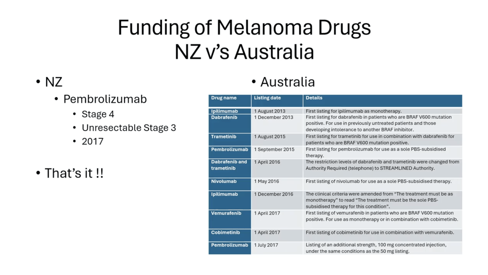
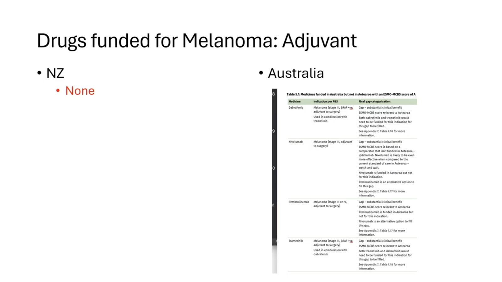
- Can consider adjuvant Pembrolizumab or Nivolumab (not funded)
- Evolving evidence to suggests benefit
Immunotherapy
PD-1 Inhibitors
- Immune checkpoint inhibitors
- PD-1: immunoregulatory cell surface receptor
- “Programmed Death” 1
- Located on surface of T-Cells
- Mechanism of action
- PD-1 Activation by PD-1 Ligand (PD-1L) on tumour cells down-regulates T-Cell activation
- Anti-PD-1 Monoclonal antibody (IgG) is directed against and blocks PD-1
- PD-1L cannot bind to PD-1 on T-Cells
- Thus, T-cells activated
- Get cell mediated immune response against tumour cells or pathogens
- In New Zealand
- Pembrolizumab (Keytruda) given in NZ
- Funded in NZ for:
- Unresectable
- Stage 3 and 4 disease
- With measurable disease on imaging
- Initially 12 weeks funded, and response assessed as per RECIST criteria
- Stage III resected disease
- As per Keynote 054 trial
- Renewal if:
- Complete or partial response or stable disease that is confirmed on imaging
- Stage 3 and 4 disease
- Resctable
- Not funded but indicated
- Stage IIB, IIC, or III melanoma who have undergone complete resection
- Not funded but indicated
- Unresectable
- Funded in NZ for:
- Pembrolizumab (Keytruda) given in NZ
Pembrolizumab
- Dose
- 2mg/kg, every 3 weeks IV
- Response
- 30% tumour shrinkage
- 5% complete response
- 55% 2 yr survival (cf 40% Ipilimumab)
- 30% progres free survival (PFS) at 12 months
- Toxicity
- Significant toxicity (10-15% risk of adverse events)
- Pneumonitis 3%
- Colitis 3%
- Hepatitis < 1%
- Immune mediated Thyroid disorders ~ 3%
- High risk of Foetal disorders - Avoid pregnancy
- Keynote 054 trial
- Phase III Double blind RCT
- Pembrolizumab (200mg every 3 weeks, for up to one year) vs placebo
- 1019 patients
- Indication
- After resection of stage III disease
- Primary endpoint
- Recurrence free survival
- Median F/U 3yrs
- Results
- 4.9 year follow up date
- Prolonged recurrence free survival (55 vs 38%)
- Longer distant metastasis-free survival (60.6% vs. 44.5%)
- 4.9 year follow up date
- Phase III Double blind RCT
CTLA-4 Inhibitors
- Immune checkpoint inhibitors
- Monoclonal IgG antibody
- Blocks CTLA-4 that suppresses T-cell function interaction with APC
- CTLA = Cytotoxic T-Lymphocyte-Associated Antigen
- Ipilimumab
- 3mg/kg every 3 weeks IV
- 20% response rate
- High Side Effect profile (Manage with steroids)
- 50%
- Enterocolitis
- Hepatitis
- Dermatitis
- Neuropathy
- Endocrinopathy
- Not funded in NZ but available in private
- CheckMate 238 - Nivolumab vs Ipilimumab - similar outcomes but Ipilimumab more toxic
- CheckMate 915 Nivolumab and Ipilimumab vs Nivolumab - no benefit but increase toxicity
Targeted therapy
BRAF inhibitors
- Up to 50% of melanoma has BRAF mutation
- BRAF is part of MAPK signalling pathway
- Pathway of cell signaling in melanoma. Mutations in this pathway lead to increased signals for cells to proliferate, survive, invade, make angiogenesis etc.
- BRAF is part of MAPK signalling pathway
- Mutation causes cell proliferation without apoptosis
- Most are V600 mutation
- More common in young, or metastatic disease
- BRAF inhibitors directly target/block mutated BRAF gene
- E.g. Dabrafenib
- NB: BRAF/MEK combination better than BRAF monotherapy, E.g. Dabrafenib and Trametinib
- Side effects minimal, include arthralgia, rash, fatigue
- NB: High rate 2° skin cancers (SCC etc.)
- NB: Resistance and recurrence can develop
- Response often short lived (6-10 months)
- Not funded - clinical trial or self-fund - $15,000/ month
In-Transit Disease
- Confirm via FNA
Definitions
- Historical Definitions
- Represents arrest of tumour emboli in the dermal or subcutaneous lymphatics
- In-transit Melanoma
- Dermal or subcutaneous recurrence arising between the primary lesion and the draining lymph node
- Satellite Lesion
- In-transit disease within 2cm of the scar
- Both classified and treated similarly by AJCC 8th
- NB: In-transits counted as Nc (add an N category)
Incidence
- Up to 10% develop in-transit metastasis
- Often 1st site of recurrence
- Median time to presentation 12-18 months from definitive excision of primary lesion
- Development of in-transit mets strongly associated with:
- Advancing age
- Increasing tumour thickness
- Primary melanoma ulceration
- Mitotic rate
- Presence of LVI
- Regional LN involvement
- Extent of in-transit recurrence, pace of disease, association with regional and distant spread is highly variable
- Makes management difficult
SNB
- Staging of In-transit mets should be by SNB
- Positive LN detected by SNB or clinically indicate poorer prognosis
- N1c 5yr Surv 81%
- No LN identified, just in-transits
- N2c 5yr Surv 69%
- Single positive LN, plus in-transits
- N3c 5yr Surv 52%
-
1 LN involved, plus in-transits
-
- N1c 5yr Surv 81%
- Retrospective analysis showed elective lymphadenectomy for in-transit mets had no impact on outcome
- Positive LN detected by SNB or clinically indicate poorer prognosis
Local Therapies
- Surgery
- Resection indicated in patients without distant metastasis
- If technically feasible
- 3 or more lesions or can’t get the skin closed primarily should warrant alternative evaluation
- If technically feasible
- Resection to clear margins
- Wider margins not necessary
- Resection indicated in patients without distant metastasis
- Local ablation
- Electrocautery
- CO2 laser – could use for local control of small lesions
- Intralesional IL-2 Injection (not available in NZ)
- Imiquimod therapy
Regional Therapies
Isolated limb perfusion and infusion
- Both a similar - perfusion requires surgery and openly accessing the vessels, wheras infusion involves cannulation of the vessels.
Indications for isolated limb perfusion/infusion
- Isolated metastasis to one limb
- A primary lesion with a poor prognosis
- Thick or ulcerated tumours
- Palpable regional nodal metastasis
Isolated limb perfusion
- The femoral vein and artery or axillary vein and artery are clamped
- The artery and vein are cannulated.
- A pump oxygenator is used to maintain oxygenation of the blood going through the limb.
- Heated cytotoxic mediation are infused - phenylalanine mustard and actinomycin D are commonly used - usually for 90 minutes
Isolated limb infusion
- The femoral artery and vein are accessed with a catheter and a torniquet is used.
- Blood is circulated at a much slower rate and only for 30 minutes
- During IRI, the limb becomes very hypoxic which leads to marked acidosis.
Response rate
- 75% response rate
- Limb loss is 0.8% and death rate 0.6%
Downsides
- Side effects are unpleasant - blistering, erythema, peeling, pain.
- Is expensive and resource intense.
Systemic Therapy
- In extensive in-transit disease
- Treating as stage IV disease with systemic therapy may be more appropriate
Management of Stage IV Disease
- Options
- Systemic
- BRAF and MEK inhibitors
- PD-1 Inhibitors
- CTLA-4 inhibitors
- Chemo doesn’t work!
- Surgical metastectomy
- Can cherry pick lesions
- May give survival advantage
Management of Melanoma Brain Metastasis
- Brain met management should be discussed at MDM
Incidence
- Melanoma has high propensity to metastasise to the brain
- Up to 50% of stage 4 disease will develop brain mets
- 25% have them at initial diagnosis
- Associated with poor prognosis
- Median overall survival 2.8 - 4 months
- Control of brain mets important
- Progression often leads to deterioration in neurologic function and quality of life
Surgical Therapy
- Brain Mets that are symptomatic or generate mass effect are best treated with surgery
- Surgery effective and rapid at relieving symptoms and improving functional outcome
- Best for single mets, > 1cm
- If multiple or small, radiotherapy may be better option
- Local control rates very high (80-93%)
- And durable
- Typically operating microscopy, neuro navigation, and en-bloc resection
- Peri-op complications 6-8%, mortality 2%
Systemic Therapy
- Systemic therapy effective against brain mets
- Combined BRAF inhibitors/MEK inhibitors inhibitors saw response in 58%
- Anti-PD-1 (Immunotherapy - Pembrolizumab) monotherapy saw response in at least 20%
- Can be considered as first line treatment
- As alternative to local brain therapy
Radiotherapy
- Radiotherapy can achieve a high rate of local control
- Could consider for patients with:
- Single or small number of brain mets
- < 1cm best, can be up to 3cm
- May give some palliative benefit for multiple mets
- Whole brain radiation therapy
- As no surgical option
- Can also consider radiotherapy after surgical resection of the lesions
Management of Special Cases
Melanoma in Pregnancy
- Principles should be the same, but consider:
- Stage of pregnancy
- Stage of melanoma
- Should involve MDM with obstetrician
- Where possible, delay general anaesthetic to 2nd trimester
- SBN should be performed with same indications
- Patent blue should not be used
- Teratogenicity
- Anaphylactic risk
- T99 sulphur colloid is safe
- Patent blue should not be used
- Radiation should be postponed until post-partum
- Unless urgent need, away from womb
- Targeted therapy and immunotherapy should be avoided
Mucosal Melanoma
- Rare tumours
- Very poor prognosis
- Most commonly
- Vagina and vulva
- Anus
- Upper aerodigestive tract
- Almost completely absent of BRAF mutations
- And response to immunotherapy also not as good
- Up to 1/3 overexpress C-Kit
- But not in sinus/nasal melanoma
- Implications for treatment with Imatinib
- All patients should undergo full staging and C-Kit molecular analysis
Anal Melanoma
- Incidence
- Rare, <1% anorectal tumours
- Age 60, slight F > M
- Presentation
- Haemorrhoidal type bleeding
- Mass
- Perianal discomfort
- Polypoid lesion, frequently ulcerated
- Most around or below dentate line
- Only small proportion from rectal mucosa
- Many do not appear dark
- Usually 3-4cm at presentation
- At least 20% present with regional LN mets
- 20-40% will have distant mets on presentation
- Work up
- Stage the patient
- Endoanal USS to help depth of invasion may be useful
- Stage the patient
- Management
- Historically APR, now reserved for cases with locoregional disease and primary tumour can’t be WLE’d
- WLE (sphincter preserving) gives good local control, and similar overall survival
- Optimal margins haven’t been investigated, most use 2cm
- Consider post-op radiotherapy after WLE, esp. if close margins
- Regional node metastasis not strongly related to survival (as high rates distant mets)
- Elective lymphadenectomy not supported
- SNB role remains undefined
- Therapeutic lymphadenectomy is indicated if proven LN involvement
- Overall survival poor
- Disease confined to anorectum – 35% 5yr survival
- Regional nodal disease – median survival 20 months
- Distant mets – rarely survive 12 months
Head and Neck Mucosal Melanoma
- Complete surgical excision is fundamental aim
- May be difficult to achieve without great morbidity
- Adjuvant radiotherapy may improve local control
- But not survival
- Primary radiotherapy has poorer outcomes compared to surgery
- Role of SNB undefined
- May have prognostic role
- Oesophagus
- Radical excision in contained disease
Vulval and Vaginal Melanoma
- Vulval
- No survival advantage for radical procedures over WLE with 1-2cm margins
- But extensive/exenteration procedures may be required, particularly if central
- No advantage for elective lymphadenectomy
- Vaginal
- Very rare
- Any suspicious lesion should be biopsied
- Usually present with advanced disease
- Compete removal frequently requires exenteration
Male Uro-Genital Melanoma
- WLE preferred over Penectomy if possible
- Overall survival poor
- Most die within 3 years
Ocular Melanoma
- Complex and uncommon form of melanoma
- Should be managed in specialist locations
- Eye-conserving therapies for ocular melanoma result in similar rates of local control to enucleation
- 1st surgery is most important
- Inappropriate primary surgery results in upstaging of disease
- Worse prognosis due to inadvertent tumour seeding
Prognosis
- NODAL STATUS, NODAL STATUS, NODAL STATUS
- Independent prognostic factors
- Breslow thickness
- Mitotic rate
- Ulceration – very important factor
- Clark’s level – only for T1 melanoma
- Age
- Site – mucosal, sole, palm, ear, subungual – poor
- Gender: F better than M
- Regression
- Tumour infiltrating lymphocytes
- Histological type; desmoplastic & acral bad
- Satellites, in-transit metastases
- Sentinel LN
- – If +ve → 56% 5YS
- – If -ve → 90% 5YS
Long Term Survival
- Melanomas of the head and neck (cf trunk / limbs)
- Increased recurrence rate
- Decreased survival
- More likely to spread to LN, but SLNBx more difficult
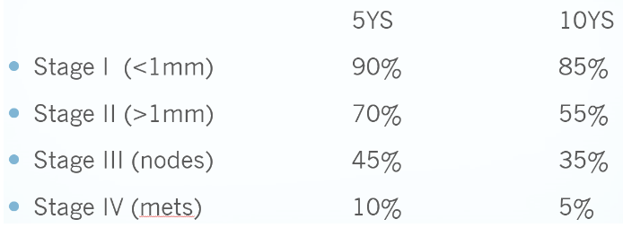
- Stage III disease → 45% 5YS
- 50% 5YS if one node +ve, 25% if > 4 nodes
- Most survive ≥ 12mths
- Stage IV → 10% 5YS
- Improved survival if all known mets are resected
- Up to 40% 5YS, if all disease in pts staged by CT & PET is excised
- Survival after resection of abdo mets:
- Median survival 8.2 months after resection of GI mets
- After resection of solid organ mets 20.9 months
- 1YS 35%, 5YS 18%
- Improved survival if all known mets are resected
Prognosis by Stage
Recurrence
- Recurrence rate
- 20% overall
- Median DFS 2.6yrs
- Can be latent
- Recurrence > 10yrs not uncommon
- 13% of SLN –ve pts will still develop recurrence somewhere by 3yrs
- Recurrence rate related to
- Stage
- Prognostic factors (thickness, ulcer, mitotic rates)
- Stage → Recurrence rate
- I 5%
- II 30%
- In melanoma with occult primary
- Prognosis is actually better than mets with known primary
Follow-up
- Sun Protection Advice
- At f/up check for
- Local Recurrence
- Satellite lesions
- LNs
- Rest of skin for new melanoma
- Signs/symptoms of metastatic disease
- Stage I (T1, T2a)
- See once for D/C then GP Annual follow up for 10 years
- Stage II (T2b-T4, NO)
- 4-6 monthly for 2 years then annually
- Stage III (Any N +)
- 3-4 monthly for 2 years, then 6 monthly until 5 years then annually
- Stage III/IV disease should get CT at 3 months, then Q6months, to 2yrs
- Except SNB+ patients, who should get nodal USS every 6months for 2 years
Stage 3 +/- 2b/c
- 6Monthly CT chest, abdo, pelvis and CT/MRI head
- 4-6 monthly USS of nodal basin if +ve SLNB and no CLND
Neoadjuvent treatment in melanoma
- Clinically node positive
- Stage IIIb and upwards
- There are recent trials including the NADINA trial (2024) and SWOG1801 (2023) trial which have shown improved EFS with neoadjuvant immunotherapy but long term data (and thus overall survival data is still pending).
- Currently, patients should only receive neoadjuvant therapy as part of a clinical trial
- The agents used in the trials have been is Ipilimumab and Nivolumab (NADINA) OR Pembrolizumab
Available in NZ vs Australia