Section: Vascular Curriculum: Curriculum, page 54
Definition
- Area of epidermal loss that has not healed within 6 weeks
- Ulcer = Persistent discontinuity of the epithelial surface
Incidence
- 1-2% of the adult population in Western world
- 3-5% of > 65yo
- Incidence ↑ with age
- F > M
Aetiology
- Lymphovascular (90%)
- Venous (50-70%)
- Mixed venous & arterial (10-20%)
- Arterial (10%)
- Lymphoedema
- DM → Microvascular disease (& secondary neuropathy)
- Blood dyscrasias
- Thalassemia, Sickle Cell disease, Polycythaemia Rubra Vera
- Inflammatory
- Vasculitis – RA/ SLE/ Polyarteritis Nodosa
- Pyoderma gangrenosum (RA/ UC)
- Infective: e.g. TB
- Underlying Osteomyelitis
- Neoplastic
- Malignancy – e.g. Squamous cell carcinoma, Basal cell carcinoma, Melanoma
- Marjolin ulcer
- Trauma
- Obesity / Pressure sores – reduced mobility – won’t use calf muscle pump as much
- Neuropathic ulcers = 2° to an inadequate sensory nervous system
- E.g. DM, nerve injuries, leprosy, spinal cord lesions
Clinical
- History
- Pain (Lack of = neuropathic)
- Systemic diseases
- Hx of trauma
- Hx of DVT/ varicose vein treatment
- Mobility
- Examine arterial system, nervous system & veins
- Distribution of trophic & ischaemic ulcers may be similar (as the pressure points are same in both – e.g. over heel; distribution dependent on whether pt is bed-bound or ambulant)
- Arterial ulcers
- Painful, punched out, often on pressure areas
- Ischemic ulceration
- Large vessel disease; atherosclerosis or embolism
- Small vessel disease: DM, Buerger’s disease, microembolism, pressure necrosis / trauma, Scleroderma, Raynaud’s
- Venous ulcers
- 90% - medial aspect of lower third of the leg, above medial malleolus = Gaiter area
- May not be very painful, especially if chronic
- Granulation tissue in base, uneven epithelial margins (peripheral healing)
- Shallow with sloping edge
- Raised/thickened edge in a chronic venous ulcer should raise the suspicion of malignant change (Marjolin ulcer)
- Frequently colonized with bacteria, but may not be infected
- Infection more likely if bacterial load is > 105 organisms/ gram of tissue or if β-haemolytic strep is present at level of 102 organisms/ gram
Pathology
- Repeated pressure & trauma contribute the cause of both ischaemic & trophic ulcers
- Microbiology
- S. aureus (≈ 40%)
- Pseudomonas Aeruginosa (≈ 25-30%)
- Of the pts with venous ulceration
- 50% have reflux in the superficial system alone
- 38% have reflux in both superficial and deep venous system
- 10% reflux in the deep system alone
Investigations
- ABI/arterial studies
- Test for glucose
- Vasculitis screen: ESR, CRP, ANA, RhF, ANCAs
- Consider punch Bx to rule out malignant ulcer / malignant change
- Culture – Strep Group A, Staph aureus – colonisation common, routine bacterial cultivation not necessary
- Imaging
- Arterial system and venous system as indicated
Management
- Consider all causes of leg ulcer (arterial disease, vasculitis, DM)
- Stop smoking
- Chronic venous ulceration
- Graduated compression stockings – Class 2 usually effective
- Elevation
- Exercise
- Dressings
- Wounds heal 50% faster when moist
- Occlusive dressing – Hydrocolloid (Duoderm, Comfeel)
- Change every 5-7 days
- Forms gelatinous barrier
- Lower infection rate, equivalent healing time, less pain
- Non-adherent - reforming epithelium does not get damaged when removed
- Debridement of necrotic tissue (prevents epithelialization & encourages infection)
- Infection treatment
- if appropriate - no evidence for routine ABs
- Manuka-honey
- Non-sig improvement
- Cochrane 2008: no benefit, except in burns?
- DVT prophylaxis
- Diuretics
- Aspirin – evidence inconclusive
- Diabetic ulcers
- Hyperbaric oxygen ↓s amputation rates but is expensive
- For mixed ulcers
- If ABI > 0.8: Compression bandaging
- If ABI 0.5 - 0.8: Modified compression ± consider for revascularization if ulcer not healing
- If ABI < 0.5 → Refer for vascular revascularization surgery (before debriding)
Surgical management of venous disesae
- Improves ulcer healing and lowers rates of recurrence
- Goals
- Definitively treat any refluxing axial veins, associated pathologic perforator veins, or associated varicose veins.
- Types
- Superficial axial venous ablation
- See Varicose veins
- Endovascular vs Surgical
- See Varicose veins
- Pathologic perforating veins
- Mostly teated endovascular now but can use open ligation and subfascial endoscopic perforator ligation
- Deep venous obstruction or insufficiency
- Iliocaval venous obstruction
- Femoral or infrainguinal obstruction
- Superficial axial venous ablation
Prognosis
- Venous
- Ulceration: Healing rates 70-80% at 24wks, ≈ 20% resistant
- 12-month ulcer recurrence rates 30% (though may be up to 70%)
- Recurrence rates ↓ in pts who have compression + surgery for their veins: At 3 yrs: recurrence rates = 10 vs 40% In those treated with compression alone
- Risk factors for poor healing
- Age
- Ulcer chronicity (> 3 months)
- Venous refill time ≤ 20s with a below knee tourniquet
- Smoking
- Ulcer size
ESCHAR
- Effect of Surgery and Compression on Healing and Recurrence study by Gohel, BMJ 2007
- RCT 500 patients with superficial and/or deep reflux and open or recently healed leg ulceration
- 3 UK centres
- Compared compression (multi-layered bandage for open ulcer, class 2 elastic stockings for healed) alone with surgery plus compression
- Showed no difference in ulcer healing at 3 years (89% in compression vs 93% in surgery group)
- Surgery group have lower recurrence rate at 3 years (31% vs 56%, NNT = 4), with most benefit for superficial and superficial plus deep segmental reflux, but no statistically significant benefit for deep reflux only
- Ulcer free time to 3 years was significantly longer in surgery group (100 vs 85 weeks)
Dressings
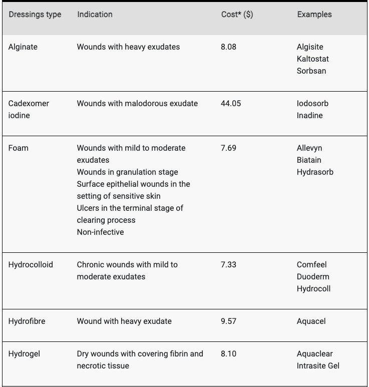
Chemical debridement
- Iodasrob
- Good for wet
- Hydrogel and Intrasite
- Good for dry