Section: Skin and soft tissue Sub-section: Skin cancer Curriculum: Curriculum, page 52
Definition
- Locally invasive cancer of skin
- Arising from progenitor cells in the basal layer of the Epidermis
- No precursor lesion
Incidence
- Most common skin cancer
- BCC > SCC 4:1
- Lifetime risk in Caucasians ≈ 25% (1:4)
- M > F
- Rare < age 40
Classification
- Nodular/Nodulocystic 70%
- Superficial (Spreading) 20%
- Aggressive variants
- Infiltrative/Morphoiec BCC
- Usually found in mid facial sties
- Waxy, scar like plaque with indistinct borders
- Wide and deep subclinical extension
- May infiltrate cutaneous nerves
- Basosquamous
- Mixed BCC and SCC features
- Infiltrative growth pattern
- Infiltrative/Morphoiec BCC
Nodular/Nodulocystic
- Well defined nodular lesion with waxy appearance
- Noduloulcerated
- Contains central ulcer (common)
- Pigmented
- Within macrophages in stroma
- Cystic
- Central degeneration of cells, can look like blue nevus
- Micronodular
- More aggressive with higher recurrence rates
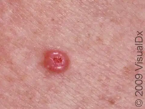
Superficial (Spreading)
- Can look like psoriasis, tinea, eczema
- Macular growth pattern
- Ill-defined margin and central non-healing crusting wound
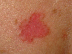
Aetiology
- Risk Factors
- UV (not as strong an association as SCC & Melanoma)
- Radiotherapy
- Arsenic
- Immune suppression
- Genetic Risk Factors:
- Gorlin Syndrome
- Genetics
- Autosomal Dominant
- Germline pathogenic variants in the human homolog of the patched (PTCH1) gene which leads to activation of the hedgehog pathway
- Clinical
- Multiple BCCs usually < 30 yrs
- Odontogenic keratocysts
- Ovarian fibromas
- Medulloblastoma
- Other features include palmar and plantar pits, craniofacial and skeletal abnormalities, ocular abnormalities, and ectopic intracranial calcifications
- Screening
- Regular skin checks
- Genetics
- Xeroderma Pigmentosum
- Gorlin Syndrome
Clinical Presentation
- No precursor lesion
- SCC & melanoma can both have a precursor
- Most on Head & Neck – Forehead/face (90%)
- Rare on back of hand (unlike SCC)
- Slowly growing
- Months – yrs
- Tend to infiltrate locally but rare to metastasize
Nodular
- Pearly nodule with surface blood vessels or diffuse spreading
- Small, translucent / shiny (pearly) elevated nodules
- Central ulceration & raised / rolled pearly edges (umbilication)
- Telangiectatic vessels are commonly seen over the surface
- Esp. infiltrating type: Ill-defined borders and telangiectasia
- Ulcerate early
- May erode deeply (Rodent Ulcer) but almost never metastasizes
Superficial
- Pink / red, scaly patch
- Often multiple
- Upper trunk, shoulders
- Bleed / ulcerate easily
- Difficult to distinguish from Bowen’s disease
High risk subtypes
- Sclerosing
- Mid-face
- Skin-colored / waxy
- Scar-like
- Perineural spread
- Micronodular
- Infiltrative
- Basosquamous
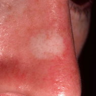
Pathology
- Abnormal growth of progenitor cells in the basal layer of the epidermis
- Deepest layer of the epidermis
- Typical microscopic features of BCC
- Clumps with “palisading nuclei” (lined up like a fence)
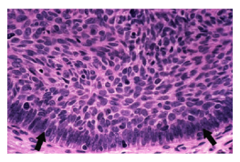
- Slow local invasion; rarely metastasize
- Lymph Node Metastasis 0.4% (Basosquamous form most likely)
- If the tumour starts as a nodule, the centre may die
- Results in a lesion with a rolled / elevated (but not inverted) edge
- Superficial BCC
- Often extends (microscopically) beyond the clinical borders
- Infiltrating and Sclerosing BCCs
- Exhibit islands of tumour extending into the tissue
- ± Perineural invasion in 3% of pts
- Finding correlates with highest recurrence rates and positive margins after excision
- Basosquamous type contains elements of both BCC & SCC
- Can metastasize
Investigation and DDx
- Excision biopsy for diagnosis
- Differential Diagnosis
- SCC
- But BCCs usually have long hx and a more typical rolled edge
- Keratoacanthoma
- Usually a short history
- SCC
Management
- Should divide into low risk BCC and high risk BCC
Low risk BCC
Definition
- <2cm in diameter
- Located on trunk or extremities (excluding genitalia and hands).
- Superficial or nodular type
- Lack of perineural invasion.
Treatment of low risk BCC
Surgical/first line therapy
- Excision with a macroscopic 2-3mm margin. This is gold standard. Adequate microscopic margin is 0.5mm
- If margins are positive - should have re-excision. If surgery not possible can consider radiation OR close clinical follow up.
Second line therapy
- Recommended for patients who wish to avoid surgery (unsatisfactory cosmetic result or difficulty with wound healing) and are low risk.
- Options include:
- Imiquimod 5% cream
- 5 days per week for 6 weeks until erythema, crusting, and scab formation develops.
- Stimulates the immune system by activating toll like receptor 7
- Topical 5-Fluorouracil 5%
- Twice daily for 6 weeks.
- Photodynamic therapy
- Imiquimod 5% cream
- Options include:
Other less commonly used options
- Cryosurgery
- Intralesional therapy with 5-FU and bleomycin.
High risk BCCs
Definition of high risk BCC
- Tumours >2cm
- Tumours on the head, neck, hands, feet, pre-tibia, and anogenital areas.
- Recurrent lesion.
- Lesions in sites of prior radiation therapy.
- Aggressive subtype.
- Perineural invasion
- Immunocompromised patient.
Treatment of high risk BCC
Surgical/first line therapy
- MOHS (mohs micrographic surgery)
- Is essentially excision, frozen section with margin examination using formalin, followed by re-excision as required.
- Standard surgical excision with 4-6mm macroscopic margin- higher recurrence rate than MOHs.
- If margins are positive can re-excise OR consider radiation therapy.
Second line therapy
- Radiation therapy - good option for large tumours where surgery would be morbid or for patients who are not surgical candidates. Higher recurrence rate.
Treatments not recommended
- Topical therapies such as imiquimod and 5-FU
Systemic therapy for locally invasive or metastatic BCC
- Metastatic BCC is extremely rare and should be managed by experience MDT.
- Targeted treatment is sonic hedgehog inhibitors – visnodegib, and sonidigeb.
- Inhibit SMO
- Bad side effects - muscle spasm, alopecia, altered taste, nausea, fatigue. 30% will stop treatment.
Cryotherapy
- Freeze for 5-15 sec to create a 1-3mm rim of freeze beyond lesion, and then thaw over 20-40 sec
- Perform 2 cycles
- Depth of freeze is 1.5x lateral spread
- Longer freeze-thaw times destroy portions of dermis
- Erythema occurs immediately
- Freezing can cause separation at the dermoepidermal junction and produce a bulla, which usually resolve in a few days
- Avoid freezing areas where nerves lie superficially
- Superficial lesions crust in 7-10 days then falloff
- NB: Major issue is no tissue for histology and clearance margin uncertain
Topical Chemotherapy
- 5-Fluorouracil (Efudix)
- Twice daily for 6 weeks.
- Blocks incorporation of thymidine into DNA
- Imiquimod (Aldara)
- 5 days per week for 6 weeks until erythema, crusting, and scab formation develops.
- Stimulates the immune system by activating toll like receptor 7
- Upregulates cytokines
- Can be used for early superficial BCC (but not nodular BCC)
Radiotherapy
- Indications:
- Difficult lesions in elderly only (may itself cause cancer)
- BCC with neural involvement
- Avoided near cartilage (necrosis risk)
- 3000-5000 cGy in 6-20 fractions
- 200-500cGy/ session
Prognosis
- If untreated will cause invasion & massive ulceration
- 95% curable with excision
- Recurrence Risk Factors
- Excision margin involved
- 60% of margin +ve BCC don’t recur - Don’t always have to re-excise
- Exception: Deep margins should be negative - difficult to monitor clinically
- Immunosuppression
- Prior radiotherapy
- Site – Head & neck (especially lip & pinna)
- Perineural involvement
- ↑ Size
-
1.5cm 10% at 5 yrs
-
3cm 25% at 5 yrs
-
- Depth of invasion
- Aggressive variants - infiltrating, multifocal, sclerosing
- 1/3 will develop a 2nd primary BCC within 5 years
- Excision margin involved