Section: Skin and soft tissue Sub-section: Skin cancer Curriculum: Curriculum, page 52
Definition
- Benign , rapidly growing, locally destructive overgrowth of hair follicle cells
- May regress spontaneously with scarring
- Arises from the infundibulum of the hair follicle
Risk
- Sun exposure
- Age
- Male
- Genetic
- Xeroderma Pigmentosum
- Muir-Torre syndrome
- Multiple self-healing squamous epithelioma (Ferguson-Smith syndrome)
- Eruptive keratoacanthoma of Grzybowski
- Incontinentia pigmenti
Clinical
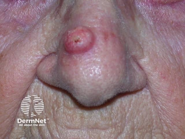
- Solitary, rapidly growing nodule on sun-exposed skin of the face and upper limbs.
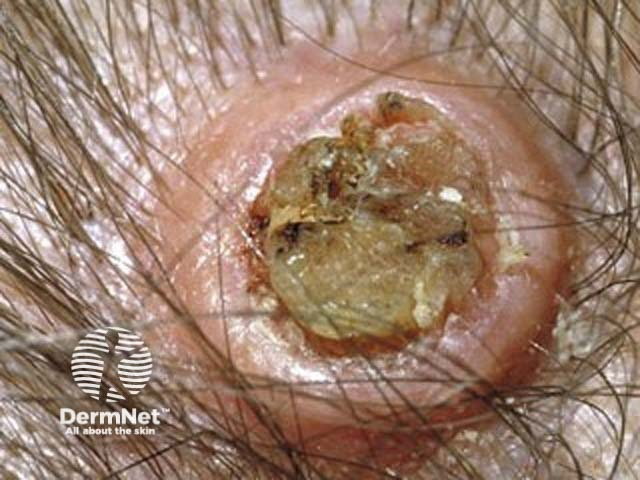
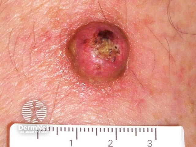
- Sharply demarcated, firm, erythematous or skin-coloured, with a classic central hyperkeratotic plug and an even shoulder.
- Removal of the keratotic core will leave a ‘crater’-like appearance to the lesion.
- 3 clinical phases:
- proliferative, maturation & involution
- Grows rapidly (2-4wks) as a ‘volcano’ / cone-shape (flesh coloured) with a dense keratin plug in its ‘crater’
- Then spontaneously regresses (over 2-3mths) → Leaves a deep, puckered scar
- proliferative, maturation & involution
Pathology
- KA arises from the pilosebaceous unit, particularly the hair follicle epithelium. It is considered a well-differentiated squamous neoplasm
Differential diagnosis
- SCC
- May be well-differentiated variants of SCC capable of spontaneous regression
- Difficult to distinguish from SCC histologically
- Usually no atypia & sharply demarcated, with an epithelial lip
- Compared to SCC – many mitotic figures, ulcerated
Investigations:
- Excision biopsy
Management
- Clinically they may be indistinguishable from well-differentiated squamous cell carcinoma
- Thus, many clinicians and pathologists prefer the term SCC, KA-type and recommend surgical excision
- Surgical management is usually preferred
- Unless there is clear evidence that regression is in progress.