Section: Skin and soft tissue Sub-section: Skin cancer Curriculum: Curriculum, page 52
Definition
- Malignancy of epidermal keratinocytes
Incidence
- Middle age – elderly
- 1/5 of non-melanoma skin cancers
- 50% chance of second in SCC in 5 yrs.
Aetiology/Risk Factors
- Sunlight most common cause in Fair skin
- UV-B (B = Bad)
- Actinic Keratoses (proliferation of keratin cells in basal layer)
- Immunosuppression (50% increased risk)
- Smoking → Oral SCC
- Chronic scars / Ulcers
- Infections:
- Chronic Granulomas (e.g. TB of the skin, Syphilis)
- Viral – HPV/ EBV
- Occupational exposures:
- Contact with Tars, Hydrocarbons, Arsenic
- Chemical & thermal burns
- Exposure to ionizing radiation / Radiotherapy
- Drugs - azathioprine, hydrochlorothiazide, BRAF inhibitors (very common
- Genetic predisposition:
- Xeroderma Pigmentosum – RR 1000 x
- Albinism
Pathogenesis
- Genes included are - TP53 and NOTCH pathway
Classification
- High Risk SCC
- Location on the ear, vermillion of the lip, central face, hands, feet, genitals.
- Histology
- Depth > 6mm, or Clark 4/5
- Poorly Differentiated
- Subtype –Desmoplastic, carcinosarcomatous, Adenosquamous type
- LVI/Perineural Invasion (>0.1mm nerve)
- Recurrent tumours
- Immunosuppression
- Site of previous radiation treatment or chronic inflammation (Marjolin ulcer)
- Rapidly growing
- Low risk
- Diameter < 2cm.
- Well or moderately differentiated tumour
- None of the above features.
Clinical Classification
Invasive SCC
- Dysplastic keratinocytes which are full thickness and invade the basement membrane to involve the dermis.
- Can be well, moderately, or poorly differentiated.
Keratotic Horn
- Hard keratin accumulation
- Arise from benign, pre-malignant or malignant SCC
Keratoacanthoma
- May be indistinguishable from well-differentiated squamous cell carcinoma
- May spontaneously regress
- Arise from hair follicle cells
Verrucous Carcinoma
- SCC induced by HPV
- Slow growing
- Usually occurs on the sole of the foot, but can be Anogenital (known as Giant Condyloma acuminatum of Buschke-Lowenstein)
- Can be on the plantar foot (Epithelioma cuniculatum)
- Fungating
- Invade deeply & extensively
- But rarely metastasis
- Surgery is mainstay of treatment
- Can progress to high grade SCC after RT
- Strongly associated with
- Smoking
- EtOH
Marjolin ulcer
- SCC which arises from sites of chronic wounds or scars.
- Malignant transformation is very slow, with average latency of 30 years.
Lymphoepithelioma-like carcinoma of the skin
- Very rare, indolent, malignant skin tumour of epithelial origin.
- Resembles nasopharyngeal SCC
Bowen’s disease
- SCC in-situ = Pre-malignant
- Pathology
- Diagnosed when keratinocyte dysplasia is full thickness of the epidermis without infiltration into the dermis (through the basement membrane)
- Keratinocytes show pleomorphism, hyperchromatic nuclei, and nuclear mitosis.
- Frequently there is associated thickening of the epidermis.
- Clinical
- Reddened area with plaque like thickening
- Can cover large areas
- Rough, scaly, erythematous macules which develop on sun-damaged skin and demonstrate keratinocyte atypia
- Progression
- The rate of transformation to an SCC is low - about 1% per year
- Progression to invasion 15-20 years (1:1000 lesion/year)
- 5% show invasion at time of removal
- Bowen’s disease of the penis
- Erythroplasia of Queyrat
Bowenoid papulosis
- Premalignant lesion which arise from genital wards - associated with HPV 16 (and others) infection
Clinical Presentation
- Sun exposed skin – nose, ears, lips, forehead
- Raised edge, often symmetrical with central keratin
- Key to diagnosis = Presence of surface keratin
- Occasionally inflammatory appearance – poor differentiation
- Forms ulcers / fungating lesions with heaped-up edges
- 75% on head; 15% hands
- Lesion grows for 1-2mths before a pt complains of it
- Grows faster than BCC
- SCC arising in a burn (or chronic wound)
- Lymphadenopathy may be
- Reactive
- Due to metastasis
Pathology
- UV radiation causes damage 2 ways:
- Direct carcinogenic effect on epidermal keratinocytes
- Depression of cutaneous immune surveillance
- p53 mutation in 90% SCC
- Arise from keratinocytes due to mutations during their progression from the basal to the cornified layer
- Most SCCs arise from Actinic keratosis
- 3-5% metastasize via lymphatics
- To LN’s, lungs, bone, or brain
- High risk lesions – 10-15% metastasize to LN
Microscopic Appearance
- Large cells with
- Intercellular bridges & keratin pearls
- Normal layering lost
- Replacement by atypical cells
- Can extend into hair follicles
- Cellular atypia seen in all layers of epidermis
- Tumour cells infiltrate epidermis, dermis & adjacent tissues
Workup
- Full history
- Examine local LNs and do full skin check
- Excision biopsy for diagnosis
- Indication for further imaging
- Patients with high risk SCC’s
- Patients with involved nodes
Evaluation of nodal metastasis
- Patients with palpable nodes should undergo FNA or lymph node biopsy.
- Generally if the FNA is negative - you should excise the node to ensure it isn’t involved (i.e. FNA has relatively high false negatives)
- CT is the modality of choice to evaluate clinically negative nodal basins.
- MRI is also good - but more expensive.
Evaluation of distant metastasis
- Most common sites are lung, liver, brain and bone.
- Modality of choice is CT
- PET-CT in New Zealand - “Staging or restaging of locally advanced cutaneous Squamous cell carcinoma for patients who are otherwise suitable for locoregional therapy with curative intent”
Staging
T Staging
- pT1: ≤ 20mm in maximum clinical dimension
- pT2: > 20mm to ≤ 40mm
- pT3: > 40mm or minimal cortical/marrow invasion
- pT4a: Tumour with gross cortical/marrow invasion
- pT4b: Tumour with axial skeleton/skull base/foraminal invasion
N Staging
- N0: No regional LN mets
- N1: Mets in a single ipsilateral LN ≤ 3cm and no Extranodal Extension (ENE)
- N2
- A - Mets in a single ipsilateral LN > 3cm but < 6cm and ENE -ve
- B - Mets in multiple ipsilateral LN, < 6cm, ENE -ve
- C - Bilateral or contralateral LN, < 6cm, ENE -ve
- N3:
- A - Mets in a LN > 6cm and ENE -ve
- B - Mets in any nodes and clinically overt ENE +ve
M Staging
- M0: No distant mets
- M1: Distant mets
Staging by TNM
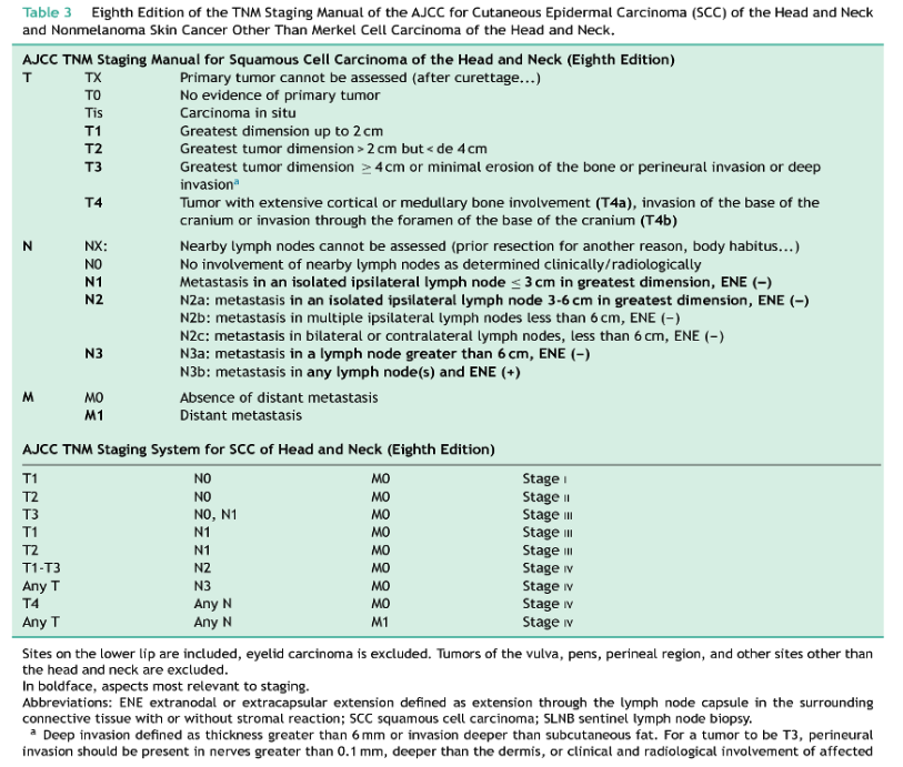
Stage
- Stage 1
- T1
- Stage 2
- T2
- Stage 3
- T3
- N1, T1-T3
- Stage 4
- T4
- N2-N3
Management
Treatment of low risk SCC
- Surgical
- Standard excision - should have 5mm clinical margin. Need to take excise down to subcutaneous fat.
-
1mm microscopic margin is acceptable.
- Mohs surgery - can be utilized as a skin-sparing technique where margin of 4mm would lead to a poor cosmetic outcome.
- Other options
- These options do not allow margin evaluation - should only be used for small low risk lesions in low risk sites.
- Curettage and electrodessication
- Cryosurgery - freezes tumours. Occurs from formation of intra and extracellular ice crystals, hypertonicity, and disruption of the phospholipid membrane.
- These options do not allow margin evaluation - should only be used for small low risk lesions in low risk sites.
- Treatment of multiple low risk lesions
- Commonly occurs on areas of sun damaged skin where there are multiple actinic keratosis.
- Options include
- 5-Fluorouracil cream
- Low dose systemic retinoids (isotretinoin)
- Options include
- Commonly occurs on areas of sun damaged skin where there are multiple actinic keratosis.
- Treatment of Bowen’s disease
- Surgery - 4mm margin
- Topical
- Topical 5-Fluorouracil 5% - BD for 4 weeks.
- Imiquimod
- Stimulate an immune response thus area will become inflammed - this indicates the cream is likely to be effective.
- Cryotherapy
- Currettage and electrodessication
Treatment of high risk SCC
- Surgery
- The recommended modality is MOHS surgery
- Standard excision is commonly performed but is inferior (higher recurrence rates)
- Treatment which doesn’t allow margin assessment is contraindicated (i.e. cryosurgery and curretage).
- Should aim for >6mm clinical margin
- A histological margin of >1mm is adequate.
- Management of nodal disease
- Aggressive surgical resection is primary treatment modality.
- If 2+ positive nodes, or one node >3cm, any node with extracapsular extension - patient should received adjuvant radiotherapy (N2 or N3)
- Radiation
- May be recommended as definitive treatment, adjunctive, or palliative treatment where complete surgical removal is not possible
- Indications for adjuvant radiation
- Tumours with positive surgical margins not amendable to further surgery
- Tumours with extensive PNI or large nerve involvement.
- Multi-focal tumour spread
- Presence of microsatellite tumours
- Lymph node involvement (N2 or N3)
- Massive local extension or intracranial involvement.
- Contraindications:
- Xeroderma Pigmentosum
- Basal Cell Naevus Syndrome
- May induce more tumours in treatment area
- Systemic therapies
- Targeted
- PD-1 inhibitors like Pembrolizumab are highly efficacious for locally advanced and metastatic SCC.
- Anti-EGFR
- Cetuximab is commonly used as immunotherapy.
- Chemotherapy
- Carboplatin, Paclitaxel are commonly used.
- Other adjunctive treatments
- Oral retinoids such as isotretinoin are commonly used
- Targeted
Unresectable Disease
- Platin-based Cisplatin with Radiotherapy
- Targeted therapy: Cetuximab (Epidermal Growth Factor Inhibitor) +/- Chemo +/- Radiotherapy
- Immunotherapy – PD-1 Inhibitor
- Cemiplimab (Australia)
Prognosis/Natural Hx
- Need to re-excise if positive margins
- Recurrence rate ≈ 50% if excision margin involved
- Tumour thickness correlates well with biologic behaviour
- Tumours that recur locally are usually ≥4mm
- Rare for tumour < 4mm to metastasize
- Tumours > 10mm thick → Mets frequent
- If LN positive disease → 15% 10YS
Recurrence
- Recurrences usually occur within 6/12
- Depends on grade & depth
- Risk factors for recurrence (Same as High risk factors)
- Patient factors:
- Immunosuppression
- Burns wound
- Previous RTx
- Patient factors:
- Tumour factors:
- Size of SCC > 2cm
- Depth of invasion >4mm
- Degree of differentiation
- Perineural / vascular involvement
- Location on head/neck