Section: UGI Sub-section: Oesophagus Curriculum: Curriculum, page 94
Definition
- Oesophageal luminal narrowing secondary to inflammation, fibrosis, or neoplasia
- Benign vs malignant
- Concerning if rapidly progressive
- Especially if associated weight loss
Aetiology
Extrinsic Causes
Vascular Rings
- Developmental abnormality
- Aberrant right subclavian artery from descending aorta
- Most common
- Travels behind oesophagus, incomplete ring
- Often asymptomatic, may cause posterior compression
- “Dysphagia Lusoria”
- Associated with non-recurrent laryngeal nerve
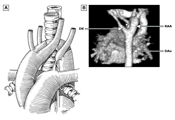
- Anomalous formation of right aortic arch with left ligamentum arteriosum and resultant retro-oesophageal left subclavian artery
- Complete ring
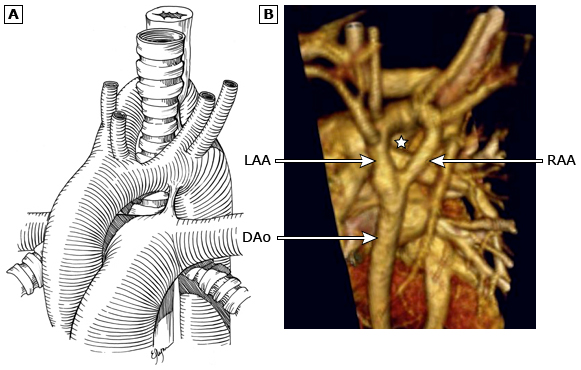
- Double aortic arch
- Fail to remodel appropriately, which results in two aortic arches both connecting the ascending and descending aortas
- Complete ring
- Other causes
- Left aortic arch with a right descending aorta and right ductus arteriosus (or ligamentum)
- Right aortic arch with a left descending aorta and left ductus arteriosus (or ligamentum)
- Left aortic arch with an aberrant right subclavian and a right ductus arteriosus (or ligamentum)
- Right aortic arch with mirror-image branching and a left ductus arteriosus (or ligamentum) arising from a retroesophageal dimple pointing to the left (also described as a double aortic arch with atretic left arch)
- Right aortic arch with retroesophageal left innominate and either a left ductus arteriosus or diverticulum
- Aberrant right subclavian artery from descending aorta
Double aortic arch
 Right aortic arch with aberrant left subclavian artery and left ligamentum arteriosum

Pulmonary artery sling
- Left pulmonary artery comes off right instead of main branch & courses between trachea & oesophagus
- Associated with other cardiac defects & foregut abnormalities
- All require repair to prevent tracheal or left PA stenosis

Innominate artery compression syndrome
- This syndrome occurs when the innominate (or brachiocephalic) artery originates later along the course of the transverse arch, resulting in takeoff to the left of the trachea.
- Anterior tracheal compression with associated tracheomalacia results when the aberrant innominate artery passes back to the right.
Intrinsic Causes
- Peptic strictures – most common (90%)
- Schatzki ring
- Rings
- Webs
- Plummer-Vinson syndrome
- Others
- Anastomotic
Peptic Strictures
- Concentric lower oesophageal ring at squamo-columnar junction
- 2-10% of GORD pts
- Due to chronic inflammation & fibrosis
- Most < 1cm in length
- Management
- Biopsy to exclude malignancy
- Must treat reflux initially – PPI’s, anti-reflux surgery
- Sequential dilation with 10mm balloon
- Perforation risk 1-2%, 90% success rate
- Division with laser of tight band
- May reduce need for repeated dilation
- Stent placement – temporary as needs to be removed
- Severe strictures may require resection.
Oesophageal Ring
- Types of rings
- A - normal smooth muscle contraction in the esophagus
- B - mucosal structure precisely at the squamocolumnar junction that is smooth and thin (≤5 mm in axial length). Owing to its location, a B ring is covered with squamous mucosa proximally and columnar epithelium distally
- Esophageal rings are usually mucosal but in rare cases may be muscular due to hypertrophy of an A ring.
- Schatzki ring, the most common type of esophageal ring, is a narrow mucosal B ring that is less than 12.5 mm in diameter
- Aetiology
- Hiatus hernia
- Schatzki rings are almost always associated with a hiatal hernia. In a study of 167 patients with a Schatzki ring, a hiatal hernia was detected in 97 percent
- Lies precisely at squamocolumnar mucosal GEJ
- Does not have a component of true oesophageal muscle
- Aetiology not understood
- ? Persistent apposition of two mucosal layers & fibrosis of submucosa.
- Almost always accompanied by a small hiatal hernia -? fibrosed proximal margin of sliding HH.
- Schatzki rings are almost always associated with a hiatal hernia. In a study of 167 patients with a Schatzki ring, a hiatal hernia was detected in 97 percent
- Eosinophilic oesophagitis
- Schatzki rings have also been associated with eosinophilic esophagitis
- However, more often patients with eosinophilic esophagitis have multiple proximal esophageal rings, long strictured segments, linear furrows in the mucosa, and eosinophilic abscesses that appear as white papules.
- Hiatus hernia
- Symptoms
- Often asymptomatic
- Episodic dysphagia (often large pieces of meat causing complete obstruction).
- Often spontaneously passes after a few minutes
- Investigations
- Barium swallow +/- endoscopy.
- Endoscopy
- Biospy ? eosinophilic esophagitis.
- Dilation in patients with eosinophilic esophagitis is usually reserved for patients with rings who have not responded to medical therapy.
- Biospy ? eosinophilic esophagitis.
- Management
- Endoscopic
- Dilation - Large caliber bougie (≥50 French) or balloon dilator (18 to 20 mm)
- Endoscopic
Oesophageal Web
- Thin, membranous structures that partially or completely compromise lumen.
- Only involve mucosa & part of the submucosa
- Aetiology
- Congenital
- Mostly lower 2/3, failure of coalescence of oesophageal vacuoles which normally leads to complete luminal patency between 25-31 days of embryologic development
- Most likely circumferential, thick & rough
- Acquired
- More common, anterior cervical oesophagus, post-cricoid, thin mucosal fold
- Associated with:
- Plummer-Vinson Syndrome
- Dermatological conditions
- Bullous pemphigoid, Epidermolysis bullosa, pemphigus vulgaris, psoriasis, Stevens-Johnson syndrome
- Congenital
- Mostly asymptomatic. Occasional solid food dysphagia.
- Investigations
- Barium endoscopy 1st investigation
- Endoscopy may miss it high in the cervical oesophagus
- Management
- Membranous disruption with endoscope or bougie or balloon dilation
- Very rarely require surgical excision with mucosectomy
- Slight increased risk of oesophageal cancer
Plummer-Vinson Syndrome
- also known as Paterson-Brown-Kelly Syndrome
- Upper oesophageal web
- Dysphagia
- Iron deficiency anaemia
- ± associated with glossitis & angular stomatitis / cheilosis
- Risk of Oesophageal cancer or pharyngeal Squamous cell carcinoma
- Rare; mostly females
- Typically seen in
- Edentulous, middle-aged, malnourished women with atrophic oral mucosa, glossitis, spoon-shaped fingernails & Fe deficiency anaemia
Other causes
- Eosinophilic oesophagitis
- Infection
- Crohn’s
- Neoplasia, post treatment strictures (radiotherapy, EMR etc)
- Don’t dilate malignant stricture – just pass feeding tube and workup
- Can stent later
- Don’t dilate malignant stricture – just pass feeding tube and workup
- Pill-injury
- Potassium, iron, beta-blocker, doxycycline, tetracycline, NSAIDs, bisphosphonates, quinidine
- Stick at 2nd constriction as there is a low peristaltic wave here
- Caustic Ingestion
Clinical Presentation
- Dysphagia
- Cnce lumen <13mm, meat/bread impaction
- Regurgitate food
- Odynophagia
- Weight loss
- Recurrent respiratory symptoms
- Difficulty breathing
- Caustic injury
Investigations
- Gastroscopy
- CXR
- Exclude pneumomediastinum/pneumothorax suggestive of perforation
- Hoarseness/stridor (caustic injury) = intubation early +/- nebulized steroids.
- Barium Swallow
- To diagnose a Schatzki ring; place prone turned slightly to right; patient takes in a large breath as bolus reaches GEJ.
- CTA for anomalous anatomy
Management
Chronic Strictures
- Endoscopic Rx
- Don’t bougie until see re-epithelialisation.
- Bougie strictures even if asymptomatic – may need daily for weeks
- Dilatation if less than 2 cm
- Remove impacted food (don’t push through ring/obstruction – perforation risk).
- Good for Schatzki ring, surgery not indicated unless dilatation fails or intractable reflux
- Savary Gillard Dilation
- Over guidewire, requires passage of endoscope beyond the stricture so wire not placed blind
- Dilate up 3 x successive sizes, bring back for repeat if needed
- Can assess force required for dilation
- Must do post procedure endoscopy check
- Ballon dilation “Through the scope dilatation”
- Don’t need to pass the stricture, can see through the balloon,
- Don’t need to pass pharynx repeatedly
- Webs
- Dilatation or piece-meal excision with biopsy forceps or laser lysis, bougie or balloon
Operative
- Slings, rings – repaired
- If pulmonary sling not repaired get LPA narrowing & tracheal stenosis
- Need open sternotomy + cardio-pulmonary bypass – anatomic re-positioning
- Webs – surgery for thick webs refractory to endoscopic Rx
- Transcervical or transthoracic approach to oesophagus
- Longitudinal myotomy & circumferential excision of web, circumferential re-approximation of mucosa with interrupted absorbable sutures followed by longitudinal closure of muscle
- Anti-reflux procedures ± Collis gastroplasty (as oesophageal shortening often present) –
- Creating a gastric tube & bringing it up into the thorax
- Resection and reconstruction- delay for at least 6 months.
- Stomach pull-up, versus jejunal loop interposition, versus long-segment colon interposition graft.
- Need to resect oesophagus/stomach – 1000 x increased risk of cancer