Section: UGI Sub-section: Oesophagus Curriculum: Curriculum, page 94
Definition
Malignant neoplasia of the oesophagus, which is mostly carcinoma
Incidence
- Uncommon (< Pancreatic Ca!)
- M > F (SCC 3:1, adenocarcinoma 7:1)
- Occurs in 5th-7th decades of life
- SCC more common than adenocarcinoma
- Incidence of adenocarcinoma increasing
- Esp. in White men → ~ 50:50
- Incidence of SCC in Western countries decreasing
- 5 / 100,000 in US/UK
- Most common in China (100 / 100,000), Singapore, Iran, Russia, S Africa
Classification
- Histologic
- SCC
- Further sub-classified on histology type
- Adenocarcinoma
- Undifferentiated
- Other
- SCC
- Location
- Cervical
- Upper-thoracic
- Mid-thoracic
- Lower-thoracic/GOJ
- GOJ adenocarcinomas classified further by Siewert & Stein Classification
Location of Tumour
- Cervical
- Between UOS and Sternal Notch
- 15-20cm from incisors
- Upper Thoracic
- Between Sternal Notch and Azygous Vein
- 20-25cm from incisors
- Between Sternal Notch and Azygous Vein
- Mid Thoracic
- Between Azygos Vein and Inferior Pulmonary Vein
- 25-30 cm from incisors
- Between Azygos Vein and Inferior Pulmonary Vein
- Lower Thoracic/GOJ
- Below Inferior Pulmonary Vein
-
30cm from incisors
- GOJ tumours further classified by Siewert & Stein Classification (See Gastric adenocarcinoma notes)
- Between UOS and Sternal Notch
Risk Factors/Aetiology
- SCC
- Smokers and drinkers, oesophageal carcinogen exposure
- Adenocarcinoma
- Fat white male with GORD

Risk Factors for Oesophageal SCC
- Smoking (RR17) and EtOH exposure (RR10) account for 90%
- Synergistic effect
- Achalasia
- Pharyngeal pouch
- Caustic Ingestion (esp. Alkali)
- Post-Radiation (Breast Ca etc.)
- Plummer-Vinson Syndrome
- Tylosis
- Autosomal dominant condition
- Hyperkeratosis of skin, Papillomas
- ? Achlorhydria from chronic PPI use
- Possible low acid leads to bacteria making carcinogenic nitrosamine compounds (not proven)
- Dietary
- Nitrosamines (fish, cured meat bacon, BBQ/pickled foods)
- Hot food/drink
- Human papillomavirus has also been implicated
Risk Factors for Oesophageal Adenocarcinoma
- Barrett’s Oesophagus (RR = 50)
- GORD (RR = 8)
- GORD ⇒ Metaplasia ⇒ Dysplasia ⇒ Ca
- Smoking (RR = 3.4)
- Obesity (RR = 2.4)
- Hx of Breast Ca with Rtx (RR = 4.8)
- Low SES
- H. Pylori
Clinical Presentation
- Progressive dysphagia 75%
- Late sign, lumen usually well narrowed by this point
- Weight loss 50%
- Voice change / hoarseness
- Very late sign
- Coughing & Aspiration
- May be due to a tracheo-oesophageal fistula
- Fatigue
- Anaemia
- Most patients are node +ve at presentation
- 80%; or 50% if tumor < 5cm
- May have palpable Virchow Node
- Look for metachronous smoking-related cancer (head, neck or lung)
Pathophysiology
- NB: Often a significant submucosal extension of tumor (cranial > caudal) beyond the visible mucosal edge
- Therefore wide margins required
- 10cm proximal & 5cm distal
Pathophysiology of Oesophageal SCC
- Found Upper/Middle/Lower Oesophagus
- 1:5:2
- Invade both horizontally and vertically
- Upper tumours spread to cervical and upper mediastinal nodes
- Significant proportion also track down to perigastric nodes
- Distant mets spread to Liver, Lung, Adrenals, Kidneys
Macroscopic
- Appearance depends on depth of invasion
- 60% exophytic or fungating
- 25% ulcerative
- 15% infiltrative
- Can use lugols iodine to stain during endoscopy
- Normal mucosa stains (glycogen content)
- SCC doesn’t, stays white

Microscopic
- Get nests of cells with
- Central Keratin Pearls
- Intercellular bridges
- Abundant eosinophilic cytoplasm
- Graded well, moderately or poorly based on
- Extent of mitotic activity
- Nuclear atypia
- Degree of squamous differentiation (keratinisation)
- NB: Histology changes dramatically after neoadjuvant therapy
- Get necrosis, inflammation, fibrosis and granulomas
- Main variants
- Verrucous Carcinoma
- Locally aggressive, usually large before clinical presentation
- Well differentiated with minimal atypia
- Spindle Cell
- Mixed SCC and high-grade spindle cell component
- Highly aggressive with 5 yr survival only 10-15%
- Basaloid
- Highly aggressive with poor prognosis
- Verrucous Carcinoma
Molecular Pathology
- 80% show mutation with loss of tumour suppressor gene p53
- Also have mutations in retinoblastoma gene RB and p16
Pathophysiology of Oesophageal Adenocarcinoma
- Found in Lower Oesophagus and GOJ (80%)
- Incidence increasing heavily
- Related to GORD and obesity
- Micro
- Glandular structures formed by irregular columnar cells with mucus secretion
- Typically papillary or tubular histology
- Macro
- 50% have macroscopically infiltrative growth pattern
- Rarely polypoid
- Graded well, moderately or poorly differentiated
- According to glandular architecture
- 10% are mucinous/signet ring
- Up to 75% have LN involvement at diagnosis
Investigations
Clinical Assessment
- Determine symptoms and duration
- Family hx
- Medical comorbidities/fitness for surgery
- Assess nutritional status
- Pt wishes
Upper GI Endoscopy
- Main diagnostic tool
- Perform on any patient with dysphagia
- Endoscopic appearance varies, but usually seen as friable, ulcerated masses
- Earlier tumours can appear as nodules or ulcers
- Need to document
- Location – distance from incisors
- Nature – friable, firm, polypoid, ulcerated (Unlikely to be T1)
- Proximal & distal extent
- Relationship to Cricopharyngeus, GOJ, cardia
- Degree of obstruction
- Describe any co-existing Barrett’s
- Distensibility of stomach
- Do multiple biopsies, gives greater sensitivity – minimum 6-8
- Use Lugol’s iodine for suspected SCC
- Do not dilate stenotic tumours at initial endoscopy until histology back and pt further staged
- Contrast swallow only recommended if endoscopy cannot be done for some reason


CT Chest/Abdomen
- Initial staging for
- Nodal and distant disease
- N – stage ~ 30-80% accuracy
- Poor for T stage; accuracy 57%
- Can not differentiate T1 + T2
- T1a – nodal involvement 1-2%
- T1b – 20%
- Nodal and distant disease
CT PET
- Routine, but should be 2nd line after CT
- Before EUS
- If distant disease, then they do not need EUS
- If suspicious local node can guide EUS location
- Before EUS
- Best M staging tool
- Upstages in 15%
- NB: 10% GOJ cancers not PET AVID
- Cancers with diffuse infiltration less PET avid
- Recommended to differentiated ‘indeterminate lesions’ dx on CT
- NB: Risk of false positives or other findings that would need workup
EUS
- Better for T & N staging than CT
- Be selective
- Early-stage disease
- ? Deeper than T1 and would need neoadjuvant therapy
- Suspect T4 (aortic or airway involvement)
- Suspect nodal disease on PET
- Early-stage disease
- Recommended for pts without obvious locoregional or distant disease on CT scan
- Determine T stage and N stage accurately
- Best T stage (accuracy 90%; sens 59-74%)
- N staging (sens 75%; spec 85-98%, *) – increased sensitivity/specificity with EUS-FNA
- Over-stages T, under-stages N
- If finding of ascites → Cancer incurable in 75%, even with no other evidence of disease
- EUS-FNA can be useful for assessing nodal mets
- EUS changes management in up to 25% of pts + should be the test of choice for diagnosing mediastinal lymphadenopathy
- NB: May be impossible if obstructing tumour to get complete EUS
Staging Laparoscopy and Peritoneal Washings
- For patients with GOJ cancer only
- Involvement of the intra-abdo portion
- If finding of metastatic disease would alter management
- Do washings and send for cytology routinely
- May see occult liver disease
- Can see extent of tumour and nodes around left gastric pedicle
- Can see extent of tumour along greater curve of stomach
- Can this still be used as a conduit?
Bronchoscopy or Endobronchial USS (EBUS)
- Consider if concerns for tracheobronchial invasion on other workup (EUS etc.)
- EBUS better than bronchoscopy
- Sensitivity 90% and spec 90% for upper/middle third Ca if tracheobronchial invasion suspected
- Presents clinically as cough
- Also useful for examining carinal/tracheal nodes
- NB: Only useful for tumours above T4/Carina
Staging
AJCC 8th Edition TNM Staging

T stage
- T1a – Into Lamina Propria/Muscularis Mucosa but not into Submucosa
- Possible endoscopic rx
- T1b – Into Submucosa
- T2 – Into Muscularis Propria
- T3 – Into Adventitia
- T4a – Into surrounding structures that are resectable, e.g. Pleura, Pericardium, Azygos Vein, Diaphragm, Peritoneum
- T4b – Into surrounding structures that are not resectable
- Aorta, Vertebral Body, Trachea
N stage
- N0: No regional LN mets
- N1: Mets to 1 or 2 regional LN
- N2: Mets to 3 to 6 regional LN
- N3: Mets in 7 or more regional LN
M stage M0: No distant mets M1: Distant mets
Staging System differs from SCC vs. Adenoca
- Staging Complex
- If no neoadjuvent treatment - also depends on grade of tumour and location
- If neoadjuvent treatment - then just TNM
- Generally:
- Stage 1 - T1
- Stage 2 - T2 or T3
- Stage 3 = LN involvement or T4
- Stage 4 = Distant mets or Significant LN involvement or T4
Pre-operative workup (for Fitness to Surgery)
- Medical Hx and clinical exam
- Bloods
- FBC, U+E, LFTs, ABGs
- ECG
- Echo
- Pulmonary Function Tests – FEV1/FVC
- Basic exercise tolerance
- Distance on flat, ? 2 flights of stairs
- CPET testing if borderline
Management
- All patients discussed at MDM
- HGD or T1a
- Consider EMR if appropriate
- T1b
- Oesophagectomy and Lymphadenectomy
- T2+
- Neoadjuvant treatment
- Adenocarcinoma
- FLOT4 as per ESOPEC trial
- SCC
- Proximal
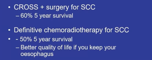
- Definitive chemoradiotherapy
- Junctional or mid
- Young
- Neoadjuvent CROSS Trial + surgery (Trimodal therapy)
- Slightly better survival and local recurrence rates then definitive chemoradiotherapy
- Neoadjuvent CROSS Trial + surgery (Trimodal therapy)
- Old
- Definitive chemoradiotherapy
- Salvage surgery can be considered
- Issue: If you give some one with a junction cancer definitive chemorads then with surgery there is a high chance of conduit necrosis as the stomach will have been in the radiation field.
- Young
- Proximal
- Adenocarcinoma
- Neoadjuvant treatment
Unresectable Disease
- T4b
- No plane between Aorta and Oesophagus
- Invading Trachea
- Distant mets
- Life expectancy < 12 months
Endoscopic Therapy
- Consider in high-grade dysplasia or T1 cancer
- Needs to be
- Unifocal, and entire lesion can be removed
- Intramucosal disease
- Healthy oesophagus
- Usually if can’t lift, will be T2
- At best, EMR will get complete resection (of T1a)
- At worst gives very good biopsy (for T1b)
- But no lymphadenectomy
- NB: T1a Adenoca should have no LN involvement
- But SCCs can have involved nodes (3.5-5.6%) due to different lymphatic drainage patterns in the Thoracic oesophagus
- Need to ablate the remaining Barrett’s (RFA, HALO)
- Complications – Bleed (1%), Perforation (0.5%), Stricture (up to 30% depending on amount excised)
- Require close ongoing surveillance, risk of metachronous Ca 20%
- T1a tumours with high risk factors (LVI, large, Poorly Differentiated) may be better served by Oesophagectomy if pt fit
Neoadjuvant Therapy
- Background
- High morbidity and mortality with surgery
- Positive radial margins in 20%
- Locoregional recurrence 40-60%
- 5Y survival poor
- Palliative studies of radiotherapy & chemotherapy shown to have benefit
- Neoadjuvant studies have show improved outcomes
- Benefits of neoadjuvant treatment:
- Improved tumour oxygenation pre-op than post-op
- Therefore more penetration of chemo/rad
- Potential to improve resectability and reduce risk of tumour spillage
- Increase chance of R0 resection and reduced risk of local recurrence
- Improved chance of treating undetectable micrometastasis
- Patient able to tolerate neoadj treatment better prior to major surgery
- May reduce tumour volume pre-op
- Improve dysphagia/nutrition prior to surgery
- Spare patients with early progression from major surgery
- Improved tumour oxygenation pre-op than post-op
- Disadvantages:
- Poorly tolerated by some pts
- May make pt physiologically less well prior to major surgery
- May have disease progression prior to surgery
- Though generally these patients would have done poorly anyway
Treatment paradigm
- Adenocarcinoma
- FLOT4 as per ESOPEC trial
- Of note prior to ESOPEC (and similar evidence prior) CROSS was the standard and prior to that MAGIC (ECF) was the standard.
- FLOT4 as per ESOPEC trial
- SCC
- Proximal
- Definitive chemoradiotherapy
- Junctional or mid
- Young
- Neoadjuvent CROSS Trial + surgery (Trimodal therapy)
- Slightly better survival and local recurrence rates then definitive chemoradiotherapy
- Neoadjuvent CROSS Trial + surgery (Trimodal therapy)
- Old
- Definitive chemoradiotherapy
- Salvage surgery can be considered
- Issue: If you give some one with a junction cancer definitive chemorads then with surgery there is a high chance of conduit necrosis as the stomach will have been in the radiation field.
- Young
- Proximal
Summary of papers
- OEO2 trial
- 2002 + updated 2009:
- Long term follow-up shows pre-op chemo improves survival
- 5yr survival 23% (chemo + surgery) and 17% (surgery alone)
- Higher R0 resections and good evidence of down-staging
- Included SCC and AC. 400 each arm
- Chemo = Cisplatin & 5FU
- Other studies
- Neoadjuvant chemo increases survival: 34 → 43% at 2YS
- Neoadjuvant chemo decreases circumferential margin positivity
- But previous studies in potentially resectable cancer have possibly shown a non-significant survival advantage in the long term
- e.g. RTOG 8991 = Intergroup 0113
- 2002 + updated 2009:
- MAGIC Trial
- NEJM 2006; 355:11
- Showed survival advantage out to 5yrs for ECF
- MAGIC trial, page 1
- Pre & post-operative ECF and surgery, vs surgery alone (503 patients)
- Inclusions:
- Any age, WHO 0-1
- Histologically confirmed adenocarcinoma
- ≥ T2
- Gastric or distal 1/3 oesophagus
- No metastatic disease, resectable
- Exclusions
- Previous chemo/rad, poor heart/kidneys
- Epirubicin, Cisplatin, 5FU (ECF)
- 3 x pre-op cycles and 3 x post-op cycles
- Surgery
- 6/52 post chemo, then restart 6-12/52 post surgery
- NB: Only 40% completed post-op cycles
- Type and extent of surgery operator dependent
- NB: 74% of study was Gastric Ca
- Results
- Decreased tumour size and stage
- Significantly improved progression free and overall survival
- 5yr survival 23% vs 36%
- NEJM 2006; 355:11
- CROSS Trial
- CROSS Trial (NEJM 2012)
- CROSS Trial, page 1
- Neoadjuvant chemoradiation vs surgery alone
- Carboplatin and Paclitaxel + radiotherapy 40g in 23F for 5 weeks
- Surgery within 4-6/52 post
- Inclusions
- T1/N1, or T2/T3 NO or N1
- Oesophageal and GOJ cancers (22% SCC, 75% adeno)
- Results
- 368 patients
- Significantly higher R0 resection (56 vs 90%)
- 5yr survival advantage
- 46% vs 36%
- Overall survival longer 48m vs 24m
- SCC 80m vs 50m
- Adenocarcinoma 40m and 28m
- Results not as good for adenocarcinoma as they are for SCC
- CROSS Trial (NEJM 2012)
- ESOPEC, page 1
- Methods
- RCT - FLOT4 vs CROSS Trial
- Inclusion: cT1 cN+, cT2–4a cN+, or cT2–4a cN0 disease
- Excluded T1 N0, T4b
- Primary end pointL overall survival
- Results
- FLOT4: 221 and CORSS 217 patients
- OS 57% FLOT vs 50% CROSS - HR 0.7 for death
- Progression free survival - 51% FLOT, 35% CROSS
- Adverse event (g3 or above) 58% FLOT vs 50% CROSS
- Mortality 3.1% FLOT vs 5.6% CROSS
- Conclusion
- FLOT led to improved survival
- Methods
Surgery
- Indicated for T1b tumours or higher
- If < 75yrs and fit
- If possible to achieve R0 resection (T3 or less, no distant mets)
- Margins need to be large due to submucosal spread
- Aim 10 proximal, and ideally 10cm distally
- R0 if > 1mm clear
- Lymphadenectomy
- Gives optimal staging and improved locoregional control
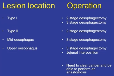
- 2 or 3 field procedure – 2 stage done in our institution
- Optimal no. of LN removed = 18
- No clear differences/survival advantage for neck dissection
- Type of Operation
- High (within 5cm of Cricopharyngeus) – Definitive chemo/rad
- Proximal 1/3 – 3 stage
- Middle 1/3 – Ivor Lewis Oesophagectomy
- Distal 1/3 – Ivor Lewis Oesophagectomy
- Gastric conduit (95%)
- Can use colon or jejunum
- Post-op jejunostomy
- Not done as a palliative operation
Lymphadenectomy Fields in Oesophagectomy
- Three-field Lymphadenectomy
- Removal of the first and second fields along with a neck dissection clearing the Brachiocephalic, deep lateral and external Cervical nodes, as well as right and left Recurrent Nerve lymphatic chains (deep anterior cervical nodes)
- Two-field Lymphadenectomy
- Removal of Para-aortic nodes, right and left pulmonary hilar, Paraoesophageal nodes, Subcarinal and Right Paratracheal nodes
- One-field Lymphadenectomy
- Removal of Diaphragmatic, Right and Left paracardiac, Lesser Curvature, Left Gastric, Coeliac, Common Hepatic and Splenic Artery nodes
Route of Reconstruction
- Pre-sternal
- Only if both other routes compromised by previous reconstructions
- Retrosternal
- Used if emergency treatment of anastomotic dehiscence causing posterior mediastinal sepsis
- Posterior mediastinal
- Preferred rout
- Preferred rout

Presternal Route
- Historical only
- Longest route
- 4cm longer than posterior mediastinal
- 2cm longer than retrosternal
- Only indication is if both other routes compromised by previous reconstructions
Retrosternal Route
- Potential space between sternum and anterior mediastinum developed
- 2cm longer than posterior mediastinal
- Unnatural position of cervical oesophagus in front of trachea, causes unpleasant sensation when swallowing
- Used for reconstruction when anastomotic leak causes posterior mediastinal sepsis
Posterior Mediastinal Route
- Preferred route of reconstruction in primary surgical excision of the oesophagus
- Shortest route between abdomen, apex of thorax and neck
- Gastric or colonic substitutes easily passed through posterior mediastinum after completion of mediastinal dissection in thorax
- No attempt made to close pleura
Organ of Reconstruction
- Determined by site of the primary lesion
- Should be kept as simple as possible
- Stomach
- Colon
- Jejunum
Reconstruction Using Stomach
-
Best conduit if anatomy allows
-
Five broad principles and practices to prepare stomach as conduit
- Use isoperistaltic stomach maintaining vascular supply
- Excision of the lesser curve
- Preservation of the intramuscular vascular arcade
- High point of the stomach
- Gastric Drainage
-
Additional methods of stomach lengthening
- Kocher manoeuvre
- Excision of lesser curve of stomach
- Incision of serosa on gastric wall
-
Isoperistaltic stomach maintaining vascular supply
- Right Gastroepiploic and Right Gastric Arteries and Veins are vital to stomach viability when used as conduit
- Greater omentum opened
- Gastroepiploic Artery is carefully identified and preserved
- Vascular arcade interrupted at point where right gastroepiploic meets Left
- Short Gastrics ligated and divided

- Excision of the Lesser Curve
- Any cancer of lower 2/3 oesophagus requires clearance of lesser curve nodes
- +Left gastric, common hepatic, and proximal splenic A nodes
- Left Gastric Artery should be ligated at its origin
- Resect proximal ½ of lesser curve, including cardia
- Preserve Right Gastric
- Maintains intramural vascular network
- Preserve Right Gastric
- Aim gastric conduit 5cm width or greater
- Less risk ischaemia
- Any cancer of lower 2/3 oesophagus requires clearance of lesser curve nodes

-
Preservation of the Intramuscular Vascular Arcade
- Extensive intramuscular anastomoses exist between greater and lesser curves
- Need to maintain this by not resecting too much of lesser curve
- Maintain right gastric supply to lesser curve
- Take care when ligating short gastrics, do away from greater curvature to avoid damage to intramural network
- Fundus blood supply from Right Gastroepiploic Artery through this intramural arcade
- Important as this is the region for anastomosis
-
High Point of the Stomach
- Stomach very flexible
- High point is the logical place for anastomosis
- Easily identified
- Traction in upward direction after all mobilisation complete

- Gastric Drainage
- Pyloroplasty or pyloromyotomy contentious
- Some evidence pyloroplasty reduces incidence of gastric outlet obstruction
- Minimal short-term complications
- Pyloroplasty routinely done by Companion series authors
- Reduces early life-threatening complications of:
- Gastric stasis and aspiration
- And late complications of
- Vomiting and bloating
- Reduces early life-threatening complications of:
- Heineke-Mikulicz Pyloroplasty
- Longitudinal incision, closed horizontally
- Pyloroplasty or pyloromyotomy contentious
Reconstruction Using Colon
-
Indications
- Tumours needing extensive oesophageal and gastric resection
- NB: With thorough staging, very few are actually suitable for resection
- Previous gastric resection
- Can consider if failed gastric conduit (necrosis) in initial attempted anastomosis
- Tumours needing extensive oesophageal and gastric resection
-
Colon recommended over jejunum as gives larger reservoir
-
Disadvantage
- Function of the conduit deteriorates over time
- Not as durable as stomach in the long term
-
Aim to use isoperistaltic segment depending on individual variations in vascular anatomy
- Transverse Colon based on Left Colic vessels
- Cannot perform if Marginal Artery not sufficient at Splenic Flexure
- Right Colon based on Middle Colic vessels
- Transverse Colon based on Left Colic vessels
-
Technical Considerations
- Colonoscopy and CT-Angio prior
- Pre-op mechanical bowel prep and PO antibiotic cover to sterilise bowel for 48hrs pre-op
- Free omentum from hepatic flexure, transverse, splenic flexure
- Mobilise entire colon to deliver outside, to inspect blood supply
- Sigmoid mobilisation gives length to tunnel transverse to chest
- Divide proximal colon, make anastomosis to oesophagus, then get rid of redundancy by anchoring sutures to hiatus
- Re-establish continuity of colon (end-to-end anastomosis)
- Perform colo-jejunostomy or colo-gastrostomy with the conduit
Reconstruction Using Jejunum
-
Third choice after stomach and colon
-
Can do
- Roux-en-Y
- Segmental interposition
- Free jejunal transfer with microvascular anastomosis
- If high in the neck
-
Anastomotic Principles
- Adequate blood supply
- Tension free
- Epithelial to epithelial approximation
- Precise layer to layer sutures
- No difference in single/double layer
- No difference in sutures/staples
- ? wrap with transposed omentum
See Oesophagectomy
Complications of Surgery
- 60% get complications
- Clavien-Dindo ≥ 3 17%
- 30-day mortality 2%
- 90-day mortality 4.5%
- Immediate
- Intra-op
- Bleeding
- Injury to other structuresàRLN, Phrenic N, thoracic duct, L)MB etc
- Anaesthetic complications
- Intra-op
- Early
- Pulmonary complications
- Aspiration
- Pneumonia
- CV complications
- AF (Common)
- MI
- Chylothorax (5%)
- Anastomotic leak (2-5%)
- Conduit already relatively ischaemic, and if in the neck then may be under a
- degree of tension o Conduit Ischaemia
- DVT/PE
- Pulmonary complications
- Late
- Functional Conduit Disorders
- Dysphagia (65%, 3-5% severe) – Need to consider stricture
- Reflux (60-80% depending on how diagnosed)
- Delayed emptying (50%, vagotomy)
- Dumping (up to 50% , improves with time)
- GOO
- Hernia
- Ventral
- Thorax
- Diaphragmatic
- Functional Conduit Disorders
Long Term Issues
- GORD 60%
- Diarrhoea 50%
- Bloating 45%
- Dysphagia 40%
- Dumping Syndrome 15%
Anastomotic Leak and Leak from Gastric Conduit
- Early leak (48-72hours)
- Technical fault
- If condition of patient good – reoperate to correct fault
- Late leak (5-10 days)
- Usually due to conduit ischaemia or tension on anastomotic line
- If leak at the oesophagogastric join
- Operating at this time is hazardous and often detrimental
- Use intensive non-operative management
- NG decompression
- Radiologically guided percutaneous drains – chest and mediastinum
- Antibiotics
- Jejunostomy feeding
- Don’t stent
- Late anastomotic leakage does not result in high mortality
- If leak at Gastric resection line dehiscence
- Need to re-operate as large amounts of leakage
- Anastomotic leak, cervical oesophagogastrostomy ≈ 20%
- Can drain
Conduit Necrosis
- Can be catastrophic complication
- Must diagnose early with endoscopy
- Resuscitate
- IV abx
- Return to theatre
- Exclude with cervical oesophagostomy
- Close viable gastric remnant
- Place feeding jejunostomy (if haven’t already)
- Once stable – colonic interposition
Chylothorax
- Transthoracic 2-3%, trans-hiatal up to 10%
- Apparent by day 7
- Increase in chest fluid after commencement of diet
- Results in massive malnutrition, immunosuppression and reduction in CD4 count due to loss of WCC/fat nutrition
- Chyle = intestinal lymphatic fluid enriched with fat
- Confirm by measuring TAG concentration of drain output
- Low output leak < 500ml/day
- Treat with enteral feed with medium chain fatty acids or TPN
- Thoracic duct should transport 4L/day
- Treat with enteral feed with medium chain fatty acids or TPN
- Large leak
- Should be managed with return to OT and ligation of thoracic duct
- Prophylactic abx with Cotrimoxazole (covers pneumocystis)
RLN Palsy
- At risk during cervical oesophagectomy or high anastomosis
- Usually only unilateral – usually contralateral nerve can compensate
- Unilateral RLN injury
- Varying degrees of hoarseness
- Microaspiration
- Coughing
- Other symptoms seriously affecting patients’ quality of life
- Bilateral RLN injuries
- May induce aphonia
- Dyspnoea
- Asphyxia
- Can be life threatening
Gastric Outlet Obstruction
- Prevented by routine pyloroplasty
- Always bring entire stomach conduit into chest
- Prevents changes of pressure across stomach
- Low dose erythromycin or metoclopramide can reduce effects
Other Complications
- Dumping Syndrome
- Relatively common but most resolves in 12 months
- Manage by avoidance of high carb loads
- Duodeno-gastro-oesophageal reflux
- Common, can be acid or alkaline reflux
- Control with motility agents and PPI
- Symptoms can be troublesome
- More common for thoracic anastomosis than cervical
- Benign anastomotic stricture
- Common
- Usually respond to single dilation
Adjuvant Therapy
Adjuvant Treatment
- Chemotherapy
- Low quality of evidence - each patient should be discussed at an MDT
- In patients who have had neoadjuvent treatment
- Residual pathological disease
- Approach undefined
- Can consider with different chemotherapy regime
- No residual pathological disease
- Not recommended
- Residual pathological disease
- If patient has not had neoadjuvent treatment
- SCC
- Not indicated after R0 resection of SCC
- Irrespective of nodal status
- Not indicated after R0 resection of SCC
- Adenocarcinoma
- Treatment can be considered in > T2 / N+
- SCC
- ChemoRTx:
- ? some survival advantage in patient who have not had neoadjuvent treatment
- based on Intergroup 0116 Trial:
- Inclusion - primaries ≥ T3 and/or node-positive, gastric / GOJ
- Med survival 27 vs 36 months
- But this study has been criticized, as only 40% of pts had a R0 resection
- based on Intergroup 0116 Trial:
- ? some survival advantage in patient who have not had neoadjuvent treatment
- Radiotherapy alone
- not recommended in one RCT
- If radiotherapy given; 60-64Gy total - 5 days a week for 7 weeks
- Immune checkpoint inhibitors
- Indicated for patients with residual disease (not funded currently in NZ)
- Nivolumab
- CheckMate 577 trial
- 794 patients who had received neoadjuvant CRT for esophageal or EGJ cancer (70 percent AC)
- Nivolumab (240 mg) or placebo every 2 weeks for 16 weeks followed by nivolumab 480 mg or placebo every 4 weeks
- Primary end point
- median disease-free survival- (22.4 versus 11 months) across all groups
- CheckMate 577 trial
Radical Chemoradiotherapy
- Consider if
- Very proximal tumour (cervical)
- Locally advanced (unable to get R0 resection)
- Patient not fit for resection
- SCC
- Chemoradiotherapy vs surgery alone:
- Survival rates are similar (German study & FFCD 9120 trial)
- But there are no major RCTs so Rx has to be tailored
- If no surgery, get increased local recurrence
- If surgery, get increased early (post-op) mortality
- 2yr survival 35-39% no different (but DFS better in surgery group)
- Survival rates are similar (German study & FFCD 9120 trial)
- ChemoRTx vs Surgery + ChemoRTx for advanced SCC: (FFCD 9102 trial)
- No diff
- Advanced disease should only be treated with CRT
- ChemoRTx vs RTx alone in SCC:
- ChemoRTx better
- Median survival 14 vs 9 months
- 5yr survival 27% vs 0
Management of Mets, Palliation, Recurrence
- Indications for palliation:
- Patient choice/comorbid
- Clinical/radiological distant metastatic disease
- T4 with invasion into airway/aorta (T4b)
- Peritoneal disease on laparoscopy
- Options depend upon previous treatment
- If patient had surgery previously
- → Chemoradiation
- If patient had surgery previously
- If patient had no previous surgery
- → Consider palliative surgery, if operable
- Palliative options = “Best supportive care”
Endoscopic Options
Self-Expanding Metal Stents (SEMS)
- Partially covered best (uncovered gets ingrowth, fully covered migrates)
- 3cm longer than tumour, prox + distal flange
- Deployed by endoscopy under fluoroscopic guidance
- Indications:
- Recurrence following chemoradiotherapy
- Contraindications to chemoradiotherapy
- Actual or impending fistula
- If Tracheo-oesophageal fistula – use covered SEMS
- Total dysphagia, incl. pts who are candidates for multimodal therapy
- Contraindications:
- Curable by multimodal therapy (unless total dysphagia)
- Stent placement < 2cm from UES
- Complete luminal occlusion
- Multiple multi-angulated lesions
- Inability to tolerate, high ECOG, or significant bleeding tendency/anticoags

Laser/Photodynamic Therapy
- Relieves dysphagia in up to 70%
- Rare as time consuming and requires multiple sessions.
- Indications:
- Ingrowth of stents
- Cervical oesophagus lesions that cannot be stented
- Duration of dysphagia free usually 4-16 weeks
- Pulses of energy that cause tissue necrosis and vaporization – tissue damage only occurs for 0.5mm beyond treatment site so rare to cause perforation
- Complications:
- Early:
- Chest pain (mucosal burn)
- Perforation – 5%
- Haemorrhage 1%
- Early:
- Late:
- Tumour regrowth – usually require monthly tx
- Delayed strictures
- Persistent dysphagia
Argon Plasma Coagulation
- Argon emitted as a jet of gas that then has high-freq electrical energy passed through it to ionize gas into ‘plasma’ which distributes a superficial ‘burn’ to tissues
- As tissue dries out and desiccates it becomes less conductive therefore limiting risk of perforation
Intra-Tumour 100% EtOH injection
- Consider if bleeding tumour etc.
- 0.5ml-1ml aliquots – can use as much as needed.
- Irregular tissue necrosis can occur
- Often need repeated treatments
Palliative Radiotherapy
- Successful in relieving dysphagia in ≈ 50% of patients
- SCC much more radiosensitive
- External beam
- 30-60Gy over 5-6 weeks
- Complications:
- Pulmonary fibrosis
- Stricture
- Fistula
- Recurrent dysphagia (only 40% pts have acceptable dysphagia)
- Brachytherapy (? Available in NZ)
- Brachytherapy applicator passed over the guidewire and positioned at the tumour by fluoroscopy.
- Pt the moved to a protected treatment room and connected to the ‘Selectron’ machine – transfers caesium-137 micropellets
- Usually given as a single dose of 10-15Gy
- RCT found this to have better effect for dysphagia with less morbidity
- NB: Initial response is for tumour to swell = Dysphagia initially worsens
- May heal with a stricture
Palliative Chemotherapy
- Improves survival from 9 → 12mths
- Cisplatin + 5-FU + Paclitaxel
- Response rates 20-50%
- Cisplatin, 5-FU, paclitaxel & anthracycline → promising response (Cochrane 2006)
- NJ or PEG
- ? Appropriateness of feeding in terminal cancer
Prognosis/Natural History

Main determinants:
- Depth of invasion
- Presence of LN mets (these correlate)
- Non-resectable Ca → 8-month average survival
- Post-op ≈ 5% mortality; morbidity 30-40%
- Post-op 5yr Survival ≈ 15-20%
- For SCC:
- 5YS after surgery =10-20%
- 35% if neoadjuvant Rx is given
- NB: Similar survival if SCC treated with chemoradiotherapy only
Follow-up
- Unit specific protocols
- ? 6/12 for 2yrs then annually
- May look at doing closer if radical chemo/rad used
- Could potentially benefit from earlier salvage surgery
- Most of discussion is about functional status and reflux symptoms
- Long term PPIs
- CT based on symptoms
- Weight loss etc.
- No evidence for routine scans
- No evidence that early diagnosis of stage 4 helps
- Follow-up routine endoscopy also contentious
Rarer Forms of Oesophageal Malignancy
- Account for approx. 2%
- Neuroendocrine
- Most common, usually distal, poor prognosis
- Carcinosarcomas
- ? Two separate tumors that have collided,
- Polypoid, lower 2/3
- Melanomas
- Lower 2/3, look for satellite lesions, poor prognosis
- Sarcomas
- Kaposi’s leiomyosarcomas
- Equal distribution along oesophagus