The following are “knows” from the syllabus
- Resection/reconstruction options
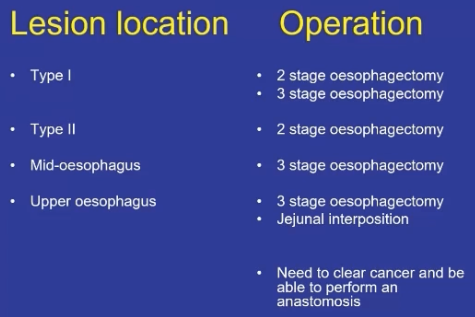
Approaches
- Two stage
- Three stage
- Trans-Hiatal
Ivor-Lewis Oesophagectomy
- Ivor-Lewis Oesophagectomy
- Laparotomy and Right Lateral Thoracotomy
- GA with single lung ventilation
- Op Viva
- Abdominal portion
- Thoracic portion
Abdominal Portion
- Laparosopic or open
- Rooftop incision, Fixed – table retraction – Adjuncts overview
- Check for peritoneal or nodal disease
- Mobilise stomach
- Greater curve
- Entering lesser sac through gastrocolic ligament, proceeding towards spleen first, then towards duodenum.
- Take care to preserve the right gastroepiploic vessel
- Divide all posterior adhesions so stomach can slide
- Divide short gastrics (place pack behind spleen to bring forward)
- Mobilise lesser curve.
- Kocherise duodenum
- Lymphadenctomy
- Divide Left gastric vessels at origin and perform lymphadenctomy
- Left Gastric, Coeliac, Common Hepatic, proximal splenic
- Divide Left gastric vessels at origin and perform lymphadenctomy
- Greater curve
- Conduit
- Create the gastric conduit with multiple firing of a stapler parallele to the GC with proximal aspect going toward apex of fundus
- Aim 5cm margin distal to GEJ. If radioRx, may need a 5cm margin from field for healing
- Suture conduit to transected oesophagus in correct orientation to allow pull through into the thorax if thoroscopic
- GOJ dissection
- Develop space between crura up into mediastinum.
- Divide a rim of diaphragm around the hiatus, including the crura themselves, so that a circumferential rim is resected with the oesophagus.
- Resect pleura bilaterally along oesophagus.
- Mobilise the lower 1/3 of oesophagus from pericardium and aorta up to level of tracheal bifurcation – all tissue from anteriorly is taken enblock up to pericardium
- Consider insertion of a feeding jejunostomy
- Consider pyloroplasty because you have interrupted vagal input (100U of botox in 4 quad at NSH)
- 2 drains.
- Close abdomen.
Thoracic Portion
- Position
- Left lateral decubitus position and break table to spread ribs – held by bean bag/supports.
- Right arm on padded rest, left arm stretched in front.
- Left lateral decubitus position and break table to spread ribs – held by bean bag/supports.
- Incision
- Right Posterolateral thoracotomy 4th/5th rib space:
- Curvilinear incision from spinous process of T3, inferior to tip of scapula, then forward to 3-4cm below right nipple
- Divide through muscles and enter 4th intercostal space. Excise 1 inch of posterior aspect of 5th rib to allow retraction of the rib cage
- Insert Finichetto retractor and ask anaesthetist to collapse lung
- Right Posterolateral thoracotomy 4th/5th rib space:
- Mobilise
- Divide inferior pulmonary ligament
- Retract lung forward to expose posterior mediastinum
- Dissection
- Pleura incised anterior to esophagus, along posterior edge of pericardium up to azygous arch and down to hiatus posteriorly along the aortic edge
- Divide pleura anterior to azygous and divide azygous vein
- Incircle the oesophagus and put a Penrose drain around it
- Dissection of thoracic oesophagus to proximal extent with en bloc resection of surrounding fatty tissue containing nodes
- Careful not to injure the Vagus, L) main bronchus, phrenic nerve, thoracic duct
- Out my institution we take the Thoracic duct just above the level of the aortic hiatus
- Transect proximal oesophagus
- Anastomose
- Anastamosis via side-to-side with purple endoGIA stapler
- Close the common enterotomy hand-sewen
- Ensure no tension, fire stapler.
- Omemntum inbetween bronuch and join
- Close opening in lesser curve and oversew entire staple line
- Alternatively do handsewn anastomosis with interrupted single all layer 3/0 vicryl
- Insert 2 chest drains between 8th and 9th intercostal spaces, anterior one to apex near anastomosis, and posterior one to lower chest.
- Approximate ribs and close wound in layers
- Pass NG tube into duodenum.
- Routine NG necessary until bowel function returns
- Prevents tracheal acid aspiration and may prevent respiratory complications
Trans-Hiatal Oesophagectomy
- Has fallen out of favour given concern of inadequate LN harvest in chest
- Trans-hiatal approach with cervical incision
- Indications:
- Carcinoma of hypopharynx/cervical oesophagus (+ neck dissection)
- Intraepithelial SCC /high grade Barrett’s
- If unable to be treated with treated with EMR
- Does not remove mediastinal LNs
- Abdominal incision and through hiatus, then stomach pulled up, left cervical incision to perform anastomosis
- Pros:
- Avoids leak into chest, quicker
- No sig difference in survival between operations
- Cons:
- Higher rate of strictures (blood supply poorer)
- Risk to great vessels & airway from blind pull of conduit through mediastinum
- Mortality 4%, leak rate 3%
Three Phase Oesophagectomy (McKeown)
- En-bloc Oesophagectomy
- Aggressive with radical Thoracic & Abdominal Lymphadenectomy & WLE
- Removes
- Azygous
- Hemiazygous
- Intercostal veins
- Thoracic Duct
- 3 incisions (left neck, right chest, abdomen)
- Cervical anastomosis
- Mortality 4.5%, morbidity 50%, leak 8%
- No improvement in 5YS but has demonstrated increased morbidity
Lymphadenectomy Fields in Oesophagectomy
- Three-field Lymphadenectomy
- Removal of the first and second fields along with a neck dissection clearing the Brachiocephalic, deep lateral and external Cervical nodes, as well as right and left Recurrent Nerve lymphatic chains (deep anterior cervical nodes)
- Two-field Lymphadenectomy
- Removal of Para-aortic nodes, right and left pulmonary hilar, Paraoesophageal nodes, Subcarinal and Right Paratracheal nodes
- One-field Lymphadenectomy
- Removal of Diaphragmatic, Right and Left paracardiac, Lesser Curvature, Left Gastric, Coeliac, Common Hepatic and Splenic Artery nodes
Route of Reconstruction
- Pre-sternal
- Only if both other routes compromised by previous reconstructions
- Retrosternal
- Used if emergency treatment of anastomotic dehiscence causing posterior mediastinal sepsis
- Posterior mediastinal
- Preferred rout
- Preferred rout

Presternal Route
- Historical only
- Longest route
- 4cm longer than posterior mediastinal
- 2cm longer than retrosternal
- Only indication is if both other routes compromised by previous reconstructions
Retrosternal Route
- Potential space between sternum and anterior mediastinum developed
- 2cm longer than posterior mediastinal
- Unnatural position of cervical oesophagus in front of trachea, causes unpleasant sensation when swallowing
- Used for reconstruction when anastomotic leak causes posterior mediastinal sepsis
Posterior Mediastinal Route
- Preferred route of reconstruction in primary surgical excision of the oesophagus
- Shortest route between abdomen, apex of thorax and neck
- Gastric or colonic substitutes easily passed through posterior mediastinum after completion of mediastinal dissection in thorax
- No attempt made to close pleura
Organ of Reconstruction
- Determined by site of the primary lesion
- Should be kept as simple as possible
- Stomach
- Colon
- Jejunum
Reconstruction Using Stomach
-
Best conduit if anatomy allows
-
Five broad principles and practices to prepare stomach as conduit
- Use isoperistaltic stomach maintaining vascular supply
- Excision of the lesser curve
- Preservation of the intramuscular vascular arcade
- High point of the stomach
- Gastric Drainage
-
Additional methods of stomach lengthening
- Kocher manoeuvre
- Excision of lesser curve of stomach
- Incision of serosa on gastric wall
-
Isoperistaltic stomach maintaining vascular supply
- Right Gastroepiploic and Right Gastric Arteries and Veins are vital to stomach viability when used as conduit
- Greater omentum opened
- Gastroepiploic Artery is carefully identified and preserved
- Vascular arcade interrupted at point where right gastroepiploic meets Left
- Short Gastrics ligated and divided

- Excision of the Lesser Curve
- Any cancer of lower 2/3 oesophagus requires clearance of lesser curve nodes
- +Left gastric, common hepatic, and proximal splenic A nodes
- Left Gastric Artery should be ligated at its origin
- Resect proximal ½ of lesser curve, including cardia
- Preserve Right Gastric
- Maintains intramural vascular network
- Preserve Right Gastric
- Aim gastric conduit 5cm width or greater
- Less risk ischaemia
- Any cancer of lower 2/3 oesophagus requires clearance of lesser curve nodes

-
Preservation of the Intramuscular Vascular Arcade
- Extensive intramuscular anastomoses exist between greater and lesser curves
- Need to maintain this by not resecting too much of lesser curve
- Maintain right gastric supply to lesser curve
- Take care when ligating short gastrics, do away from greater curvature to avoid damage to intramural network
- Fundus blood supply from Right Gastroepiploic Artery through this intramural arcade
- Important as this is the region for anastomosis
-
High Point of the Stomach
- Stomach very flexible
- High point is the logical place for anastomosis
- Easily identified
- Traction in upward direction after all mobilisation complete

- Gastric Drainage
- Pyloroplasty or pyloromyotomy contentious
- Some evidence pyloroplasty reduces incidence of gastric outlet obstruction
- Minimal short-term complications
- Pyloroplasty routinely done by Companion series authors
- Reduces early life-threatening complications of:
- Gastric stasis and aspiration
- And late complications of
- Vomiting and bloating
- Reduces early life-threatening complications of:
- Heineke-Mikulicz Pyloroplasty
- Longitudinal incision, closed horizontally
- Pyloroplasty or pyloromyotomy contentious
Reconstruction Using Colon
-
Indications
- Tumours needing extensive oesophageal and gastric resection
- NB: With thorough staging, very few are actually suitable for resection
- Previous gastric resection
- Can consider if failed gastric conduit (necrosis) in initial attempted anastomosis
- Tumours needing extensive oesophageal and gastric resection
-
Colon recommended over jejunum as gives larger reservoir
-
Disadvantage
- Function of the conduit deteriorates over time
- Not as durable as stomach in the long term
-
Aim to use isoperistaltic segment depending on individual variations in vascular anatomy
- Transverse Colon based on Left Colic vessels
- Cannot perform if Marginal Artery not sufficient at Splenic Flexure
- Right Colon based on Middle Colic vessels
- Transverse Colon based on Left Colic vessels
-
Technical Considerations
- Colonoscopy and CT-Angio prior
- Pre-op mechanical bowel prep and PO antibiotic cover to sterilise bowel for 48hrs pre-op
- Free omentum from hepatic flexure, transverse, splenic flexure
- Mobilise entire colon to deliver outside, to inspect blood supply
- Sigmoid mobilisation gives length to tunnel transverse to chest
- Divide proximal colon, make anastomosis to oesophagus, then get rid of redundancy by anchoring sutures to hiatus
- Re-establish continuity of colon (end-to-end anastomosis)
- Perform colo-jejunostomy or colo-gastrostomy with the conduit
Reconstruction Using Jejunum
-
Third choice after stomach and colon
-
Can do
- Roux-en-Y
- Segmental interposition
- Free jejunal transfer with microvascular anastomosis
- If high in the neck
-
Anastomotic Principles
- Adequate blood supply
- Tension free
- Epithelial to epithelial approximation
- Precise layer to layer sutures
- No difference in single/double layer
- No difference in sutures/staples
- ? wrap with transposed omentum
Post-operative care
- Standardised unit pathway/ERAS
- HDU/ICU
- Fluid balance
- Epidural analgesia
- Early, intensive physiotherapy
- Antithrombotic measures
- Routine PPI
- Early nutrition from jejunostomy (day 1)
- Antibiotics 48hrs
- Gastrograffin swallow check after 4-5 days
- NB: No evidence for this to be done routinely
- Companion series suggest only do if sepsis, effusion or haemodynamic instability
- But ⇒ Most people do anyway
- Begin PO feeding after this, COFs > FOFS
Complications of Surgery
- 60% get complications
- Clavien-Dindo ≥ 3 17%
- 30-day mortality 2%
- 90-day mortality 4.5%
- Generic
- Pneumonia 14%
- Cardiac 17%
- AF 15%
- DVT/PE
- CVA
- Haemorrhage - rare
- Specific
- Anastomotic leak 12%
- 3% get anastomotic leak needing intervention
- Conduit necrosis 1.2 %
- Chyle leak 4-5%
- RLN palsy 4%
- Trache-oesophageal fistula 1%
- Gastric outlet obstruction
- Dumping syndrome
- Duodeno-gastro-oesophageal reflux
- Benign anastomotic stricture
Long Term Issues
- GORD 60%
- Diarrhoea 50%
- Bloating 45%
- Dysphagia 40%
- Dumping Syndrome 15%
Anastomotic Leak and Leak from Gastric Conduit
- Early leak (48-72hours)
- Technical fault
- If condition of patient good – reoperate to correct fault
- Late leak (5-10 days)
- Usually due to conduit ischaemia or tension on anastomotic line
- If leak at the oesophagogastric join
- Operating at this time is hazardous and often detrimental
- Use intensive non-operative management
- NG decompression
- Radiologically guided percutaneous drains – chest and mediastinum
- Antibiotics
- Jejunostomy feeding
- Don’t stent
- Late anastomotic leakage does not result in high mortality
- If leak at Gastric resection line dehiscence
- Need to re-operate as large amounts of leakage
- Anastomotic leak, cervical oesophagogastrostomy ≈ 20%
- Can drain
Conduit Necrosis
- Can be catastrophic complication
- Must diagnose early with endoscopy
- Resuscitate
- IV abx
- Return to theatre
- Exclude with cervical oesophagostomy
- Close viable gastric remnant
- Place feeding jejunostomy (if haven’t already)
- Once stable – colonic interposition
Chylothorax
- Fat absorption
- Transthoracic 2-3%, trans-hiatal up to 10%
- Apparent by day 7
- Increase in chest fluid after commencement of diet
- Results in massive malnutrition, immunosuppression and reduction in CD4 count due to loss of WCC/fat nutrition
- Chyle = intestinal lymphatic fluid enriched with fat
- Confirm by measuring TAG concentration of drain output
- Low output leak < 500ml/day
- Treat with enteral feed with medium chain fatty acids or TPN
- Thoracic duct should transport 4L/day
- Treat with enteral feed with medium chain fatty acids or TPN
- Large leak
- Should be managed with return to OT and ligation of thoracic duct
- Prophylactic abx with Cotrimoxazole (covers pneumocystis)
RLN Palsy
- At risk during cervical oesophagectomy or high anastomosis
- Usually only unilateral – usually contralateral nerve can compensate
- Unilateral RLN injury
- Varying degrees of hoarseness
- Microaspiration
- Coughing
- Other symptoms seriously affecting patients’ quality of life
- Bilateral RLN injuries
- May induce aphonia
- Dyspnoea
- Asphyxia
- Can be life threatening
Gastric Outlet Obstruction
- Prevented by routine pyloroplasty
- Always bring entire stomach conduit into chest
- Prevents changes of pressure across stomach
- Low dose erythromycin or metoclopramide can reduce effects
Other Complications
- Dumping Syndrome
- Relatively common but most resolves in 12 months
- Manage by avoidance of high carb loads
- Duodeno-gastro-oesophageal reflux
- Common, can be acid or alkaline reflux
- Control with motility agents and PPI
- Symptoms can be troublesome
- More common for thoracic anastomosis than cervical
- Benign anastomotic stricture
- Common
- Usually respond to single dilation