Section: UGI Sub-section: Oesophagus Curriculum: Curriculum, page 95
Definition
- Achalasia is a primary oesophageal motility disorder due to impaired/absent relaxation of LOS
- Thought to be due to loss of ganglion cells in the myenteric plexus to the LES leading to degeneration of neuromuscular function and failure of vagal induced relaxation with HTN of LES
- Leads to:
- Decreased/absent peristalsis of the oesophageal body
- Increased pressure of the oesophagus
- Chronic dilation of oesophagus
- Pre-malignant condition
- 10% chance of developing cancer over 20yrs
- SCC most common
- Due to mucosal irritation due to undigested fermenting food
- Even patients with treated achalasia have ongoing ca risk
Incidence
- 6/100,000 person years
- Young women and middle-aged men/women
- Age at presentation:
- Bimodal distribution - 20-40 & > 60yo
Classification
- Primary
- Due to loss of ganglion cells in myenteric (Auerbach) plexus – more common
- ? Idiopathic ? Infectious neurogenic degeneration
- Chicago Classification into 3 types:
- Type 1 = Classic Achalasia
- Abnormal median IRP and absent contractility (100% failed peristalsis)
- Type 2 = Failed peristalsis but pan-oesophageal pressurization
- Abnormal median IRP and absent contractility (100% failed peristalsis)
-
- with panesophageal pressurization in 20% or more swallows
- Type 3 = Spastic achalasia
- Abnormal IRP and evidence of spasm (20% or more swallows with premature contraction) with no evidence of peristalsis
- Type 1 = Classic Achalasia
- Due to loss of ganglion cells in myenteric (Auerbach) plexus – more common
- Secondary
- Malignancy → Pseudo-achalasia = rare presentation of CA @ GOJ
- Chagas disease – parasitic infection with Trypanosoma cruzi
- South America, Brazil
- Trypanosoma cruzi infection (bug bite) → Initially acute conjunctivitis, preauricular lymphadenitis, fever, anorexia → Long-term neurological disorders with diffuse ganglion cell involvement (includes destruction of myenteric plexus) → Abnormal motility → Disease in multiple organs, includes cardiomyopathy, megacolon, megaureter, & megaoesophagus
- Dx: serological tests
- DDx: DM, Polio, Sarcoid, Amyloidosis
Aetiology
- Unknown
- ? Degenerative disease of neurons
- ? Infections of neurons by virus
Pathology
- Degeneration of ganglion cells in the Myenteric Plexus of Auerbach, esp. body & distal oesophagus
- Cholinergic innervation of sphincter is intact
- Non-cholinergic noradrenergic innervation is lost
- Usually mediates LOS relaxation via post-ganglionic inhibitory neurons
- Through NO / VIP etc
- ? May be degeneration in Vagus Nerve ± Dorsal Motor Nucleus of Vagus
- Net result = Unopposed Acetylcholine stimulation and contraction
- Non-cholinergic noradrenergic innervation is lost
- Mucosa develops changes of chronic stasis, includes oesophagitis and ulcers
- Chronic stasis → Ulceration → Continuous irritation of the mucosa
- ↑ Bacteria → ↑ Nitrosamine
- Leads to dysplasia → SCC
Differential Diagnosis
- Secondary causes
- Malignancy → Pseudoachalasia = rare presentation of CA @ GOJ
- Chagas’ disease – parasitic infection with trypanosoma cruzi
- Diffuse oesophageal spasm
- Strictures
- Peptic
- Scleroderma related etc
- Caustic
Clinical Presentation
- Triad
- Progressive dysphagia (90%)
- Regurgitation of undigested food (60%)
- Weight loss (60%)
- Heart burn (40%), post prandial choking and nocturnal cough common.
- (Aspiration) pneumonias / respiratory complications (10%)
- Patients usually present late in the disease course
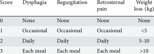
Eckardt score
Investigations
- CXR
- Barium Swallow
- Oesophageal Manometry – Gold Standard
- Endoscopy
- CT – usually not necessary
- Need to R/O pseudoachalasia
- Malignancy or post-surgery
- Need to R/O pseudoachalasia
CXR
- Often demonstrates a homogeneous, usually R sided, paramediastinal soft-tissue opacity ± mediastinal widening
- Air-fluid level behind heart
- Absence of gastric air bubble
- Tight LES not allowing air in
- Respiratory complications
- e.g. Aspiration pneumonia or lung abscess


Barium Swallow
- Dilated oesophagus
- Bird-beak sign in > 85%
- Abrupt, smooth tapering of distal oesophagus
- 1.5-4.5cm distal narrowing
- Various degrees of proximal oesophagus dilatation
- Mild < 4cm
- Severe > 6cm = Mega-oesophagus/sigmoid oesophagus
- Non-propulsive tertiary (dysfunctional) oesophageal contractions
- Failure of contrast to enter stomach

Manometry
- Gold standard test to diagnose achalasia, must have positive manometry before surgical management
- 3 subtypes
- 5 findings: 2 x LOS, 3 x Oesophagus
-
- Hypertensive LOS sphincter
-
35mmHg (normal = 10-20)
-
- LOS doesn’t relax
-
10mmHg during active swallow
-
- High oesophageal baseline pressures
- Relative to gastric baseline
-
- Low-amplitude waveforms
-
- No evidence of progressive peristalsis
- No evidence of progressive peristalsis
-


Endoscopy
- To detect complications
- Exclude malignancy, oesophagitis, ulcers, strictures
- Will find closed LOS but little/no resistance to endoscopy
- C.F. Malignant strictures
- Likely to see food/fluid residue
- High risk of aspiration
- Liquid diet few days prior, elevate head of bed
- Liquid diet few days prior, elevate head of bed

Management
Aim
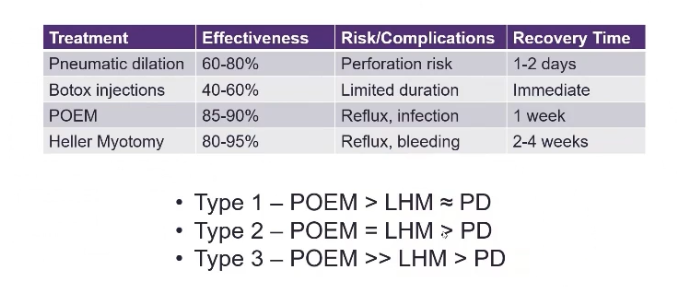
- Treat the functional obstruction at the LOS, while minimising the risk of reflux
- Medical
- Endoscopic
- Surgery

Medical Management
- Effective in a few patients only
- Mostly used in patients unsuitable for endoscopy/surgery
- Provides temporary relief only
- Ca channel blockers
- Sublingual nitrates
Endoscopic Management
- Balloon/pneumatic dilation
- Botox injection
- Per oral endoscopic myotomy (POEM)
Balloon/Pneumatic Dilatation
- Not for megaoesophagus
- Perforation risk is too high
- Advantages
- No surgery, lower cost
- Good if failed cardiomyotomy, or super obese etc
- Disadvantages:
- 20-35% incidence of GORD
- 5% risk of perforation
- 70% effective
- 70% of pts experience a relapse within 2 yrs
- Many more after 4 yrs
- More effective than Botox in the long term (>6 month)
- (Cochrane 2006)
Botox Injection
- Inferior treatment
- Blocks Ach release and prevents smooth muscle contraction
- Results in LES relaxation
- Overcomes unopposed Ach stimulation that results from loss of inhibitory neurons
- Effective in 85% of patients, but…
- 50% have recurrence by 6 months
- 30% of pts have on-going relief at 1-2.5yr f/up
- (even after multiple Rx)
- Causes inflammation at GOJ
- Can make subsequent myotomy more difficult, especially of multiple rounds of botox
- Therefore mostly used in elderly / poor surgical candidates
Per Oral Endoscopic Myotomy (POEM)
- Technique:
- Mucosal incision to make submucosal tunnel
- Inject adrenaline, indocarmine and saline to make bleb
- Hybrid knife used to develop submucosal tunnel extend to 2-3cm beyond GOJ
- Myotomy performed – circular muscle fibres divided
- Closure of mucosa with endoclips – distal to proximal
- Contra-indications
- Severe oesophagitis, coagulation disorders, portal HTN (varices), previous radiation to oesophagus
- Oesophogram Day 1 to exclude leak
- Soft diet progressed, BD PPI and soft diet for 2/52
- Risks:
- Pneumoperitoneum up to 50% - generally resolves
- Pneumothorax <5%
- Mucosal tears – require closure
- Bleeding in submucosa – urgent endoscopy to coagulate bleeding vessel
- GORD 20-60%
- No option of doing anti-reflux procedure with the technique - one of the main quoted downsides
Surgical Management
- Surgery is the preferred treatment
- Safest and most effective rx
- 85% have excellent symptomatic response
- Only 1% risk of perforation (cf 4% with dilation)
- Less than 0.5% mortality (cf 5% mortality for dilation)
- Procedure of choice:
- Modified Heller Myotomy ± Partial Fundoplication
- Gives better results to pneumatic therapy at 2yrs (prospective data)
- Oesophagectomy
- Occasionally required for end-stage disease – megaoesophagus or sigmoid oesophagus, failure of myotomy, or stricture not amenable to dilation
- Eliminates risk of malignancy
- Occasionally required for end-stage disease – megaoesophagus or sigmoid oesophagus, failure of myotomy, or stricture not amenable to dilation
- Lap surgery
- 5% complication rate (perf / leaks)
- 4% conversion to open surgery
- 80% pts had minimal or no Sx; men had poorer dysphagia Sx outcome (ANZ 06 76:558)
- Controversy re whether Fundoplication is necessary & which type
- Dor (Ant 180): may help seal the area of myotomy & prevent leak
- Toupet (Post 270): may help hold the myotomy open
- Nissen – get recurrent dysphagia
- Thoracic (L thoracotomy) / thoracoscopic route: useful for redo op (virgin territory)
- Modified Heller Myotomy ± Partial Fundoplication
- Complications:
- Mucosal perforation
- Recognize intra-op & repair
- Pneumonia
- Pleural effusion
- Para-oesophageal hernia
- Subphrenic abscess
- DVT
- Mucosal perforation
Laparoscopic Heller Myotomy
- Steep reverse Trendelenberg
- Port placement
- Retract liver with Nathanson
- Open phrenoesophageal membrane
- Excise gastroesophageal fat pad to expose GEJ, identifying and preserving anterior vagal
- trunk
- Dissect out anterior oesophagus; don’t go posterior.
- Myotomy
- Split longitudinal fibres and divide circular with harmonic scalpel (or scissors)
- Avoid too much electrocautery to prevent going too deep
- Enter submucosal plane, carry proximally 5-6cm from the GEJ and distally 2-3cm
- onto anterior gastric wall.
- Separate muscle edges for 50% of circumference.
- Endoscopy to rule out perforation and show obstruction is cleared
- Fundoplication
- 60% have reflux and positive pH studies without o Avoid 360 degree because of dysphagia
Prognosis
- Natural History
- Without tx pts will develop progressive dilation and can progress to end-stage achalasia with megaoesophagus (diameter > 6cm)
- 5% may require oesophagectomy
- Cancer risk:
- Risk increased 30-fold but still rare
- Annual incidence still < 1%
- Over 10 yr period risk ~ 2-3%
- Dx on average 15 yrs post dx of achalasia
- Typically SCC
- Pts have ↑ed incidence of malignancy: ≈ 3-5% risk of SCC (usually mid-oesophagus)
- Adenocarcinoma can also occur, but SCC much more common
- Surveillance not recommended
- Would require ~ 500 men and 2200 women to detect 1 cancer for each gender.
- After surgery:
- Good result in 80-90%; mortality <1%
- Cancer risk persists, especially in males (BJS 08; 95;1488)
- Adenocarcinoma can occur in pts who develop GORD (≈ 15%) after myotomy
- Recurrent dysphagia can be treated with dilatation or a second myotomy
- NB: Aperistalsis in the body of the oesophagus is usually irreversible
Follow-up
- Be conservative
- Surveillance endoscopy 3-5yrs