Section: UGI Sub-section: Gastric Curriculum: Curriculum, page 96
Definition
- Malignant epithelial tumours of the gastric mucosa with glandular differentiation
- Includes Siewert & Stein III
- Anything > 2cm from GOJ
- Excludes cardia (which is treated as GOJ/Oesophageal)
- Includes Siewert & Stein III
Epidemiology
- Incidence
- 7/100,000 in NZ
- Māori & PI > European 20/100,000
- M > F 2:1
- Except diffuse type F>M 1.2:1
- Rare < Age 50
- 2nd most common cancer world-wide
- Incidence decreasing, esp. in West (? due to H. pylori management)
- Incidence highest in Japan (>40/100,000)
- In migrants: subsequent generations (after 1st) acquire risk rate of their new environment
Risk factors

- Male
- Lifestyle factors
- Environmental (dietary) factors
- Infectious
- Genetics
- Blood group A
- Increased risk (RR ≈ 1.2) (Diffuse type)
- Blood group A
- Other Risk Factors
- Hypertrophic gastropathy (Menetrier’s disease)
- Pernicious anaemia
- Previous gastric surgery
- Adenomatous gastric polyps
- Hyperplastic polyps
Lifestyle Risk Factors
- Cigarette smokers:
- 2-3x increased risk of proximal Gastric Ca
- High EtOH intake
- Low socioeconomic status
- Obesity
Environmental (Dietary) Risk Factors
- High salt, smoked meats, poorly preserved food
- Nitrates in the food converted to carcinogenic N-nitroso compounds by bacteria in the stomach
- Note N-nitroso compounds also in cigarette smoke
- Synergism with H. pylori
- H. pylori promotes growth of bacteria that increase N-nitroso compounds
- H. pylori inhibits secretion of ascorbic acid, (see below)
- Nitrates in the food converted to carcinogenic N-nitroso compounds by bacteria in the stomach
- Fruit & vegetables → Protective
- Release ascorbic acid
- Ascorbic acid removes carcinogenic N-nitroso compounds and scavenges free radicals.
- Calcium, Vitamin A, Vitamin C (anti-oxidant activity) → Protective
- Stop conversion of nitrates to nitrites
Infectious Risk Factors
- H. Pylori
- EBV associated Gastric Ca
- Present in carcinoma cells
Genetic Risk Factors
- 90% sporadic
- 10% have an inherited familial component
Hereditary diffuse gastric carcinoma syndrome
- Autosomal dominant, Penetrance 70-80%
- Germline mutations in CDH1 (16q) gene
- Encodes the cell adhesion protein E-cadherin
- Predisposition to
- Diffuse-type Gastric Cancer
- Lobular Breast Cancer
- Signet Ring Colon Cancer
- Management
- Prophylactic Total Gastrectomy after age 20 recommended
- Surveillance Gastroscopy from 16
- Lifetime risk 80%, average age of cancer 38
- Breast screen from age 35
- Cumulative lifetime risk 50%
- Recommend colonic screening
- Prophylactic Total Gastrectomy after age 20 recommended
- Diagnosis
- For diagnosis (Familial condition) must have at least
- 2 family members with one being diagnosed < 50
- OR 3 family members
Other Genetic Risk Factors
-
Familial adenomatous polyposis
- 85% of patients have Fundic Gland Polyps
- 40% of these have dysplasia
- Also much higher frequency of potentially malignant duodenal polyps
- Surveillance Gastroscopy recommended
- 85% of patients have Fundic Gland Polyps
-
- Overview
- Hypoproteinaemic Hypertrophic Gastropathy
- Rare, acquired disease, Pre-malignant
- Characterised by massive gastric folds in the fundus and body
- Gives mucosa cobblestone or cerebriform appearance (looks like brain!)
- Pathology
- Foveolar Hyperplasia
- Expansion of surface mucous cells, and loss of parietal cells
- Typical changes seen on endoscopy
- Protein loss from the stomach
- Excessive mucus production
- Hypo or achlorhydria (gland atrophy)
- Foveolar Hyperplasia
- Aetiology
- Unknown - ? CMV infection in children, ? H.Pylori in adults
- Presentation
- Epigastric pain, vomiting, weight loss, anorexia, peripheral oedema
- Investigations
- Gastroscopy and biopsy to R/O gastric carcinoma or lymphoma
- Can do 24hr pH monitoring to detect low acid levels
- Management
- Anticholinergics, PPIs, Octreotide, H.pylori eradication
- Inconsistent results
- Consider total gastrectomy if
- Massive ongoing protein loss despite medical therapy
- Dysplasia or carcinoma on biopsy
- Anticholinergics, PPIs, Octreotide, H.pylori eradication
- Overview
-
Genes implicated in Gastric Ca:
- C-met, c-erbB2, K-sam, E-cadherin
- Microsatellite instability seen in 20-30% of intestinal type Gastric Ca
- E-Cadherin reduction/absence seen in 50% of diffuse type Gastric Ca
Other Risk Factors
- Pernicious Anaemia – 3-4x increased risk
- Achlorhydria and mucosal atrophy with antral/intestinal metaplasia
- Previous gastric surgery → RR 2
- Roux-en-Y or previous peptic ulcer disease surgery
- Due to
- Alkaline duodenal gastric reflux
- Or Bacterial overgrowth (increased conversion of nitrates)
Adenomatous Gastric polyps
- 10-20% risk of developing carcinoma
- Increases with size of polyp
- Polypectomy sufficient if no in-situ or malignant cancer
- Often has atypia
- If > 2cm, sessile or proven cancer then should be treated as per Gastric cancer
- Hyperplastic polyps associated increased risk
- Probably because they form in stomach with established gastritis
Classification Systems
Systems
- Siewert and Stein
- For GOJ cancers
- Lauren Classification
- Microscopic/Histological
- WHO Classification
- Bormann’s Morphologic Classification
- Paris classification
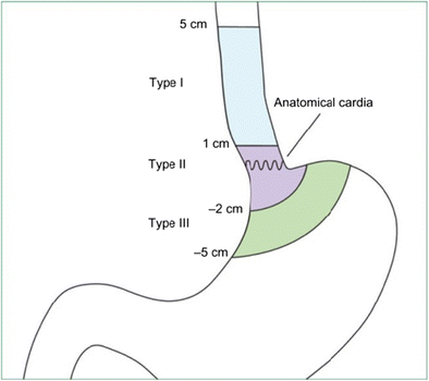
Siewert & Stein Classification
- System of anatomical classification used for adenocarcinomas of the GOJ
- Type I:
- Tumour center 1-5cm above GOJ
- Usually arises from Barrett’s
- Lymphatic spread to Mediastinal and Coeliac LNs
- Type II:
- Arises 1cm proximal or 2cm distal to the GOJ
- Type III: Proximal Stomach
- Center of tumor lies 2-5cm below the junction, but invades the cardia area
- Type I:
Lauren Classification
- Microscopic/Histological Classification system
- Intestinal Type
- Environmental → Gastric atrophy/intestinal metaplasia
- M > W
- Increasing age
- Haematogenous spread
- Microsatellite instability/APC gene mutation/P16/P53 inactivation
- Diffuse type
- More commonly familial and with blood group A
- W>M, younger pts
- Poorly differentiated, signet ring cell, transmural/lymphatic spread
- Decreased E-Cadherin
- Intestinal Type
WHO Classification
- Histological Classification
- Based on degree of resemblance to metaplastic intestinal tissue
- Adenocarcinoma
- Papillary
- Micropapillary
- Tubular
- Mucinous
- Signet Ring Cell
- Contains > 50% Signet Ring Cells
Bormann’s Classification
- Morphologic classification based on gross appearance of tumour
- Type 0: Superficial (15%)
- Usually seen as early gastric CA Protruded / superficial elevated / flat / superficial depressed / excavated types
- Classified according to Murakami classification (similar to Paris)
- Type 1: Polypoid (25%)
- Large bulky growth
- Type 2: Flat lesion with borders (25%)
- Type 3: Ulcerated but flat borders
- Type 4: Diffusely infiltrating/lateral spreading
- Linitis plastica (10%)
- Type 4 that involves whole stomach
- Linitis plastica (10%)
- Type 5: Doesn’t fit into categories above
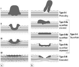
Paris classification
Morphologic system for superficial gastric cancers/ Early Gastric Cancers
Clinical
- Often asymptomatic, until advanced
- Most present late
- 65% T3/T4 stage
- 85% LN mets at diagnosis
- 50% present with unresectable locally advanced or metastatic
- 95% with anorexia & weight loss ± vague abdo pain, N+V, early satiety (linitis plastica)
- 50% non-specific symptoms, e.g. dyspepsia
- NB: Only 1-2% of pts with dyspepsia have Gastric Ca finding
- Ulcerated tumours may cause bleeding → hematemesis, melena, UGIB
- 40% have some form of GI bleeding – anaemia or haematemesis
- Advanced tumours may present with:
- Palpable abdo mass
- Cachexia
- Bowel obstruction
- Ascites / oedema
- Hepatomegaly
- Involvement of other organs from peritoneal seeding:
- Ovaries: Krukenberg tumour
- Pelvic cul-de-sac “Blumer’s shelf”
- LN Metastasis
- Supraclavicular LN = Virchow’s node ≡ Troisier’s sign
- Left Axillary LN (Irish’s node)
- Periumbilical node / metastatic deposit (Sister Mary-Joseph’s nodule)
- Trousseau’s sign (migratory thrombophlebitis) – more common with Pancreatic Cancer
- Acanthosis nigricans
- Sign of Leser-Trélat
Pathology
Carcinoma Sequence
- Invasive gastric carcinoma preceded by a cascade of precancerous lesions
Tumour Pathogenesis
-
95% of Gastric Cancer = Adenocarcinoma
-
Location
- Antrum (50-60%) > Cardia (near GOJ) > Body
- Trend is for more proximal location
- Most cancers found on the lesser curve
- As are benign gastric ulcers
- However an ulcer on the greater curve is more likely to be malignant
- Antrum (50-60%) > Cardia (near GOJ) > Body
-
Intestinal Type Gastric Ca
- M > F (2:1), typically older age
- Gland formation: well to poorly differentiated tumours
- Haematogenous spread more common (than Diffuse)
- Grow in expanding, rather than infiltrative, patterns
- Occur secondary to chronic atrophic gastritis & intestinal metaplasia
- H. pylori infection & Infiltration with MALT and neutrophils
- Leads to damage to epithelial cells
- Alterations in cell cycle with increased apoptosis and cellular proliferation
- Results in atrophic gastritis
- Nuclear and architectural abnormalities with progressive mutations leading to carcinoma.
- Associated with cancer of cardia & distal oesophagus + antrum
- Better prognosis than diffuse
- Common genetic mutations:
- KRAS, P53, APC, MMR genes, HER2
-
Diffuse Type Gastric Ca
- M = F, younger, incidence increasing
- Familial (Hereditary Diffuse Gastric Carcinoma) version
- Less related to environmental influences
- More associated with blood group A
- Histology:
- Poorly differentiated, lacks gland formation, composed of signet ring cells
- Scattered, poorly cohesive cells with diffuse infiltration
- Often exhibiting deep infiltration of stomach wall
- May exhibit pronounced desmoplasia & associated inflammation with relative sparing of overlying mucosa
- Metastasizes early via lymphatics > LN and transperitoneal spread
- May arise from single-cell mutations within normal gastric glands
- Linitis plastica more common
- Prognosis poorer
- Common gene mutations:
- E-cadherin (50%), P53, C-met amplification, N-cadherin over exposure
-
Pathological Grade
- Probably has less prognostic implication than in other cancers
- Well differentiated
- Moderately differentiated
- Poorly differentiated
- Anaplastic
- Predominant patterns: Papillary, tubular, poorly differentiated, signet-ring cell, mucinous
- Cytokeratin positive immunohistochemistry
- Probably has less prognostic implication than in other cancers
Molecular pathology
- Adenocarcinoma of stomach identified by histopathological markers. Positive for CK7, CDX2 (usually intestinal type). Negative for CK20, CK5/6, CK14, p63 and p40.
- HER2 testing – metastatic gastric cancer could be eligible for Trastuzumab
- VEGFR – could be eligible for ramucirumab
- c-MET is a transmembrane tyrosine kinase receptor, amplified at higher frequency in diffuse-type gastric cancer. FGFR2 amplification also detected. Trials of these inhibitors have not been successful.
- p53 mutation in 60% gastric carcinoma
- APC 30-40% of moderately & well differentiated intestinal type. <2% of poorly differentiated diffuse-type gastric carcinoma
- MSI 15-38%. MSH2, MLH1, MSH6, PMS1 & PMS2.
Stage
Early Gastric Cancer
- 90% 5-yr survival
- T1 = Limited to mucosa / submucosa
- Regardless of LN status
- NB: Lymphatics run in submucosa
- Important to differentiate intramucosal from high grade dysplasia and intramucosal from submucosal
- LN metastasis in 2-3% for intramucosal
- Poor predictors for LN involvement:
- LVI
- Ulceration
- Tumour > 30mm
- With none of above, < 1% had LN involvement
- Poor predictors for LN involvement:
- LN metastasis in 20-30% for submucosal lesions
- Rarely diagnosed at this early stage in Western countries
Advanced Gastric Cancer
- T2-T4
- Invasion into muscularis propria or beyond
- Classified by Borrmann classification
- 5-yr survival 20-30% in western population
- Usually ulcerated with raised everted edge (Borrmann I-III)
- cf Punched out benign peptic ulcer
- Linitis plastica (Leather-bottle stomach)
- Borrmann IV
- Diffuse infiltration producing a thickened shrunken stomach with no visible change of lining mucosa
Modalities of Spread
- Direct spread through wall (local invasion)
- Lymphatic spread to regional LN
- Risk factors:
- Age at diagnosis, > 20mm tumour, depressed type, undifferentiated, LVI or submucosal invasion
- Risk factors:
- Haematological spread
- Surprisingly differentiated tumours at higher risk than poorly differentiated.
- Transperitoneal spread:
- e.g. Krukenberg tumour = ovarian infiltration by signet ring cells
TNM Staging
T stage
| Category | T criteria |
|---|---|
| TX | Primary tumor cannot be assessed |
| T0 | No evidence of primary tumor |
| Tis | Carcinoma in situ: Intraepithelial tumor without invasion of the lamina propria, high-grade dysplasia |
| T1 | Tumor invades the lamina propria, muscularis mucosae, or submucosa |
| T1a | Tumor invades the lamina propria or muscularis mucosae |
| T1b | Tumor invades the submucosa |
| T2 | Tumor invades the muscularis propria* |
| T3 | Tumor penetrates the subserosal connective tissue without invasion of the visceral peritoneum or adjacent structures¶Δ |
| T4 | Tumor invades the serosa (visceral peritoneum) or adjacent structures¶Δ |
| T4a | Tumor invades the serosa (visceral peritoneum) |
| T4b | Tumor invades adjacent structures/organs |
| N stage |
| NX | Regional lymph node(s) cannot be assessed |
| N0 | No regional lymph node metastasis |
| N1 | Metastases in 1 or 2 regional lymph nodes |
| N2 | Metastases in 3 to 6 regional lymph nodes |
| N3 | Metastases in 7 or more regional lymph nodes |
| N3a | Metastases in 7 to 15 regional lymph nodes |
| N3b | Metastases in 16 or more regional lymph nodes |
| M Stage |
| M0 | No distant metastasis |
| M1 | Distant metastasis |
- Invasive adenocarcinoma = Invasion into lamina propria
- Resectable via EMR = T1a
- If invasion into submucosa (T1b), need surgery
Distal Nodes (i.e. M1 Disease)
- Retropancreatic
- Pancreaticoduodenal
- Peri-pancreatic
- Superior Mesenteric
- Middle Colic
- Para-Aortic
- Retroperitoneal
Staging by TNM
| When T is… | And N is… | And M is… | Then the stage group is… |
| Tis | N0 | M0 | 0 |
| T1 | N0 | M0 | I |
| T2 | N0 | M0 | I |
| T1 | N1, N2, or N3 | M0 | IIA |
| T2 | N1, N2, or N3 | M0 | IIA |
| T3 | N0 | M0 | IIB |
| T4a | N0 | M0 | IIB |
| T3 | N1, N2, or N3 | M0 | III |
| T4a | N1, N2, or N3 | M0 | III |
| T4b | Any N | M0 | IVA |
| Any T | Any N | M1 | IVB |
Screening
- Mass screening in high incidence regions
- E.g. Japan
- High Risk patients should be screened 1-2yrly
- American Society for Gastrointestinal Endoscopy recommendation
- History gastric cancer
- FAP
- Peutz-Jeghers
- Menetrier’s disease
- American Society for Gastrointestinal Endoscopy recommendation
- If chronic atrophic gastritis and Gastric intestinal metaplasia
- Surveillance scope every 3yrs
BPAC Guidelines Screening for Hereditary Diffuse Gastric Ca https://bpac.org.nz/2018/gastric-cancer.aspx#main-nav
Investigations

Endoscopy
- Most sensitive and specific diagnostic method
- EMR as both staging and treatment
CT scan
- Do in all
- Identification of LNs or metastases
- Good for liver metastasis detection
- Only able to detect ≈ 25% of peritoneal mets
- vs ≈ 85% by laparoscopy
EUS
- Recommended in pts without metastatic disease
- Gold standard for T staging
- Most reliable non-surgical method for evaluating depth of invasion of gastric cancer
- Particularly T1 lesions
- Use in consideration for EMR vs surgery
- Also good for detecting regional lymph node involvement
- EUS guided FNA helps to determine if reactive or metastatic nodes
Staging Laparoscopy and Peritoneal Washings
- Consider in
- All fit patients being considered for resection
-
T1
- Without metastatic disease on CT
- Who would not otherwise need a gastrectomy for symptoms
- Do in any patient being considered for neoadjuvant therapy
- Esp. useful assessment if suggestion of locoregional spread, large tumour or at GOJ
- Detects peritoneal/metastatic disease in 20-30% of pts with disease beyond T1
- Pts best palliated by chemo, so will change management
- Higher in T4 disease and linitis plastica
- Peritoneal cytology
- At least 200ml of normal saline instilled
- Placed across different quadrants of abdo, i.e. left and right sub-phrenic space, Pouch of Douglas
- Fluid should be dispersed
- Aspirations from different parts of the body
- 50ml should be sent for analysis
- NB: Positive washings only does not mean patient is incurable
- +ve cells → ↓ prognosis (but not in Stage IA)
- Median survival 10-13 months (or 15 months with chemo)
- cf patients with overt mets (9 month overall survival)
- Indication for neoadjuvant treatment if no gross mets but washings positive
- Should repeat laparoscopy and washings at completion of treatment
FDG-PET CT
- Controversial, does not replace staging laparoscopy
- Not funded in NZ
- Issues with loss of GLUT1 receptors
- Unhelpful if primary not FDG avid
- Funded for GOJ (mandatory part of that staging)
- Gastric cancers not as PET avid as oesphgeal
Peri-op Assessment and Management
- Balance between patient (comorbidities/fitness) and disease (stage) factors.
- Need to identify factors that will change treatment options available and factors that can be improved to optimize outcomes
- Cardiac testing for any patients with symptoms/risk factors
- Pulmonary function testing
- Renal function
- Nutritional Assessment
- Other Optimisation
Cardiac Testing
- Do cardiac testing for any patients with symptoms/risk factors
- 10% of patients undergoing oesophagogastric surgery will have a cardiac event
- ECG
- Stress testing – exercise or pharmacological
- Assess for exercise induced ischemia or cardiac function that limits exercise (valvular, cardiomyopathy etc)
- Cardio/respiratory fitness testing (CPEX)
- Provides a safe, reliable, repeatable, non-invasive, objective, individual assessment of combined pulmonary, cardiac, and circulatory function.
- Anaerobic threshold
- Usually about ½ way through test, where 02 demand exceeds supply and muscles generate ATP anaerobically
- Detected by increased CO2 as HCO3 used to buffer excess H+ generated
- VO2 max been found to closely correlate with occurrence of post-operative cardiopulmonary complications.
- Target > 800mL/m2
Respiratory and Renal Testing
- Pulmonary Function Testing
- Post op pulmonary complications = 15-50%
- Can be optimized by early physiotherapy, early mobilization and incentive spirometry
- Spirometry best test to determine lung function
- Low FEV1 and FVC has been shown to correlate with poor pulmonary outcome
Renal function
- Pts on dialysis are not appropriate surgical candidates
- Patients with pre-existing renal disease should be optimised pre-op and involvement of nephrologist
Nutritional Assessment
- Pre-operative malnutrition and associated immunosuppression shown to have increased risk of adverse outcomes
- Esp. risk of infection/mortality
- Best adjunct to improve nutrition is surgically placed feeding jejunostomy
- No compromise to gastric conduit
- No risk of stent migration/perforation or disruption of radiotherapy
- Well tolerated by patients compared to NJ tube
- Malnutrition Definition
- BMI < 18.5
- Weight loss > 10% over the last 3-6 months
- BMI < 20 and weight loss 5% over last 3-6 months
- Enteral feeding preferred
- Improved gut oxygenation and colonization with healthy flora
- Reduces risk of septic complications
- Also cheaper
- Improved gut oxygenation and colonization with healthy flora
Other Optimisation
- Anticoagulation:
- Increased risk, although not much can do to change
- Smoking cessation for a minimum 8 weeks prior
- Optimise Hb with iron supplementation or transfusion
- Encourage regular cardiovascular exercise
- Pre-op exercise shown to improve function however not definitively shown to reduce complications
- Encourage weight loss if obese
- B-Blocker
- May improve ventricular dysfunction and reduce risk of perioperative arrhythmia
Intra/Post Operative Care
- Lung isolation techniques
- Single lung ventilation using double lumen tube
- Tidal volume should be minimized and PEEP added to minimize lung injury
- Early extubation recommended if possible
- Minimise intraoperative blood loss and fluids given
- Goal directed therapy
- Adequate post op analgesia
- Thoracic epidural highly effective
- Should avoid hypotension with epidural
- Minimize bolus doses, reduce rate and ensure patient euvolemic + vasopressors when required.
- Thoracic epidural highly effective
Care pathways

- Important for post-op care as common goals for all involved and targets for patients
Management
General Principles
- Always involve an MDM
- Surgeon, radiologist, pathologist, oncologist,
- Early Gastric Cancer = T1 N0
- Advanced Gastric Cancer = Anything else
- Goal of surgery
- Excise cancer with clear longitudinal and circumferential margins
- 3cm intestinal type
- 5cm diffuse type
- Prophylactic gastrectomy in patients with E-cadherin mutation
Example Management Algorithm

Endoscopic Management
Three stages:
- Endoscopy assessment + biopsy +/- EUS
- Paris classification to assess deeper involvement
- Presence of ulceration
- Histology
- Degree of differentiation/LVI
- If poor or undifferentiated > surgery
- Classified by ‘Vienna classification system’ to guide management of GI neoplasia
- Histology
- Presence of ulceration
- Paris classification to assess deeper involvement
- Endoscopic resection attempt (ESD)
- Lifting sign
- If > 2cm – Piecemeal resection
- ESD (Endoscopic submucosal dissection) can resect en-block
- Surveillance if complete resection
- 6/12 1st year then annually
- Pioneered in Japan
- Suitable for Early Gastric Ca
- If none of the factors suggesting increased risk of metastasis are present
- Poor differentiation
- LVI
- Ulceration
-
30mm
- Perforation rate ≈ 4%
- Can be repaired by endoscopic clipping or omental patch
- No evidence of increased dissemination risk if perforation
- If +ve margin after EMR → Surgery required
- USA guidelines suggest EMR for Tis or T1a
- i.e. limited to muscularis mucosae only (not into submucosa)
- Formal surgery (without preop chemo) for T1b
Neoadjuvant Therapy
-
Neoadjuvant chemotherapy is standard of care for all EXCEPT
- NCCN guidelines:
- Tis or T1a → ER or surgery (if medically fit)
- T1b → Surgery
- T2 or higher, any N → Perioperative chemotherapy (preferred) or surgery alone
- NCCN guidelines:
-
MAGIC Trial 2006 NEMJ
- Inclusion: Adenocarcinoma of stomach, GOJ, or lower oesophagus, T2+, any N
- ECF (Epirubicin, Cisplatin, 5FU) 3 x before and 3 x courses after surgery
- Only 40% received post-op course
- Increased R0 resection
- 10% higher
- Reduction in T stage and nodal status
- Improved overall survival at 5 yrs – 36% vs 23%
- Issues – Staging issues, R2 resection rate 32% vs 17%
-
FLOT4-AIO Trial
-
FLOT if good performance status
-
ECF if lesser performance status
Adjuvant Therapy
- Adjuvant Chemotherapy
- Adjuvant Chemoradiotherapy
- Radiotherapy after surgery:
- Considered if ≥ R1 resection (NCCN 2009)
Adjuvant Chemotherapy
Given as part of Magic protocol
- 3 x pre-op and 3x post-op cycles ECF
- Epirubicin, Cisplatin, 5-FU
- NB: Significant difficulty with post-op compliance
- Only 30% complete entire 3 cycles
- Epirubicin, Cisplatin, 5-FU
- Given as part of FLOT4
- If no neoadjuvent treatment then FOLFOX or FLOT4 or CAPOX
- For T2N0 with high risk features and above
- High histologic grade or the presence of lymphovascular or perineural invasion, or age <50) who have had an adequate lymphadenectomy
- For T2N0 with high risk features and above
Adjuvant Chemo/Radiotherapy
- Indications for Adjuvant Chemo/radio therapy (NCCN )
- Controversial
- T3-4
- Node-positive disease if they have undergone less than a D2 dissection
- Incomplete (microscopically incomplete or macroscopically incomplete resection
- Intergroup 0116 Trial
- For ≥T3 / N1 tumours of stomach (80%) and GOJ (20%)
- Treated with post-op Chemoradiotherapy
- 5-FU + Leucovorin + radiotherapy 5/52 (“MacDonald Protocol”)
- Median survival 27 vs 36 months
- Survival at 3yrs = 40% vs 50%
- Improved DFS (OR=1.5) and mortality (OR=1.35) compared to surgery alone
- Distant mets = most common form of recurrence in adjuvant group (33 vs 18%) cf LR more common in surgery only group (30 vs 20%)
- But this study has been criticized as 40% of pts had a D0 resection, only 10% had D2 resection
- Greatest benefit may be in high-risk pts treated with inadequate resections, ? just overcoming inadequate surgery
- Herceptin/ Trastuzumab
- Approved for HER-2 overexpression in metastatic gastric or gastro-oesophageal cancer
- 10-30% +ve for HER2
Surgery
Resection
- Early gastric cancer in middle of stomach (T1) can be treated with pylorus preserving sub-total gastrectomy
- 3cm prox to pylorus and small upper gastric remnant (for reservoir)
- Need to preserve Anterior Vagal n.
- Aim to achieve 3-5cm margins in intestinal-type
- At least 5cm margins in diffuse-type
- Total Gastrectomy (whole stomach and cardia) for proximal tumours
- Distal Gastrectomy (2/3 stomach and pylorus) for lower middle and distal tumours
- Minimum 2cm margin to GOJ (maintain function)
- Short Gastric Arteries preserved
- No survival benefit for total over distal gastrectomy for distal cancers
- Only resect adjacent pancreas if directly involved in tumour in order to achieve R0 resection
- Should have careful patient selection for this as high morbidity and may not strongly improve survival as generally poor prognostic tumours anyway
- No benefit of liver resection for isolated mets as high recurrence or distant spread
- Spleen not routinely resected, only if hilum involved
Lymphadenectomy in gastric cancer
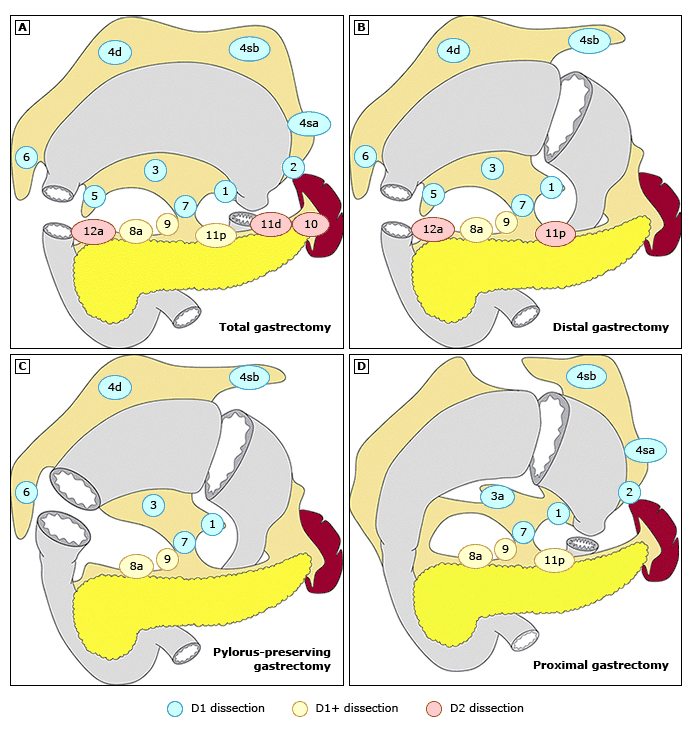
At NSH we perform a D1 plus - 1-7 + 8 and 9 as standard
- At least 16 LN are necessary to accurately stage a tumour
- T1a tumours 3% LN involvement
- T1b = 18% involvement
- T4a = 80% involvement
- Controversy re: limited vs extended LN dissection
- Removal of at least the Station 1-6 -level nodes (D1) improves 5-yr survival (20 → 34%)
- D3 = D2 + D3 – not recommended!
- Dutch Gastric Cancer Group Trial phase III:
- D1 vs D2 5yr survival rates similar (~ 45%)
- D2 Disadvantages
- More post-op complications (45 vs 30%)
- More post-op deaths (10% vs 5%)
- Longer hospital stays
- D2 Advantages
- Survival difference in patients with N2 / stage II & IIIA disease (D2 better)
- Risk of recurrence greater in D1 than D2 group (40 vs 30%)
- Unfortunately, ‘N2’ disease cannot be reliably identified pre-op
- Greatest contributing factor to post-op M&M in D2 group was addition of pancreaticosplenectomy
- “D2 resection without pancreaticosplenectomy may be better than a standard D1”
- Other Western studies
- Survival benefit for D2 without pancreaticosplenectomy
- Italian Gastric Cancer study Group (IGCSG), Austrian study, Spain - Post-op mortality after D2 ≈ 3-5%
- ANZGOSA recommendation
- Splenectomy if greater curvature involved with tumour
- Distal pancreatectomy only if direct tumour involvement
D1 Resection
- Early gastric cancer (T1) should be treated with D1 gastrectomy
- D1 dissection entails gastrectomy and the resection of both the greater and lesser omenta
- Stations 1 to 6 (+ now includes 7, left gastric)
- Includes the lymph nodes along
- Right (1) and Left (2) cardiac
- Lesser (3) and Greater (4) Curvature
- Suprapyloric (5) along the Right Gastric Artery
- Infrapyloric area (6)
D1 Plus Resection
- D1 plus
- Levels 7, 8a, 9
- Standard of care for Stages II & IIIa because
- Safe (provided pancreaticosplenectomy not done)
- Gives prognostic information
- Japanese survival stats – D2 mortality <1%
D2 Resection
- D2 dissection is a D1 plus all the nodes along the left gastric artery, common hepatic artery, celiac artery, splenic hilum, and splenic artery
- Station 7
- Left Gastric Artery (N1) – should be removed in all gastrectomy (including distal)
- Station 8
- Common Hepatic Artery (N2)
- Station 9
- Coeliac Trunk (N2)
- Station 10
- Splenic Hilum (N2)
- Not necessary to resect for all cancers, as often diff to resect without splenectomy
- Recommended for proximal gastric cancers on greater curvature
- Station 11
- Splenic Artery
- Station 12
- Hepatoduodenal nodes
- Hepatoduodenal nodes
Reconstruction
- Factors to Consider
- For high-risk patients (patient or disease factors)
- Choose safety over function
- For low-risk patients (disease factors and well patient)
- Given longer life expectancy and lower chance of recurrence consider functional quality of reconstruction
- Consider feeding jejunostomy
- NB: ANZGOSA lec says not to routinely for subtotals
- For high-risk patients (patient or disease factors)
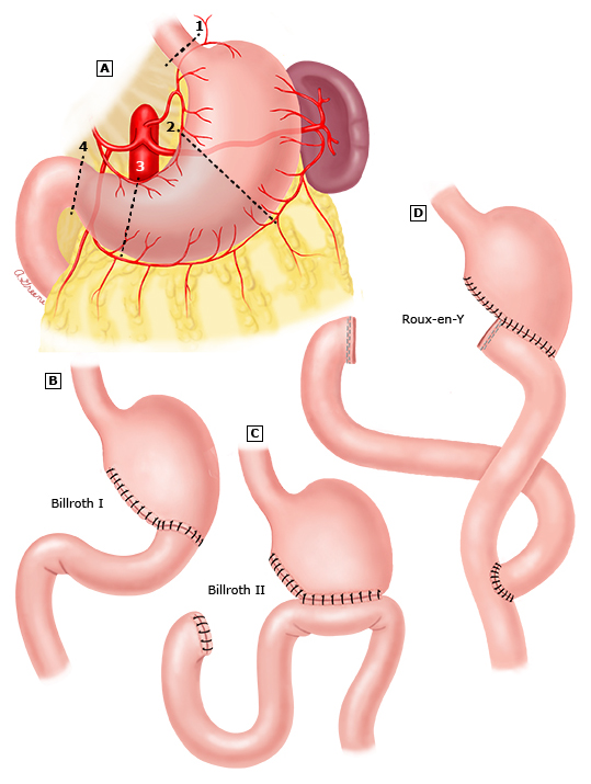
Reconstruction After Distal Gastrectomy
-
Best reconstruction option
-
Pros:
- No bile reflux
- Relatively safe anastomosis
- Low risk of obstruction if reoccurrence
-
Cons:
- Difficult to ERCP/Duodenoscopy
- Nutritional problems
- Dumping syndrome
- Two anastomosis Billroth I
-
Pros:
- Physiologic passage
- Simple anastomosis
- Preserved endoscopic access
- Preferred for distal small tumours with sufficient residual stomach volume and low chance of recurrence
-
Cons:
- Duodenal reflux
- Risk of leak reasonable (some tension on anastomosis)
- Obstruction if recurrence Billroth II
-
Pros:
- Only one anastomoses
- Can still access proximal duodenum endoscopically
-
Cons:
- Risk of bile reflux
- Afferent Loop Syndrome
-
Reconstruction After Total Gastrectomy
- Roux-en-y is the standard reconstruction
- Oesophagojejunostomy performed
- Safe, simple and good functional outcomes
- Cons = Early satiety, risk of nutritional deficiencies
Palliative Therapy
- Locally advance or metastatic disease
- Chemotherapy:
- Monoclonal antibodies
- Trastuzumab (Herceptin)
- for HER2 over-expression
- Antibody against human epidermal growth factor receptor 2 (HER2, aka ERBB2)
- ToGA trial: Survival benefit of 13.8 (trastuzumab + chemo) vs 11.1 months (chemo alone)
- dMMR/MSI-H tumors
- Check point inhibitors improves OS and can induce durable treatment responses
- Pembrolizumab etc
- Ramucirumab (VEGRF-2 inhibitor) used as part of second line with paclitaxel
- Trastuzumab (Herceptin)
Palliative Interventions
- Main indications for procedures are complications of advanced gastric cancer
- Treatment should be focussed on maximal palliation and minimal morbidity
- Perforation
- Requires surgical intervention
- Primary closure of perforation usually not possible
- Graham patch may be an option
- Especially given likely poor functional status of patient
- Gastrectomy is an option if it can be done with minimal morbidity
- Bleeding
- Endoscopic measures first line (as any other UGIB)
- Multiple attempts reasonable if patient is stable
- Could consider angiography
- If unstable, and other means haven’t worked, surgery indicated
- Tailor resection to situation
- Limited resection to grossly negative margins if life expectancy limited
- Obstruction
- Endoscopic dilatation and stent placement can provide good short-term palliation
- Tumour progression and stent migration limit efficacy long-term
- Chemo-radiotherapy can be used to attempt to relieve obstruction
- Response rates 50%
- If longer survival predicted, consider palliative bypass with gastrojejunostomy
Prognosis/Natural History
- Diffuse-type worse than intestinal-type
- Early Gastric Ca → Excellent prognosis: 10YS 90% (or ≈ 70% if LN +ve)
- Four patterns of metastasis:
- Lymphatic
- Haematological
- Peritoneal
- Direct invasion (T4b)
- Localized, node -ve Gastric Ca treated with surgery alone → 75% 5YS
- cf. node +ve Gastric Ca → 10-30% 5YS
- Advanced disease – M1 or unresectable = 6-12 months
- Overall 5YS: 20-30%
- In advanced stage with curative intent surgery: med survival = 24mths (20-30% 5YS)
- Following palliative procedures: median survival = 10 months
- In pts without palliative treatment: median survival = 5 months
- Surgical morbidity ≈ 32% & mortality ≈ 3.5%
- Recurrence rate 50% in patients resected with curative intent
- Rate of locoregional recurrence is related to T stage; rate of distant mets to N stage
- Most common peritoneal disease in T4 cancers
- Higher risk in diffuse than intestinal type gastric cancer
Prognostic Factors
- LN status
- Number provides a better prognostic indication than anatomic location
- LN +ve >20% (i.e. +ve / removed nodes) = Single most important prognostic factor
- Increased size
- Associated with LN metastasis & decreased 5Yr survival
- T category (serosal penetration most important)
- Tumour location
- Proximal 1/3 may represent a distinct entity with poorer prognosis
- GOJ tumours 5YS ≈ 5%
- Tumours tend to be larger, have extensive wall penetration, venous invasion, nodal mets & more advanced stage
- May require a different surgical approach based on a potentially different biological behaviour
- Resection margins (R0 vs R1/R2)
- LVI
- Age >70yrs
- ? genetic markers (p53, E-cadherin, CD-34, c-ErbB2, CA 72-4, CEA)
- Pathologic grade (well/mod/ poor differentiated / anaplastic) has little prognostic significance
Follow-up
- After Subtotal Gastrectomy
- Surveillance of gastric remnant recommended for 10-15yrs
- Cancer of gastric remnant
- Usually at the anastomotic site
- May be due to irritation from exposure to duodenal contents
- Eradication of H. pylori post-op decreases RR
Oesophageal + Gastric Comparison

Post-operative Complications
Anastomotic Leak
- Most commonly at oesophagojejunostomy
- Due to ischemia/tension
- Diagnose with gastrograffin swallow
- Drain and conservative management if stable
- NBM/NJ tube
- If no drain or patient septic
- Return to OT
- Drain + repair if possible
- Feeding jejunostomy if not already placed
Duodenal stump leak
- Rare
- Risks – ischemia, afferent limb obstruction
- Easily diagnosed if prophylactic drain placed (If bile ++)
- Needs return to OT
- Place Foley in duodenum to control fistula
- Or suction drain adjacent to small defect
- Decompression NJ then up duodenum to decompress
Pancreatic Fistula
- Increased risk with
- D2 resection/bursectomy, pancreatectomy or splenectomy
- Always place prophylactic drains during above surgery over pancreatic border
- Diagnose by amylase in drain fluid
- Usually dark red wine colour
- No evidence octreotide prevents fistula formation
- Management
- Adequate drainage +/- irrigation of abscess cavity once walled off
Haemorrhage
- Early
- Return to OT
- Late
- Usually due to intra-abdominal infection
- Leads to pseudoaneurysm of splenic artery
- Manage with Angiography + Embolisation
- Preferred as repeat surgery after infection is risky
Late Complications
Early Dumping Syndrome
- Occurs within 20-30mins after meal
- More common than late dumping
- More GI and less cardiovascular effects.
- Symptoms related to serotonin and bradykinin release
- GI
- Nausea/vomiting, epigastric discomfort, crampy pains + explosive diarrhoea
- Cardio
- Tachycardia, palpitations, diaphoresis, fainting, dizziness, flushing, blurred vision
- Pathophysiology
- Rapid transit of high osmotic load into small bowel
- Draws fluid in → Visceral distension → Release of vasomotor mediators
- Vasomotor (serotonin & bradykinin) release causes
- Weakness, dizziness, faint, pallor, sweat
- GI
- Most common after Billroth II (especially if >2/3 resected)
- Investigations
- Contrast study to demonstrate rapid emptying or provocation test by ingesting 200ml of 50% glucose & water
- Management
- Dietary change
- Avoid sugary foods, small frequent meals, separate liquids from solids, high protein/fat meals
- Octreotide can help – inhibits hormonal response & delays gastric emptying
- Surgery – consider interposition of 10-20cm jejunum
Late Dumping Syndrome
- Occurs 2-3 hours post-meal
- Related to high carbohydrate load into small bowel
- Get rapid glucose absorption and profound insulin release that overshoots glucose
- Rebound hypoglycaemia
- Triggers Adrenal Gland to secrete Catecholamines
- Diaphoresis/tremors/tachycardia
- Just like hypoglycaemic shock symptoms
- Management
- Small frequent meals & less carbs
- Surgical option of jejunal interposition
Nutritional Issues
- Fat malabsorption
- Lack of mixing with duodenal bile/pancreatic enzymes
- Get steatorrhea
- Treat with Creon
- Vit B12 deficiency
- Lack of IF produced by Parietal Cells
- Slow decline in body stores
- Megaloblastic anaemia at 24 months if not replaced
- Treat with 1mg Hydroxocobalamin IM 3-monthly
- Vit D deficiency
- Due to fat malabsorption
- Exacerbated by Ca2+ malabsorption (proximal duo)
- Increased risk of osteoporosis
- Treat with oral calcium and calcitriol
- Iron
- Predominantly absorbed duodenum and proximal jejunum
- Requires deoxidization to Fe2+ form (from Fe3+) for best absorption
- Tx with oral or IV supplementation
Post-Gastrectomy Syndromes
- Physiologic derangements cause by
- Loss of reservoir function
- Interruption of pyloric sphincter mechanism
- Vagal nerve transection
- 25% of patients may develop some symptoms
- Only approx 1% permanently disabled by symptoms
- Metabolic Disturbance
- Afferent Loop Syndrome
- Efferent Loop Obstruction
- Alkaline (Bile) Reflux Gastritis
- Gastric Atony
- Retained Antrum Syndrome
Other Metabolic Disturbances
- Billroth II worst
- Anaemia
- Seen in 30% by 5yrs, due to ↓intake & malabsorption of:
- B12
- Due to ↓ IF ± bacterial overgrowth
- Iron
- Due to ↓ gastric acid & ↓ duodenal absorption (= optimal site)
- Folate
- Achlorhydria → ↑ gastric pH, above optimal for folate absorption
- Osteoporosis/osteomalacia
- ↓ Ca – also absorbed primarily in duodenum (+/- ?↓Vit D)→ Bone disease
- Fat malabsorption causes fatty acids to bind to calcium in GI tract and inhibit absorption
- Increased gallstones – calcium malabsorption
- Fat malabsorption
- In Billroth 2 fats bypass duodenum
- Treat with pancreatic enzymes
Afferent Loop Syndrome
- Typically after a Billroth II but can also be seen in the BP limp of a [R&Y
- Mechanical partial obstruction of long afferent limb
- Cannot empty its contents
- Fills with biliary & pancreatic secretions
- Leading to pain
- Build-up eventually decompresses into stomach
- Leading to vomiting and relief of symptoms
- Usually related to long (>30-40cm) and antecolic afferent limb
- Symptoms
- Epigastric pain
- If severe may reflux into stomach and cause bilious vomiting
- If chronic can get Blind Loop Syndrome
- Bacterial overgrowth, binding to B12 and deconjugated bile acids
- Resulting in
- Megaloblastic Anaemia and Fat Malabsorption
- If chronic can get Blind Loop Syndrome
- Requires surgical intervention – convert to Roux-en-Y
Efferent Loop Syndrome
- Rare
- Usually due to herniation of limb behind the anastomosis from R to L
- Get colicky LUQ pain, bilious vomiting, abdominal distension
- Diagnosis with contrast study
- Failure of barium to enter efferent limb
- Management
- Operative - reduce the hernia and close the mesenteric defect
Bile Reflux Gastritis
- Common especially for Billroth 2
- Investigations
- HIDA – see biliary reflux into stomach
- Endoscopy – beefy, friable red mucosa
- Management
- Medical to control symptoms though generally poor outcomes
- Surgical - convert to Roux-en-Y
- Should preferentially do Roux-en-Y in first operation
Gastric Atony
- After truncal vagotomy, can occur even with pyloroplasty or gastrectomy
- Need to exclude other causes
- Diabetic gastropathy
- NM disorder
- Post op adhesions
- Internal hernia
- Afferent loop obstruction
- Investigations
- Scintigraphic assessment of gastric emptying/fluoroscopy study
- Management – usually medical
- Metoclopramide
- Prokinetic effects - dopamine antagonist and cholinergic enhancing activity
- Facilitates release of acetylcholine from enteric cholinergic neurons
- Erythromycin
- Binds to motilin receptors on GI smooth muscle cells, causing increased motility
- If atony refractory to medical therapy, may need gastrectomy
- Metoclopramide
Retained Antrum Syndrome
- Usually, antrum left in duodenal stump
- Get recurrent ulcers
- Investigation
- Technetium scan + SPECT
- Treatment
- Resect antral tissue that is retained