Definition
-
Autosomal dominant, inherited familial colorectal cancer syndrome due to mutation in APC gene on chromosome 5q.
- ~75% is inherited; which means 25% arise without family history, as first mutation
-
Risk of colorectal cancer is nearly 100%
-
Gardner syndrome is a sub-type
- Polyps + desmoid/osteomas etc
Characterised by:
- Hundreds of colorectal adenomatous polyps at young age
- Duodenal adenomatous polyps
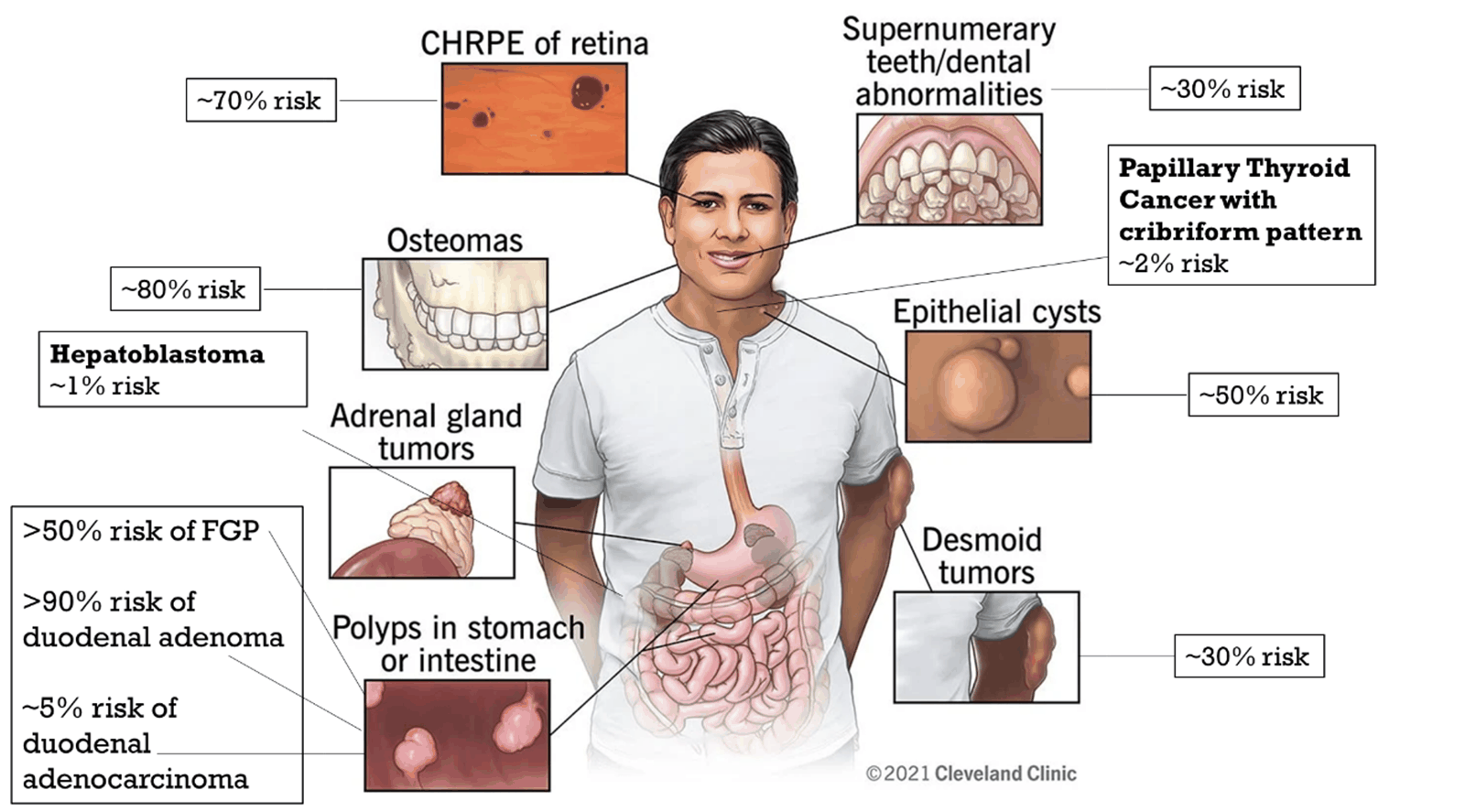
- Multiple extraintestinal manifestations
- Ectoderm: epidermoid cyst, pilomatrixoma, CNS tumours, congenital hypertrophy of retinal pigment epithelium (CHRPE)
- Mesoderm: connective tissue – (desmoid tumour, excessive adhesions), bone – (osteoma, exostosis, sclerosis), dental – dentigerous cyst, odontoma, supernumerary teeth, unerupted teeth)
- Endoderm: adenomas and carcinomas of duodenum, stomach, small intestine, biliary tract, thyroid, adrenal cortex, fundic gland polyps, hepatoblastoma
Diagnosis
- Clinical/endoscopic – over 100 colorectal adenomas.
- Chromoendoscopy with dye spray, and random biopsies to look for microadenomas (hallmark of FAP and MAP, not in Lynch)
- Gastroscopy may show fundic gland polyps or duodenal adenomas
- Genetic – mutation of APC gene (only 80% of polyps affected)
- Family history. However, 20% will be new mutations with no family history.
- Ddx: MAP (MUTYH-associated polyposis)
Genetic testing
- Affected family member tested first. Once mutation identified, at risk family offered simple blood test.
- Genotype-phenotype correlation:
- Different levels of expression, due to different site of mutation in APC gene, along with modifier genes and environment
Surveillance
- Colonoscopy - from age 10-12 (AFAP 18-20) until colectomy at which point the rectal cuff/J pouch requires annual flexible sigmoidoscopy
- Gastroscopy (including side viewing scope) - from age 25 and then as per the Spigelman stage
- Thyroid USS - age 18 then eery 2-5 years
Treatment
Lower GI
Surgery
- Should be done as early as possible. Depends on social and educational factors.
- Options:
- Colectomy + ileorectal anastomosis (IRA)
- Slightly better functional outcome
- Colectomy + ileorectal anastomosis (IRA)
- Restorative proctocolectomy (RPC) with ileal pouch-anal anastomosis
- Benefit of no rectum
- Good option of high polyp burden in rectum
- Small cuff of rectal mucosa retained at anastomosis – small risk of cancer here
- Risk of infertility in women and sexual dysfunction in men
- Total proctocolectomy and end ileostomy
Surveillance
- Both procedures need annual PR and flexi sig
- No long term evidence for NSAID or COX-2 inhibitor
Upper GI
- Fundic gland polyps
- Duodenal adenomas occur in nearly all patients with FAP, but malignant only in 5%
- severity of duodenal polyposis in FAP
| Points allocated | |||
|---|---|---|---|
| 1 | 2 | 3 | |
| Number of polyps | 1–4 | 5–20 | > 20 |
| Polyp size (mm) | 1–4 | 5–10 | > 10 |
| Histological type | Tubular | Tubulovillous | Villous |
| Degree of dysplasia | Mild | Moderate | Severe |
| Total points | Spigelman stage | Recommended follow-up interval |
|---|---|---|
| 0 | 0 | 5 years |
| 1–4 | I | 5 years |
| 5–6 | II | 3 years |
| 7–8 | III | 1 year and consider endoscopic therapy |
| 9–12 | IV | Consider prophylactic duodenectomy |
Treatment
- Endoscopic removal
- Whipples in stage IV may be required
Desmoid tumour
- Fibromatous lesions consisting of clonal proliferations of myofibroblasts
- 15% of FAP, mostly intraabdominal or abdominal wall, can be on extremities or trunk.
- Histologically benign (lack nuclear atypia & features of malignancy), however can cause SBO, ureteric obstruction, intestinal ischaemia or perforation, which could be fatal.
- Aetiology: multifactorial. Trauma e.g. operative, oestrogens, specific APC mutations
- FAP related due to inactivation of APC and subsequent accumulation of beta-catenin in cells.
- Sporadic desmoid usually mutation in gene for beta-catenin (uncommon for APC mutation).
- Investigations: CT is best. MRI may be useful. USS to monitor ureters.
Treatment:
- Observation usually preferred for asymptomatic, stable desmoids.
- Surgical – excision (1st line for abdominal wall and extra-abdominal)
- High recurrence rates
- Avoided for intra-abdominal desmoids due to significant morbidity/mortality unless desmoid-related complications develop
- Medical – NSAIDS, antioestrogens (tamoxifen), cytotoxic chemotherapy, non-cytotoxic approach – tyrosine kinase inhibitors (sorafenib, sunitinib)
- Radiotherapy may be used.