Sub-section: Thyroid Section: Endocrine
Overview of Work-up
- A discrete and radiographically definable lesion within the thyroid
- Many thyroid nodules are not palpable
- Not all palpable thyroid lesions correspond to a distinct radiographically definable lesion
- Only those truly definable radiographically may be classified as a thyroid nodule
- Common finding
- Most do not require any intervention, and only a few require thyroid resection
3 classes of indications for Thyroid resection:
- Local compressive or inflammatory symptoms
- Hyperfunctioning
- Malignancy or concern for malignancy
- Most patients with a solitary thyroid nodule have an asymptomatic, non-functioning, benign lesion
Incidence
- Increasing numbers of thyroid nodules are being found incidentally
- Possibly due to increasing availability and sophistication of imaging
- Palpable thyroid nodules are present in 1% of men and 5% of women
- Ultrasound-detectable thyroid nodules are present in 19% to 67% of unselected patients
- Frequency of palpable and non-palpable thyroid nodules increases with age
- Most of these nodules are benign
- Overall approximately 5% are thyroid cancers
Initial Evaluation
History
- Local symptoms including dysphagia, subjective dyspnoea, positional dyspnoea, pressure or choking sensation, pain, globus sensation, or symptoms precipitated by raising the arms over the head (a subjective Pemberton sign)
- Symptoms of hypo/hyperthyroidism
- Risk factors for malignancy
- Children, male patients, adults younger than 30 or older than 60 years
- Exposure to radiation (occupational sources or irradiation of the head/neck, esp. childhood)
- Personal and family hx of specific endocrine disorders
- Familial medullary thyroid carcinoma, MEN 2B syndrome/MEN 2A syndrome, PTC, polyposis, including Gardner syndrome or Cowden’s syndrome or Carney complex
Exam
- Palpate the cricoid ring because the isthmus is reliably palpable immediately inferior to this
- The bilateral lobes may be palpated directly lateral to this
- Sip of water causes the thyroid to move under the examining fingers with swallowing
- Can reveal lesions not appreciable by static palpation
- Also can palpate substernal goitre
- Anterior and posterior cervical triangles should be assessed for pathologic lymphadenopathy
- ? Fixed, ? Single, ? Firm, ? Rapid growth
- Pre op
- Check vocal cords
- Check symmetry
- Check vocal cords
Lab tests
- Thyroid function tests
- Aim to identify hyperthyroid patients
- Measure TSH if a thyroid nodule is 1 cm or larger
- Low serum TSH = Overt or subclinical hyperthyroidism
- Radioisotope scan generally indicated
- Also correlates with a lower likelihood of malignancy in a thyroid nodule, and thyroid cancers are rarely thyrotoxic
- High serum TSH suggests hypothyroidism, most commonly the result of Hashimoto thyroiditis
- Low serum TSH = Overt or subclinical hyperthyroidism
- Thyroglobulin
- Important in the follow-up of patients after initial treatment of thyroid cancer but should not be checked routinely in the initial evaluation of a thyroid nodule
- Calcitonin
- If clinical suspicion of Medullary Carcinoma, either by family history or by FNA
- Otherwise don’t do routinely
- Check calcium prior to surgery
Imaging
Ultrasound
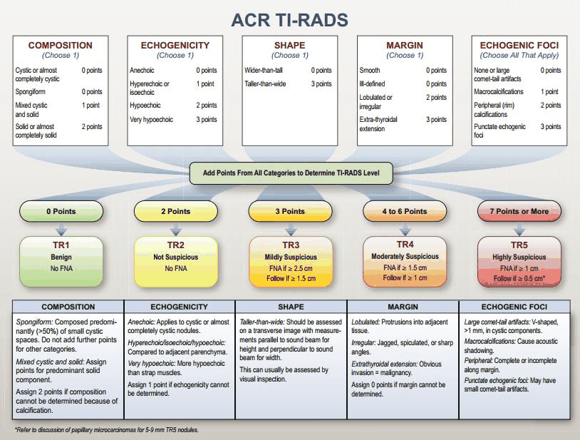
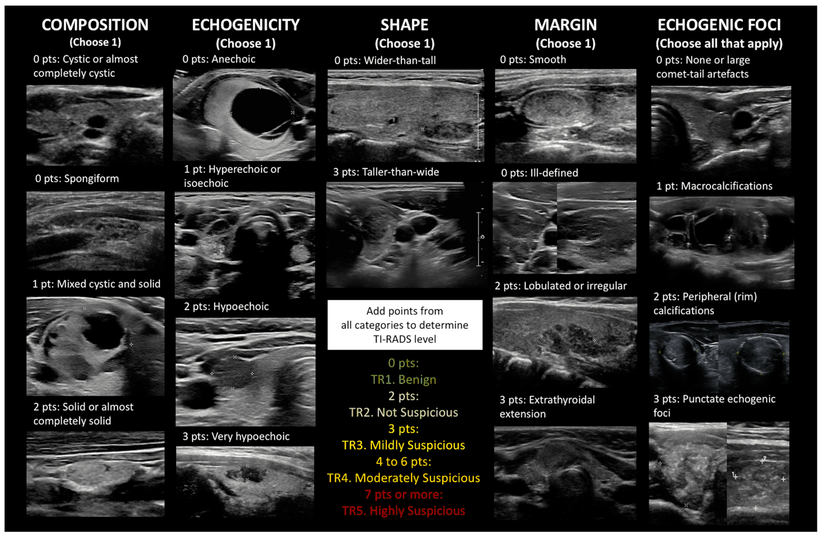
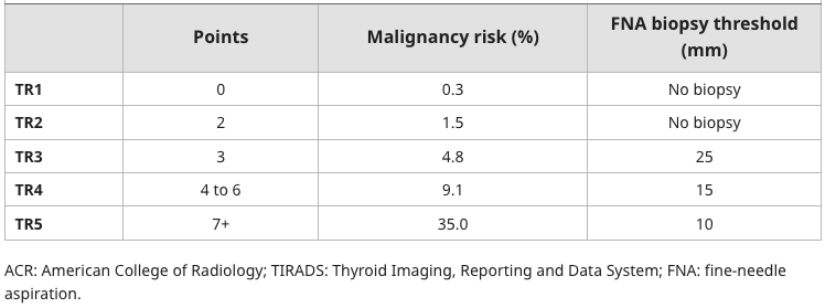
TI-RADS
- The TIRADS score is an ultrasound assessment of a thyroid nodule. It provides guidance on
- Looks at 5 different areas – composition, echogenicity, shape, margin, presence of echogenic foci.
Composition
- Cystic lesions are more likely to be benign.
- Solid lesions are more likely to be malignant
Echogenicity
- Anechoic means “black” or “fluid filled” - which are obviously benign features.
- Dense tissue/tumours absorbs USS waves and doesn’t return them thus are hypo-echoic (same as breast tumours)
Shape
- Taller than wide is more likely to be malignant as it indicates the tissue is not compressible.
Margin
- Smooth margins or ill-defined are more likely to benign.
- Lobulated margins or with extra-thyroidal extension - more likely to be malignant.
Echogenic foci
- Macrocalcifications are more likely to be benign
Peripheral calcifications and punctate echogenic foci correspond with psammoma bodies - which are collections of calcium which can occur in tumours where necrosis has occurred - in particular papillary thyroid cancer
S - Solid M - Micro/macro calcifications I - Irregular T - Tall H - Hypoechoic
Monitoring of nodules which doe not meet FNA criteria
- Nodules who do not meet criteria should be assessed in 6 months to 3 years based on their risk.
- If nodules are growing then they generally should be FNA’d
Lymph nodes
Lymph node findings suggestive of malignancy (thus require an FNA)
- Microcalcifications/Bright hyper-echoic spots.
- Cystic degeneration
- Round shape
- Loss of the fatty hilum
- Peripheral vascularization
- Hypoechogenicity
Lymph nodes if sampled should be tested for presence of thyroglobulin, if this is positive – the malignancy is highly likely
Radioisotope Scans
- Allows functional evaluation
- Indications
- Indicated if dominant nodule > 1cm with supressed TSH
- Not indicated in euthyroid or hypothyroid initial workup
- Two options
- Tc-99 Pertechnetate scan
- Taken up rapidly by normal activity of follicular cells trapped by not organified
- Short T1/2 and low radiation dose
- Allows rapid uptake of hyperfunctioning (hot) or hypofunctioning (cold) area
- Also will show uptake in salivary glands and major vessels
- Iodine scintigraphy
- I123 and I131 trapped and organified by follicular cells
- I123 has short T1/2 (12hrs) and low dose radiation
- Good for suspected lingual thyroids or substernal goitre
- I131 longer T1/2 (8 days) and higher radiation
- Good for imaging thyroid carcinoma and screening for distal mets
- Tc-99 Pertechnetate scan
- Malignancy present in 15-20% cold nodules, and < 5% hot nodules
- But activity of nodule cannot confirm or exclude malignancy
- In hot nodules biopsy can be misleading as due to the increased cell activity, findings can be mistaken for a malignancy
- FDG-PET
- Not indicated in initial workup, can incidentally identify hot nodules
- If avid nodules >1cm– USS and FNA
- If avid with suppressed TSH then radioiodine scan ?functioning
- If diffuse thyroid avidity – USS,? Lymphocytic thyroiditis
- Not indicated in initial workup, can incidentally identify hot nodules
CT and MRI
- Do not add much to initial workup of uncomplicated nodules
- Indications
- Evaluating local extension of Advanced Thyroid Ca
- If suspicious mass and bulky LN
- Pre-op planning of retro/substernal goiter
- Post-op follow-up if concerned about recurrence
FNA
- Generally shouldn’t FNA nodules < 1cm unless
- Suspicious characteristics on USS
- Suspicious LNs
- Fhx PTC
- Hx of Radiation
- Hx of Thyroid Ca
- Avid lesion seen on FDG-PET
- Can forego FNA if wouldn’t change plan to operate or extent of resection
- Method
- Use 23-27g needle
- If palpable may not need USS
- USS guidance recommended if
- Non-palpable, posterior, or cystic component (to reduce non-diagnostic cytology)
Bethesda Classification
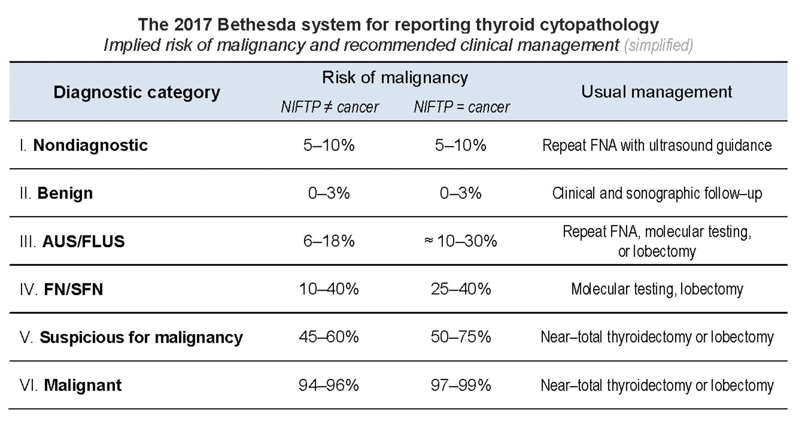
Key: AUS – Atypia of undetermined significance, FLUS – Follicular Lesion of undetermined significance, FN – Follicular Neoplasm, SFN – Suspicious for a Follicular Neoplasm
Bethesda 2023
- 1 - 13
- 2 - 4
- 3- 22
- 4- 30
- 5- 74
- 6 - 97
Management
Bethesda 1
- Cytologically inadequate
- Should be repeat FNA - if this is still inadequate then consider core biopsy.
Benign nodules/Bethesda II
- Need a repeat USS
- If high sonography suspicion initially
- Repeat USS 3-6 months
- If low/intermediate suspicion
- Repeat USS 12-24 months
- If high sonography suspicion initially
- If the lesion grows by at least 2mm, or it still looks suspicious then repeat the FNA.
- If the second FNA is benign after follow up - can be discharged.
Indeterminate cytology - Bethesda III/IV
- Atypia of uncertain significant/follicular lesion of uncertain significance of follicular neoplasm are indeterminate lesions
- These patients should undergo a repeat FNA initially.
If repeat FNA shows Bethesda III/IV
- Should proceed to mutation analysis molecular testing. If a likely malignant result is given - need diagnostic lobectomy. If likely benign - then repeat USS in 12 months.
- If mutation analysis molecular testing is not available - should have a diagnostic lobectomy.
Bethesda V and VI
- Should go forward for surgery - thyroidectomy or lobectomy.
Autonomous nodules.
- Optimal therapy of autonomous nodules is controversial - can be treated with radioiodine (low dose), surgery, of long-term anti-thyroid drugs.
- Patient preference
Stage
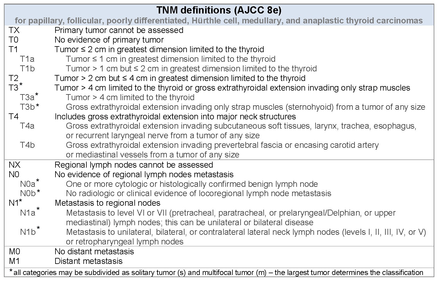
TNM simplified
T1 - tumour <2cm - T1a - <1cm - T1b - 1-2cm T2 - tumour 2-4cm. T3 - tumour >4cm cm which can only involve the strap muscles T4 - tumour which invades into major structures of the neck i.e. trachea, oesophagus, RLN, carotid artery, pre-vertebral fascia.
N1a - metastasis to level VI or VII (midline neck nodes) N1b - metastasis to another station beyond the above.
Stage
- Age <55
- Stage I
- M0 (anyT, anyN)
- Stage II
- M1 (anyT, anyN)
- Stage I
- Age >55
- Stage I
- T1-T2, N0
- Stage II
- T1-T2, N1
- T3, N0-N1
- Stage III
- T4a (anyN)
- Stage IV
- T4b (anyN)
- M1 (anyT, anyN)
- Stage I
Anaplastic stage
All stage 4
MTC stage
| T1 | N0 | M0 | I |
| T2 | N0 | M0 | II |
| T3 | N0 | M0 | II |
| T1-3 | N1a | M0 | III |
| T4a | Any N | M0 | IVA |
| T1-3 | N1b | M0 | IVA |
| T4b | Any N | M0 | IVB |
| Any T | Any N | M1 | IVC |