Section: Colorectal Sub-section: Colorectal cancer Curriculum:
Family History
Assessment:
- History of personal symptoms, previous large bowel polyps, large bowel cancers, cancers at other sites, other risk factors e.g. IBD, acromegaly.
- Family history
Risk categories
As per: Update on Surveillance Recommendations for Individuals with a Family/Whānau History of Colorectal Cancer July 2023
Slightly increased:
- Criteria
- One first-degree relative diagnosed at 55 years or older
- Advice
- Strongly advise these individuals to participate in the NBSP when they become eligible.
- Individuals should make healthy lifestyle choices and report any bowel symptoms to their health care provider.
Moderately increased:
- Criteria
- one first-degree relative diagnosed with CRC under the age of 55 years
- two first-degree relatives on the same side of the family/whānau diagnosed with CRC at any age
- Advice
- Individuals have a colonoscopy every five years from age 50, or from an age 10 years before the earliest age at which colorectal cancer was diagnosed in the family/whānau, whichever comes first.
- Provided they have had a high-quality colonoscopy within the previous five years, individuals then participate in the NBSP from the age of 60 years.
Potentially high
- Criteria
- Family history of inherited syndrome
- Suggestion of possible inherited syndrome
- one first-degree relative plus two or more first- or second-degree relatives all on the same side of the family/whānau with a diagnosis of CRC at any age
- two first-degree relatives, or one first-degree relative plus one or more second-degree relatives, and one such relative:
- was diagnosed with CRC when aged 54 years or under, or
- developed two or more bowel cancers, or
- developed an extracolonic tumour suggestive of Lynch syndrome (ie, endometrial, ovarian, stomach, small bowel, renal pelvis, pancreas or brain)
- at least one first- or second-degree family/whānau member diagnosed with CRC in association with multiple bowel polyps or a polyposis syndrome
- a first-degree relative with multiple colonic polyps if the relative is aged 30 years or younger).
- Advice
- Referred to genetic service or NZ familial GI cancer registry for accurate risk assessment.
- Will then need surveillance plan.
Molecular pathway
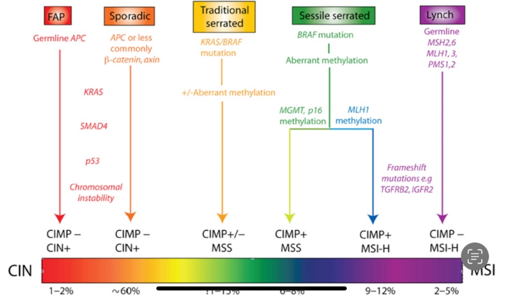
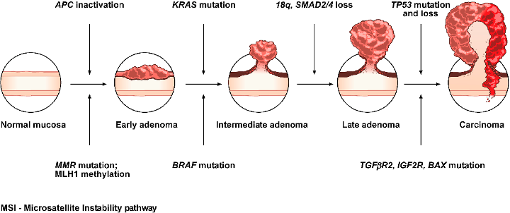
- Chromosomal instability (CIN)
- The mutator phenotype/mismatch repair pathway
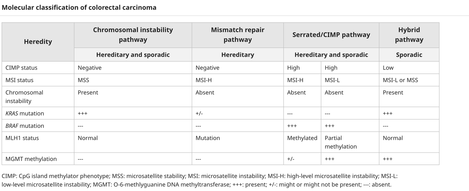
- Serrated/Hypermethylation phenotype pathway - Epigenetic pathways e.g. CpG Island Methylator Phenotype (CIMP)
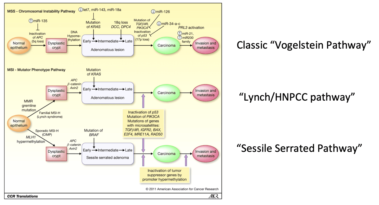
Chromosomal instability (CIN)
- Classic pathway - > Adenoma to carcinoma sequence
- Randomly acquired somatic mutations in several genes affected in hereditary cancers, due to allelic losses, chromosomal amplifications and translocations,
- APC mutation
- K-RAS; 12p12, oncogene, deletion in ~50% CRC, 50% late adenomas, <10% early adenomas
- 18q21 (SMAD4) – suppressor, mediator in TGFB,
- hereditary defect responsible for Cowden’s, JP,
- P53; 17p12, suppressor, controller of apoptosis,
- inherited defect = Li-Fraumeni syndrome
- Deletion in 75% of carcinoma
The mutator phenotype/mismatch repair pathway
- Lynch syndrome/HNPCC
- Germline mutations in one of several different DNA mismatch repair (MMR) genes, most commonly MLH1, MSH2, MSH6 and PMS2, which leads to dysfunction of the DNA MMR enzymes.
- The biologic “footprint” is the accumulation of abnormalities in short sequences of nucleotide bases that are repeated dozens to hundreds of times within the genome; these are called microsatellites, and the tumors are described as having high levels of microsatellite instability (MSI-H).
- High levels of MSI are also found in approximately 15 percent of sporadic CRCs. However, in most of these cases, gene silencing is not due to a specific MMR mutation, but to an epigenetic phenomenon as below
Serrated/Hypermethylation phenotype pathway
- Epigenetic pathways e.g. CpG Island Methylator Phenotype (CIMP)
- Activating mutations in the BRAF gene (most of which are in the V600E codon) occur almost exclusively in MSI-H, CIMP+ cancers that do not carry mutations in KRAS. Lynch-related CRCs present only with KRAS and not BRAF mutations
- Followed by hypermethylation of the gene promoter for a DNA MMR gene
Familial adenomatous polyposis
Autosomal dominant, inherited familial colorectal cancer syndrome due to mutation in APC gene on chromosome 5q.
- ~75% is inherited; which means 25% arise without family history, as first mutation Risk of colorectal cancer is nearly 100%
Characterised by:
- Hundreds of colorectal adenomatous polyps at young age
- Duodenal adenomatous polyps
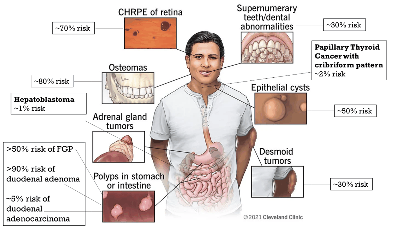
- Multiple extraintestinal manifestations
- Ectoderm: epidermoid cyst, pilomatrixoma, CNS tumours, congenital hypertrophy of retinal pigment epithelium (CHRPE)
- Mesoderm: connective tissue – (desmoid tumour, excessive adhesions), bone – (osteoma, exostosis, sclerosis), dental – dentigerous cyst, odontoma, supernumerary teeth, unerupted teeth)
- Endoderm: adenomas and carcinomas of duodenum, stomach, small intestine, biliary tract, thyroid, adrenal cortex, fundic gland polyps, hepatoblastoma
Diagnosis
- Clinical/endoscopic – over 100 colorectal adenomas.
- Chromoendoscopy with dye spray, and random biopsies to look for microadenomas (hallmark of FAP and MAP, not in Lynch)
- Gastroscopy may show fundic gland polyps or duodenal adenomas
- Genetic – mutation of APC gene (only 80% of polyps affected)
- Family history. However, 20% will be new mutations with no family history.
- Ddx: MAP (MUTYH-associated polyposis)
Genetic testing
- Affected family member tested first. Once mutation identified, at risk family offered simple blood test.
- Genotype-phenotype correlation:
- Different levels of expression, due to different site of mutation in APC gene, along with modifier genes and environment
Surveillance
- Colonoscopy - from age 10-12 (AFAP 18-20) until colectomy at which point the rectal cuff/J pouch requires annual flexible sigmoidoscopy
- Gastroscopy (including side viewing scope) - from age 25 and then as per the Spigelman stage
- Thyroid USS - age 18 then eery 2-5 years
Treatment
Lower GI
Surgery
- Should be done as early as possible. Depends on social and educational factors.
- Options:
- Colectomy + ileorectal anastomosis (IRA)
- Slightly better functional outcome
- Colectomy + ileorectal anastomosis (IRA)
- Restorative proctocolectomy (RPC) with ileal pouch-anal anastomosis
- Benefit of no rectum
- Good option of high polyp burden in rectum
- Small cuff of rectal mucosa retained at anastomosis – small risk of cancer here
- Risk of infertility in women and sexual dysfunction in men
- Total proctocolectomy and end ileostomy
Surveillance
- Both procedures need annual PR and flexi sig
- No long term evidence for NSAID or COX-2 inhibitor
Upper GI
- Fundic gland polyps are common with malignant potential
- Duodenal adenomas occur in nearly all patients with FAP, but malignant only in 5%
Spigelman staging
- Severity of duodenal polyposis in FAP
| Points allocated | |||
|---|---|---|---|
| 1 | 2 | 3 | |
| Number of polyps | 1–4 | 5–20 | > 20 |
| Polyp size (mm) | 1–4 | 5–10 | > 10 |
| Histological type | Tubular | Tubulovillous | Villous |
| Degree of dysplasia | Mild | Moderate | Severe |
| Total points | Spigelman stage | Recommended follow-up interval |
|---|---|---|
| 0 | 0 | 5 years |
| 1–4 | I | 5 years |
| 5–6 | II | 3 years |
| 7–8 | III | 1 year and consider endoscopic therapy |
| 9–12 | IV | Consider prophylactic duodenectomy |
Treatment
- Endoscopic removal
- Whipples in stage IV may be required
See: Desmoid tumour
MUTYH-associated polyposis (MAP)
- Genetics: Autosomal recessive, MYH gene on chromosome 1p
- Management: similar to FAP, annual colonoscopy can be started slightly later. Gastroscopy from age 25.
- Phenotype of FAP, half have 100’s of polyps, half have fewer than 100 polyps.
- Other manifestations:
- Duodenal adenomas – but less of a problem than in FAP
- Association with breast cancer (18%)
- Osteomas and dental cysts
- No desmoid
Lynch syndrome
Autosomal dominant inherited bowel cancer syndrome due to mutations in mismatch repair genes.
- Hereditary non- polyposis colorectal cancer (HNPCC)
- Epidemiology: 3% of colorectal cancers
- Commonest inherited bowel syndromes.
Genetics:
- Due to germline mutation in MMR – majority are MLH1, MSH2, MSH6, PMS2
- MMR are tumour-suppressor genes.
- When defective, DNA mismatches are no longer repaired, resulting in accumulation of mutations in other genes, leading to tumour formation.
- Microsatellite instability (MSI) (only 15% of sporadic show MSI)
Pathology:
- LAMPS:
- Lymphocytes/lymphoid
- Associated cancers
- Mucinous/metachronous
- Poorly differentiated/proximal
- Signet cell/synchronous
Clinical features:
- Average age at diagnosis 45 years, mostly proximal colon.
- Often multiple (synchronous or metachronous).
- Tend to be mucinous, poorly differentiated, signetring appearance, with infiltration by lymphocytes and lymphoid aggregation at margins.
- Cancers associated with Lynch syndrome:
- Large bowel 30-75%
- Endometrium 30-70% (of women)
- Stomach 5-10%
- Ovary 5-10% (of women)
- Urothelium 5%
- Other – small bowel, pancreas, brain < 5%
pedigree/family history
- Amsterdam criteria II (50% who meet criteria)
- At least 3 relatives with Lynch syndrome associated cancer, one of whom should be a first-degree relative of the other two
- At least two successive generations affected
- At least one cancer diagnosed before 50
- FAP excluded
- Tumours verified by pathological examination Pathological testing
- Histologically Bethesda criteria (determine whether to test for MSI) was used to determine who to test
- NZ Guidelines now suggest
- Test all newly diagnosed cases of colorectal cancer for mismatch repair deficiency preferably on initial biopsy
- Immunohistochemistry for MMR proteins or MSI testing are appropriate methodologies.
- If performing immunohistochemistry, then use a four-panel test for MLH1, PMS2, MSH2 and MSH6.
- If MSH2, MSH6 or PMS2 are abnormal (deficient), then the patient will need genetic testing. For this testing, refer them to the New Zealand Familial Gastrointestinal Cancer Service.
- If MLH1 is abnormal (deficient), then you must perform further testing to differentiate a sporadic cancer from a cancer associated with Lynch syndrome .
- BRAF V600E mutation analysis → if demonstrates mutant then sporadic
- If BRAF V600E is wild-type then perform promoter hypermethylation testing and if negative, then refer the patient to the New Zealand Familial Gastrointestinal Cancer Service.
- Conduct BRAF V600E mutation analysis in all newly diagnosed stage IV colorectal cancers
- Conduct extended RAS mutation testing before considering treatment with EGFR-targeted monoclonal antibodies
- Test all newly diagnosed cases of colorectal cancer for mismatch repair deficiency preferably on initial biopsy
Genetic testing
- Costly.
- Based on patient factors, family history and tumour factors
- Need consent
Surveillance
- Reduces risk of colorectal cancer by 63%
- Chromoendoscopy increases pick up
- Every 2 years from age 25 or 5 years younger than affected relative.
- Gastroscopy every 2 years from age 30
- No evidence for TBUS or CA 125 surveillance - https://www.eviq.org.au/cancer-genetics/adult/risk-management/1410-mmr-genes-lynch-syndrome-risk-management#cancer-tumour-risk-management-guidelines
Surgery
- Prophylactic
- Prophylactic colectomy should be discussed.
- Subtotal with ileorectal or proctocolectomy. Risk of rectal cancer about 12% at 12 years. Should have annual flexible sigmoidoscopy if ileorectal.
- Similar hysterectomy and BSO could be discussed once completed family.
- No significant advantage of prophylactic surgery over good-quality surveillance
- Treatment
- Once diagnosed, decision of segmental colectomy (better function but increased risk of metachronous cancer and need for colonoscopy surveillance)
- vs colectomy + ileorectal anastomosis (reduce metachronous risk, easier surveillance)
- Risk of metachronous cancer of 16% at 10 years
- Medical
- MSI reduced when exposed to NSAIDs - give 100-300mg daily Aspirin (exact dosing still being established)
- Adjuvant therapy – only for stage III not stage II.
Peutz-Jeghers syndrome
Overview
- Autosomal dominant, STK11 (LKB1) gene on chromosome 19p13.
- 25% have denovo mutation (no Fhx)
- Prevalence – 1/50,000 – 1/200,000
Clinical Characteristic
- Characterised by mucocutaneous pigmentation with multiple GI hamartomatous polyps + cancers of GI, breast, lung and genitals.
- Hamartoma – abnormal benign growth of tissue
- If 2 characteristics, considered to have PJS:
- At least 2 Peutz-Jeghers type hamartomatous polyps in small intestine
- Characteristic freckling of mouth, lips, fingers or toes
- At least 1 relative diagnosed with PJS
- Commonest polyp complication is SBO, often caused by intussusception. Incidence of subsequent SBO reduced by intraoperative small bowel enteroscopy + polypectom.
- Manifestations – BCHOMPT:
- Beast Ca
- Colorectal, cervical Ca
- Hamartomatous
- Ovarian cancer
- Mucocutaneous lesions
- Pancreatic cancer
- Testicular cancer (sertoli)
- Cancer risk high but not quantified.
Surveillance:
- Annual physical exam, haemoglobin.
- Upper and lower GI endoscopies + capsule endoscopy (from age 8). MRI enerography is an option
- Annual cervical smear from 18 or annual testicular exam
- Annual breast MRI and mamm from age 30
Juvenile polyposis
- Autosomal dominant
- Multiple characteristic juvenile polyps mostly colon, also upper GI.
- Other features sometimes associated are macrocephaly, hereditary haemorrhagic telangiectasia and congenital heart disease.
- Some have SMAD4 or BMPR1A gene mutations
- Risk of colorectal cancer ~40%. Increased risk of gastric cancer.
- Surveillance: regular upper and lower GI scopes + polypectomy.
- Occasionally prophylactic colectomy or gastrectomy required
Serrated polyposis syndrome
- Likely genetic component. Some families display AD pattern.
- Some associated with SMAD4, PTEN, MUTYH
- Majority do not have altered genes.
- WHO Criteria – one of the following:
- At least 5 histologically diagnosed serrated polyps proximal to rectum (two >10 mm)
-
20 serrated lesions of any size throughout the colon
- Any number of serrated polyps proximal to sigmoid with 1st degree relative with serrated polyposis
- Surveillance:
- 1-3 yearly from age 40 (or 10 years earlier than family presentation of SPS)
- Intervals based on number/size of polyps. High risk of CRC (50%).
- Surgery:
- Indications:
- CRC or HGD
- Severe symptoms related to neoplasia
- Multiple adenomas > 6 mm
- Significant increase in polyps since previous scope
- Patient preference to avoid surveillance
- If surgery required, likely IRA as opposed to RPC, but determined by polyp distribution and burden, age/comorbidity and functional outcome.
- Indications:
Cowden’s syndrome
- PTEN mutation
- GI Hamartomas and cancers, increased risk of cancer of breast, thyroid, endometrium and cervix, benign fibrocystic breast disease, non-toxic goitre and varied benign mucocutaneous lesions particularly trichilemmomas (benign tumour in outer root sheath of hair follicle)
- Screening: Colonoscopy (age 35), Breast MRI - age 30, Renal USS - age 40, TVUS - Age 35, Thyroid USS - age 7