Section: Surgical Oncology Curriculum: Curriculum, page 65
Definition
- Sarcomas are a rare and heterogenous group of malignant tumours of mesenchymal origi
Incidence
- Comprise 1% of malignancies in adults and 10% in children.
Risk Factors
- Genetic syndromes
- Non genetic
- Lymphoedema (of all causes
- Leads to Angiosarcoma
- Arm post-mastectomy – Stewart-Treves Syndrome
- Leads to Angiosarcoma
- Infection
- Human Herpesviruses 8 (HHV-8) implicated in the pathogenesis of Karposi ѕarϲοmа in HIV positive patients
- EBV in immunosuppressed/AIDS patients.
- Radiation
- Esp. if have RB-1 mutation
- Radiotx for Breast Ca – 0.5% risk of Sarcoma at 15 years
- High dose radiotx (50Gy) - Duration to sarcoma ~ 10yrs
- Vinyl chloride and arsenic
- Cause hepatic angiosarcoma
- Lymphoedema (of all causes
Subtypes
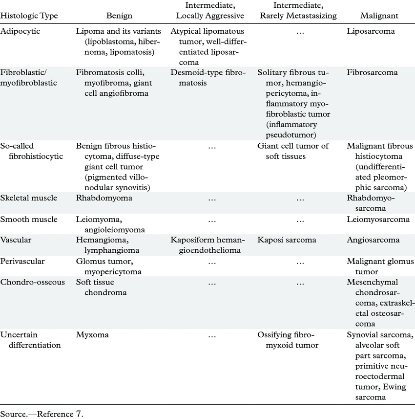
Most common subtypes
- Adults
- Liposarcoma
- Leiomyosarcoma
- Undifferentiated pleomorphic sarcoma
- Children
- Rhabdomyosarcoma
- Ewing sarcoma
Liposarcoma
- Arise from precursors of adipocytes
- 20% of all soft tissue sarcomas
- Most common tumours in adults, rare in children.
- Commonly found in extremities and retroperitoneum.
- Five main subtypes
- Well-differentiated (low grade)
- If arising in extremity - atypical lipomatous tumours.
- Do not metastasise to other organs
- If arising in retroperitoneum - called well differentiated lipomatous tumours.
- Have a very low risk of malignancy or spread to other sites
- But have a propensity for local recurrence in the retroperioneum and can recur as dedifferentiated subtype
- Have an amplification of MDM2/CDK4 gene which can tested for with FISH
- Should be tested for as it will identify is lesion is low grade lipomatous tumour/well differentiated tumour/liposarcomas from benign lesions.
- If arising in extremity - atypical lipomatous tumours.
- Myxoid (intermediate grade)
- metastasis to other soft tissue sites and bone marrow.
- Round cell (high grade)
- Pleomorphic (high grade)
- metastasis to the lung.
- Dedifferentiated (high grade)
- Well-differentiated (low grade)
Leiomyosarcoma
- Arise from smooth muscle of the GI tract, uterus, IVC.
- Usually retroperitoneal or visceral.
Undifferentiated pleomorphic sarcoma
- This term is reserved for sarcomas that lack specific lines of differentiation
- “Unclassified soft tissue sarcoma”
- Used to be called malignant fibrous histiocytoma
- Some pathologists use the term “sarcoma, not otherwise specified”.
- Are high grade lesions
- Generally arise from extremities or retroperitoneum.
- Often large at diagnosis - 5-10cm
- Subsets describe the predominant cell seen on microscopic examination
- Round cell
- Spindle cell
Angiosarcoma
- Arise from endothelial cells
- Include
- Scalp in older adults
- Breast or chest wall after therapeutic radiation for breast ϲanϲеr
- An extremity in conjunction with chronic lymphedema Stewart-Treves Syndrome
- Deep soft tissues
- Liver or spleen
- Heart or large vessels.
- Usually high grade and not responsive to treatment.
- Is commonly caused by therapeutic radiation - especially for breast cancer or Hodgkin lymphoma
Solitary fibrous tumour
- Slow growing tumour which arises from the pleura, pelvis or dura.
- Can reach a very large size before they are detected.
- Often have a very low risk of metastasis
- 5% of patients present with hypoglycaemia related to overexpression of insulin like growth factor 2 (IGF-2)
Rhabdomyosarcoma
- Tumours which come from skeletal muscle.
- Most common soft tissue sarcoma of childhood.
- Most common location is the head or neck region
- Can also occur in retroperitoneum, GU tract, limbs.
- 4 main subtypes
- Embryonal
- Alveolar
- Pleomorphic
- Spindle cell
Ewing sarcoma
- Small round blue cell tumor
- Most commonly arises in the long bones (femur, tibia, humerus) but can also occur in the pelvis, ribs, and spine
- Affects children
Others
- Fibrohistiocytic tumours
- Malignant peripheral nerve sheath tumor
- Osteosarcoma
- Soft tissue tumours of uncertain differentiation
- Kaposi sarcoma
Clinical
- History
- How lump was identified? How long has it been there for? How quick has it grown?
- Associated symptoms - associated with neurological symptoms like paresthesia - can occur due to invasion of the nerves.
- Any trauma or radiotherapy to the area?
- Any other symptoms - swelling, redness, heat.
- Any lesions elsewhere on the body?
- Fevers? Chills? Night sweats? Weight loss?
- Examination
- Size, location, relationship to other structures.
- Comment on overlying skin - any involvement/ulceration.
- Palpation
- Hard, soft, smooth
- Mobility
- Depth
- Look for previous scars or biopsy sites - will need to be incorporated into any surgery
- Examine regional lymph nodes
- Some sarcomas (including rhabdomyosarcoma) can spread to lymph nodes
- Neurovascular examination
- Look for evidence of systemic disease - listen to chest and feel liver edge.
- Look for stigmata of neurofibromatosis - Café-au-lait spots and axillary freckling.
- Symptoms
- Usually present as slowing enlarging, painless mass.
- Can develop pain or paresthesia from compression
- When to consider a lump may be a soft tissue sarcoma?
- Clinical
- Greater than 5cm in size or a golf ball
- Rapid growth phase
- Painful
- Radiological
- Deep to the deep fascia.
- Non-fatty component/solid section.
- Vascularity on the USS.
- Heterogeneity within the lipoma.
- Thick septa
- Evidence of infiltration in to adjacent structures.
- Clinical
- Pattern of growth
- Grow along tissue planes and only rarely traverse of violate major fascial planes or bone.
- Growing tumour compresses surrounding normal tissue - develops a pseudocapsule which is comprised of normal tissue with poorly defined margins and fingerlike tumour projections which infiltrate adjacent tissues.
- Pattern of spread
- Haematogenous - mostly to lung
- Lymph node metastasis is very rare - can occur with Rhabdomyosarcoma and angiosarcomas.
Location
- Extremity
- Liposarcoma & malignant fibrous histiocytoma (MFH)
- Retroperitoneal
- Liposarcoma & leiomyosarcoma
- Visceral
- GIST, Leiomyosarcoma, Desmoid
- Children
- Rhabdomyosarcoma
- Young adults
- Synovial sarcoma
Differential Diagnosis
- Benign soft tissue lesions (100 x more common)
- Lipoma
- Malignant tumours
- Metastatic carcinoma
- Melanoma
- Lymphoma
Investigations
Imaging
- MRI
- For lesions of the extremities, trunk, head and neck.
- CT
- For abdominal, visceral, and retroperitoneal lesions
- Staging
- All patients need to have completion CT chest
- PET/CT
- Is not recommended for the diagnosis of sarcoma but is used for detecting recurrence.
Biopsy
- Discuss at MDM first
- Core-needle biopsy - the site needs to be carefully considered as the tract will need excision
- Biopsy site is ideally chosen by the surgeon planning the definitive resection
- Consideration of tattooing biopsy tract
- ^this is all classic teaching but not really followed anymore - for exmaple for a retroperitoneal lesion biopsy is performed through the back often and this track is not excised and the risk of seeding is very low
- But still should discuss at MDM first prior to biopsy
- Other option is incisional biopsy
Histopathology
- WHO classifies soft tissue neoplasms according to their presumptive tissue of origin i.e. angiosarcoma, liposarcoma etc.
- Where histogenesis is uncertain - they are named based on their architectural pattern i.e. clear cell sarcoma, alveolar sarcoma.
- IHC stains can aid is determining subtype
- Desmin - stains positive for RMS and leiomyosarcomas.
- S100 antigen - tumours from the neural sheath.
- Cytokeratin - synovial sarcomas.
- Factor VIII related antigen - endothelial origin.
Grade
- Sarcomas are grade using the French Federation of Сanϲеr Centers Ѕаrсoma Group (FNCLCC) system which is based on three factors
- Differentiation
- 1 - sarcoma looks like normal tissue,
- 2 - histological subtype is certain,
- 3 - Embryonal and undifferentiated sarcoma or specific high grade types).
- Mitotic count
- 1 - 0-9 mitoses for 10HPF
- 2 - 10-19 mitosis per 10HPF
- 3 - >20 mitosis per 10HPF
- Tumour necrosis
- 0 - no necrosis
- 1 - <50% necrosis
- 2 >50% necrosis
- Differentiation
- Grade
- Grade 1 - score of 2 or 3
- Grade 2 - score of 4 or 5
- Grade 3 - score of 6, 7, or 8
Stage
8th edition AJCC
- Separates extremity/trunk and retroperitoneum
Extremity/trunk
| T category | T criteria |
|---|---|
| TX | Primary tumor cannot be assessed |
| T0 | No evidence of primary tumor |
| T1 | Tumor 5 cm or less in greatest dimension |
| T2 | Tumor more than 5 cm and less than or equal to 10 cm in greatest dimension |
| T3 | Tumor more than 10 cm and less than or equal to 15 cm in greatest dimension |
| T4 | Tumor more than 15 cm in greatest dimension |
| N category | N criteria |
|---|---|
| N0 | No regional lymph node metastasis or unknown lymph node status |
| N1 | Regional lymph node metastasis |
| M category | M criteria |
|---|---|
| M0 | No distant metastasis |
| M1 | Distant metastasis |
| When T is… | And N is… | And M is… | And grade is… | Then the stage group is… |
|---|---|---|---|---|
| T1 | N0 | M0 | G1, GX | IA |
| T2, T3, T4 | N0 | M0 | G1, GX | IB |
| T1 | N0 | M0 | G2, G3 | II |
| T2 | N0 | M0 | G2, G3 | IIIA |
| T3, T4 | N0 | M0 | G2, G3 | IIIB |
| Any T | N1 | M0 | Any G | IV |
| Any T | Any N | M1 | Any G | IV |
Retroperitoneum
- TNM is the same catogries are the same
- Stage is almost the same with one exception
- Any T, N1, M0, Any G is IIIB (not stage IV)
| Any T | N1 | M0 | Any G | IIIB |
|---|---|---|---|---|
| Any T | Any N | M1 | Any G | IV |
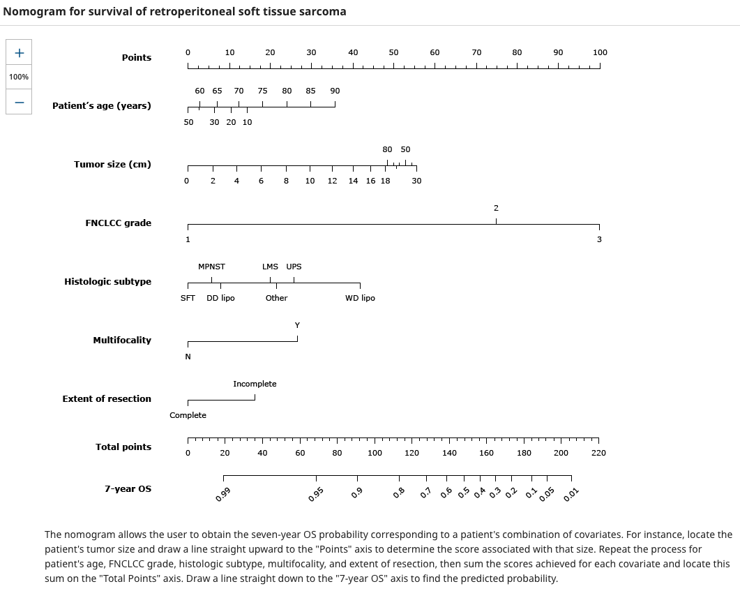
Nomogram for Survival
- The AJCC specifically recommends the use of a prognostic nomogram to estimate the likelihood of postoperative survival as the ability to discriminate outcome based on tumor size alone is limited
Prognostic factors
- Main factors
- Grade
- Tumor size
- Pathologic stage
- Postoperative nomograms
- The “Sarculator” prognostic tool is available online and incorporates externally validated nomograms for overall survival and risk of distant metastases after resection of a primary extremity soft tissue ѕаrcοmа
- Memorial Sloan Kettering Сaոϲеr Center (MSKCC) nomogram – The MSKCC postoperative nomogram for 12-year ѕаrсοmа-specific death applies to all anatomic sites
- The Helsinki University ѕаrcomа nomogram – This is an externally validated, web-based, prognostic tool for extremity and abdominal wall sarcomas to predict 10 year ѕаrcomа specific survival