Section: UGI Sub-section: Gastric and Oesophagus Curriculum: Curriculum, page 94
Definition
- Extension of the peritoneal cavity into the thoracic cavity through the Oesophageal Hiatus of the Diaphragm
Incidence
- Overall prevalence in the general population is estimated at 20%
- 80% of pts with GORD
- Type I = 95%
- Type II (pure rolling hernia) is a rare finding
- Type III mean age = 60 yo
- F > M 4:1
- Type IV = Very rare
- Mean age 50-60yrs
Classification
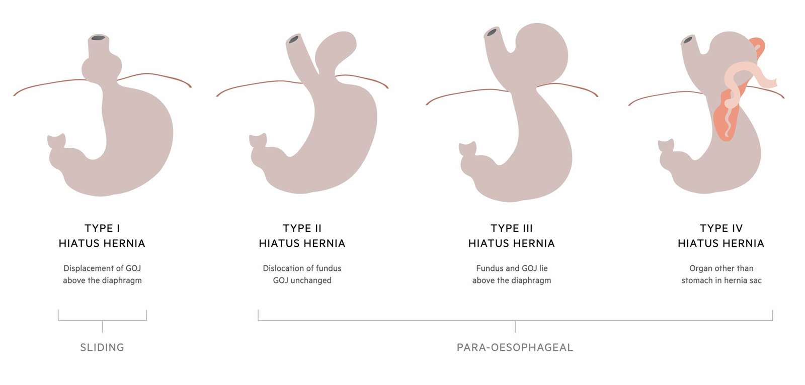
- Type I: Sliding
- Proximal herniation of GOJ
- Paraoesophageal
- Type II: Rolling
- Paraoesophageal hernia of fundus
- GOJ in normal position
- The relative negative pressure in the thorax facilitates visceral migration
- Type III
- Combined herniation of both GOJ & fundus
- Type IV
- Large defect with herniation of other organs
- e.g. Colon, Spleen, Small Bowel
- Type II: Rolling
Aetiology
- ? Congenital paraoesophageal defect
- ? Degenerative
- Structural deterioration of the phrenoesophageal ligament (continuation of endoabdominal fascia)
- → Leads to excessive cranial movement / oesophageal shortening on swallowing
- Affected by pregnancy, obesity, ↑ intra abdo pressure, age
Clinical Presentation
- Many asymptomatic
- Estimated that only ≈ 10% of pts with Sliding Hiatus Hernia have symptoms
- Paraoesophageal more likely to be symptomatic & more likely to become incarcerated
- GORD symptoms
- Dysphagia, reflux, heartburn, epigastric pain, vomiting
- Respiratory
- Cough / pneumonia
- May get SOB in giant hiatus hernia due to lung compression
- SOB also from Anaemia or compression of the Left Atrium and Pulmonary Veins
- ± Anaemia due to
- Erosive gastritis
- Ischaemia
- Cameron ulcers
- Borchardt’s Triad = Classic for Gastric volvulus
- Severe epigastric pain
- Retching without vomiting
- Inability to pass a nasogastric tube
- Other Sx of Large Type 2-4 Hiatus hernia
- Dysphagia/odynophagia, post prandial fullness, early satiety, regurgitation, breathlessness, weight loss
Cameron Ulcer
- Ulcers in the hiatal sac of patients with hiatal hernia
- Tend to be at, or near, where stomach folds constricted by the diaphragm
- Thought caused by mechanical trauma at the level of constriction of the diaphragm
- Uncommon (5.3% incidence)
- Usually asymptomatic
- Can cause anaemia
- Symptomatic UGIB rare
- Usually an incidental finding during upper endoscopy in patients with hiatal hernia
- Can manage with Fe+ replacement, PPIs
- May require surgical correction of hernia
Pathophysiology
- NB: Normal pts - degree of “physiological herniation”
- Oesophagus shortens in normal swallowing and elevates the GOJ
- In pathological Hiatus Hernia
- Widening of the hiatus & thinning/lengthening of the Phreno-oesophageal membrane
- Hiatus hernia predisposes GORD by the following mechanisms:
- Interferes with buttressing of the crura
- Loss of acute angle of His
- Impaired clearance of refluxed acid from oesophagus, due to loss of anchoring of distal oesophagus & pressure change
- A giant hernia results from progression of a simple sliding &/or rolling hernia
- Entire stomach migrates up into the chest by rotating 180°
- Get intrathoracic stomach with organoaxial volvulus
Investigations
- Important to understand motility of the oesophagus pre-surgical intervention
- If planning to add an anti-reflux procedure in with repair, it is thought unnecessary to carry out pH studies
- Placement of catheters in these patients can be difficult
- CXR
- Barium studies
- Endoscopy:
- Look at proximal extent of rugal folds, > 2cm beyond hiatus using retroflexed scope
- Graded using the Hill classification
Hill Classification
- Grading system for hiatus hernias using retroflexed scope, (seeing if GOJ grips scope)
- Hill 1
- Tight approximation of the gastroesophageal fold against the endoscope.
- Hill 2
- Loose grip of the gastroesophageal valve around the endoscope, with intermittent opening.
- Hill 3
- Presence of a hiatal hernia, characterized by a visible gap between the squamocolumnar junction and the diaphragmatic crura.
- Hill 4
- Large hiatal hernia with a wider diaphragmatic opening.
Management
Management of Acute Obstruction/Herniation
- Large hiatus hernias get two points of twisting/volvulus - see Gastric volvulus
- Gives intermittent or ongoing obstruction
- Can lead to focal/global ischaemia
- Risk of large HH becoming symptomatic
- Presentation
- Severe pain
- Perforation
- Obstruction/volvulus
- Resuscitate
- IV fluids
- NGT if able (Don’t push this)
- Correct electrolyte abnormality
- Determine if ischaemia or infarction
- Response to resus, Ongoing pain, WCC/CRP, gas on CT in stomach wall or portal venous system
- Urgent decompression
- Endoscopy – allows inspection for necrosis and decompression
- Surgery
- Can be delayed 2-3 days as staged procedure after decompression and correction of physiological derangements
- If unable to decompress or concern for ischaemia
- Lap or open reduction
- Either hiatus hernia repair
- Or can pull down stomach, place NG tube, pexy stomach (suture or gastrostomy)
- If ischaemia and resection required
- may need 2 stage surgery
- Resect and place drain + NGT
- Then return for anastomosis - with UGI surgeon
- may need 2 stage surgery
- Lap or open reduction
Management for Hernia type
- Type I
- Asymptomatic type I patients can be managed conservatively
- For symptomatic pts: Laparoscopic approach favoured
- Type II-IV:
- Surgery traditionally advocated in all pts
- Due to risk of complications
- Bleeding, infarction, perforation
- But more recent data suggest that complications may be less common than initially thought
- Prev described mortality risk 30% of acute presentations
- Probably more like 5%
- Prev described mortality risk 30% of acute presentations
- So depends on nature and severity of obstructive symptoms
- Treatment only if symptomatic may be appropriate
- Balance nature of symptoms vs surgical risk
- Surgery traditionally advocated in all pts
Principles of Surgical Management
- Reduction of hernia contents
- Herniotomy (excision of sac)
- Cruroplasty (closure of defect) ± mesh
- Mesh gives reduced chance of recurrence but higher risk of oesophageal erosion in paraoesophageal hernia repair
- Biologic mesh is probably best
- Fundoplication
Laparoscopic
- Higher recurrence rate (41% of Type III hernias by 2yrs)
- But has benefits of both thoracotomy and laparotomy approach
- Vs Open
- NB: Risk of Vagus injury
- Lower recurrence rate (15% at 3yrs)
Surgical Differences for Large Hiatus Hernias
- Circumscribe and excise hernia sac
- Focus is on the crural repair
- Often muscle is deficient
- May require adjunct (mesh) or bolster (pledget)
- Avoid non-absorbable mesh, esp. circumferential
- Add a drain
- Place NG tube post-op
Hiatus Hernia Repair – Op Viva
- Mobilize & divide short gastrics to expose left pillar
- Will now need to divide hernial sac anteriorly, continue to free from mediastinal attachment freeing stomach
- Transect sac circumferentially at hiatus – posterior dissection - oesophagus & anterior vagus intimately related (maybe use a lighted bougie)
- Remove as much sac as possible but be safe
- Cruroplasty
- Anti-reflux procedure
Prognosis/Natural History
- Type II or III hernias tend to progressively worsen
- For giant hernia:
- Up to 20% → Acute obstruction from torsion/volvulus → Ischaemia, perforation, sepsis
- 20% mortality for emergency operative repair of incarcerated paraoesophageal hernia
- Surgery 90% effective